R E S E A R C H A R T I C L E
Open Access
Insulin glycation by methylglyoxal results in
native-like aggregation and inhibition of fibril
formation
Luis MA Oliveira
1,2,3†, Ana Lages
1†, Ricardo A Gomes
1,4, Henrique Neves
1, Carlos Família
1, Ana V Coelho
4and
Alexandre Quintas
1*Abstract
Background: Insulin is a hormone that regulates blood glucose homeostasis and is a central protein in a medical condition termed insulin injection amyloidosis. It is intimately associated with glycaemia and is vulnerable to glycation by glucose and other highly reactive carbonyls like methylglyoxal, especially in diabetic conditions. Protein glycation is involved in structure and stability changes that impair protein functionality, and is associated with several human diseases, such as diabetes and neurodegenerative diseases like Alzheimer’s disease, Parkinson’s disease and Familiar Amyloidotic Polyneuropathy. In the present work, methylglyoxal was investigated for their effects on the structure, stability and fibril formation of insulin.
Results: Methylglyoxal was found to induce the formation of insulin native-like aggregates and reduce protein fibrillation by blocking the formation of the seeding nuclei. Equilibrium-unfolding experiments using chaotropic agents showed that glycated insulin has a small conformational stability and a weaker dependence on denaturant concentration (smaller m-value). Our observations suggest that methylglyoxal modification of insulin leads to a less compact and less stable structure that may be associated to an increased protein dynamics.
Conclusions: We propose that higher dynamics in glycated insulin could prevent the formation of the rigid cross-b core structure found in amyloid ficross-brils, therecross-by contricross-buting to the reduction in the across-bility to form ficross-brils and to the population of different aggregation pathways like the formation of native-like aggregates.
Background
Insulin is a small protein hormone that is crucial for the control of glucose metabolism. It regulates blood glu-cose levels by indirectly stimulating gluglu-cose transport across the cell membrane and by down regulation of enzymes involved in gluconeogenesis. External adminis-tration of insulin is critical in Diabetes type I, where autoimmune response causes a progressive and perma-nent destruction of the insulin-producing cells in the pancreas due to an interplay of environmental and genetic factors [1-3]. Insulin is composed of two poly-peptide chains, the A-chain (21 residues) and the
B-chain (30-residues) linked together by two disulfide bonds [4,5]. In the secretory vesicles of the pancreas the predominant form of insulin is a zinc-coordinated hex-amer, formed by the association of three dimers, and stabilized by two to four zinc ions. However, when released into the blood stream, insulin is present in its biologically active form,i. e. the monomer [6,7]. Mono-meric insulin is an amyloid protein forming amyloid-like fibrils in vitro, which are promoted by elevated tempera-tures, low pH, and increased ionic strength [8,9]. Insulin amyloid-like fibrils are the hallmark of a clinical condi-tion observed in insulin-dependent diabetic patients, called insulin injection amyloidosis [10]. In this patholo-gical condition, full-length insulin molecules are found in fibrillar form at the site of frequent insulin injections [9,11,12]. Additionally it was recently shown that serum samples from Parkinson’s disease patients display an autoimmune response to insulin oligomers and fibrils
* Correspondence: [email protected] † Contributed equally
1Centro de Investigação Interdisciplinar Egas Moniz, Instituto Superior das
Ciências da Saúde Egas Moniz, Campus Universitário, Monte da Caparica 2829-511 Caparica, Portugal
Full list of author information is available at the end of the article
© 2011 Oliveira et al; licensee BioMed Central Ltd. This is an Open Access article distributed under the terms of the Creative Commons Attribution License (http://creativecommons.org/licenses/by/2.0), which permits unrestricted use, distribution, and reproduction in any medium, provided the original work is properly cited.
[13], possibly indicating the presence of insulin aggre-gates in this disease as well. Insulin fibril formation has also been a limiting factor in long-term storage of insu-lin for treatment of diabetes. Thus, better understanding of insulin fibrillation mechanisms could lead to new therapeutic strategies, safer handling and more cost-effective storage of insulin. Upon fibrillation, insulin undergoes structural changes from a predominantly a-helical state to ab- sheet rich conformation. The a- to b-transition appears only to occur upon fibril assembly [14], and recently Vestergaard and co-workers proposed that insulin oligomers have an overall helical shape [15]. Being intimately related with glycaemia, it is likely that insulin may be modified by reactive a-ketoaldehydes such as 3-deoxyglucosone, glyoxal and methylglyoxal. These highly reactive compounds have been considered the most accountable for toxicity at high glucose con-centrations [16]. In fact, hyperglycemia induces the gly-cation of insulin in pancreaticb cells [17] and glycated insulin is unable to regulate glucose homeostasis in vivo and to stimulate glucose transport and adipose tissue lipogenesis [17]. Protein glycation is apost-folding mod-ification whereby amino groups in lysine and arginine side chains react irreversibly with carbonyl molecules forming advanced glycation end-products (AGE). Glyca-tion exerts profound effects on protein structure, stabi-lity and function. AGE formation in proteins is associated to the clinical complications of diabetes melli-tus [18], cataracts [19], uraemia [20], atherosclerosis [21] and age-related disorders [22]. Glycated proteins are present in b-amyloid (Ab) deposits in Alzheimer’s dis-ease [23-25], in Lewy inclusion bodies ofa-synuclein in Parkinson’s disease [26] and in transthyretin amyloid deposits in familial amyloidotic polyneuropathy (FAP) [27]. In all these amyloid pathologies, b-sheet fibril structure and the presence of AGE are common fea-tures, suggesting a possible role for glycation in amyloid formation pathogenesis. Methylglyoxal is the most sig-nificant glycation agent in vivo, being one of the most reactive dicarbonyl molecules in living cells. This com-pound is an unavoidable by-product of glycolysis, arising from the non-enzymatic b-elimination reaction of the phosphate group of dihydroxyacetone phosphate andD
-glyceraldehyde 3-phosphate [28]. Methylglyoxal irrever-sibly reacts with amino groups in lipids, nucleic acids and proteins, forming methylglyoxal-derived advanced glycation end-products (MAGE). In Ab, glycation by methylglyoxal promotes the formation ofb-sheets, oligo-mers and protofibrils and also increases the size of the aggregates [29]. Argpyrimidine is a specific methyl-glyoxal modification occurring in arginine residues, and was associated with amyloid diseases [27]. However, lit-tle is known about the effects of methylglyoxal glycation on the fibrillation of insulin. The aim of this work is to
detail the molecular mechanisms of insulin fibril forma-tion in the presence of methylglyoxal, which may be related to insulin toxicity and/or malfunction. We ana-lyzed the effects of methylglyoxal on the structure, stabi-lity and fibrillation of insulin in a concentration-dependent manner. Full glycation pattern analysis of insulin showed that a single residue modification reduces insulin fibrillation by blocking the formation of the seeding nuclei and that by contrast, methylglyoxal glycation stabilizes soluble aggregates that retain native-like structure as showed by circular dichroism experiments.
Results
Characterization of insulin glycation by methylglyoxal
Prior to mass spectrometry analysis, non-glycated and glycated insulin were probed using a specific antibody towards methylglyoxal-derived glycation adducts. As shown in Figure 1A, a dose and time dependent glyca-tion is clearly detected. To unequivocally identify gly-cated peptides and amino acid residues, non-glygly-cated and glycated insulin were digested using chymotrypsin followed by MS and MS/MS analysis. A modified gly-cated peptide should be exclusively present in the MS spectrum of glycated insulin with a mass value corre-sponding to the insulin peptide plus the specific mass increment characteristic of a MAGE modification (72 Da for the lysine specific MAGE CEL and 54, 80 and 144 Da for the arginine-specific MAGE hydroimidazo-lones, argpyrimidine and tetrahydropirimidine respec-tively). This information was used to construct an inclusion list of modified peptides to be fragmented by an additional MS/MS experiment using the MALDI-TOF/TOF instrument. The sequence information thus obtained allowed the unequivocal identification of MAGE-modified peptides and also assignment of speci-fic modified amino acid.
A comparative analysis of peptide mass spectra from the glycated and unmodified insulin reveals noticeable differences with several new peptides appearing exclu-sively in the glycated insulin (Figure 1B and Table 1). To identify MAGE-modified peptides and assign the gly-cated amino acid residues, the theoretical digestion was performed considering up to three chymotrypsin mis-scleavages (PeptideMass, Expasy, http://www.expasy.ch/ tools/peptide-mass.html) and added to the resulting peptide masses the mass increment imposed by a MAGE modification (72, 54, 80 and 144 Da). Using this approach, several peptides, appearing only in the peptide mass spectrum of glycated insulin with a specific MAGE mass increment were observed (Figure 1B). For example, the species atm/z of 991.4788 may correspond to the B-chain peptide 41-48 (LVCa|qGERGF) withm/z 937.4603 plus 54.018 Da, a mass increase characteristic of a
hydroimidazolone (MGH) modification. This strongly suggests that the arginine residue 46 is glycated by methylglyoxal with the formation of a hydroimidazolone. In agreement, the observed peptide with an m/z 934.4578 corresponds to the same peptide with a hydro-imidazolone at R46 but without cysteine alquilation (Ca|
q). To unequivocally confirm these data, MS/MS
experi-ments were performed to provide sequence information. When using the CID fragmentation technique, bond breakage mainly occurs through the lowest energy path-way, that is, the peptide bond, leading to b-ions (when the charge is retained by the amino-terminal fragment) or y-ion (when it is retained by the carboxy-terminal fragment). Thus, if an amino acid residue is modified, the particular y and complementary b ions, which encompasses the modification, will have the particular amino acid mass value plus 54.018 Da for
Figure 1 Detection and location of MAGE-modified peptides. (A) Dot-blot analysis with a specific antibody towards methylglyoxal-derived glycation adducts. A dose and time-dependent glycation is clearly detected. (B) The panels show representative sections of the MALDI-TOF/TOF spectra of peptides from unmodified and glycated insulin. New m/z peaks, absent from the control, are clearly detected in the mass spectra of the glycated insulin (highlighted in red). These new m/z values correspond to the mass of an insulin peptide plus the mass increment characteristic of a hydroimidazolone modification (54 Da). These peptides were analyzed by MS/MS, confirming the glycation of the arginine residue 46. (C) MS/MS spectrum of a glycated insulin peptide with m/z 991.4788, showing the y and b fragment ions. The detected fragment ions arise from the amino acid sequence LVCALQGERGF, with a hydroimidazolone modification on the arginine residues. All the reported glycated peptides were confirmed by MS/MS data.
Table 1 Assignment of glycated amino acid residues Observed mass (Da) Theoretical mass (Da) Peptide sequence Mass Increase (Da) MAGE Glycated residue 934.496 880.435 LVCGERGF (41-48) 54.061 MGH R46 991.521 937.456 LVC*GERGF (41-48) 54.040 MGH R46 1138.590 1084.524 LVCGERGFF (41-49) 54.066 MGH R46 1171.590 1027.503 LVCGERGFF (41-49) 144.087 THP R46 1228.616 1084.524 LVC*GERGFF (41-49) 144.092 THP R46
In all cases, the observed peptide mass has a mass increment specific of a methylglyoxal-derived AGE modification. The specific MAGE are indicated in the table and the modified amino acid residues are highlighted in the peptide sequence. (MGH - hydroimidazolone; THP - tetrahydropyrimidine)
hydroimidazolone. Taken the peptide with m/z of 991.4788 [LVCa|qGER(MGH)GF] (Figure 1C), we observed that the mass difference between y1 and y2
ions corresponds to an F residue, and the mass differ-ence between y2 and y3 ions corresponds not to the
addition of an G and R residue (57 + 156 Da) but to the addition of G and R residue plus the MGH modification on arginine (267 Da in total). The remaining mass dif-ferences between consecutive y ions also show this mass increment. The same feature is observed for the b ions. This clearly confirms that the amino acid residue R46 is modified by methylglyoxal. Only modified amino acid residues with confirmed sequence information were considered. Results are summarized in Table 1.
In the end, only the arginine residue in insulin was found to be glycated with the formation of either hydro-imidazolone or tetrahydropirimidine. Similar results were observed in a previous study that characterized methylglyoxal modification of insulin. In that study the modification of the arginine residue with a 54 Da mass increase was detected [30]. Even though authors claimed that this mass increase corresponds to a Schiff base for-mation, this mass increment is characteristic of an MGH advanced glycation end-product. The glycation reactions by methylglyoxal are very fast [31] so the for-mation of advanced glycation products are expected. In this work, we observed that the arginine residue may also be modified with the formation of a tetrahydropiri-midine (mass increment of 144 Da). This result is in agreement with our previous data showing the inherent heterogeneity of in vitro methylglyoxal glycation reac-tions [32]. In contrast, no evidences of glycation in the N-terminal and the lysine residue were observed by our mass spectrometry analysis. Although the N-terminal of insulin was found to be the major glycation target when using glucose as glycation agent [33,34], it is well known that methylglyoxal preferentially reacts and modify argi-nine residues [35].
Methylglyoxal reduces insulin fibril formation
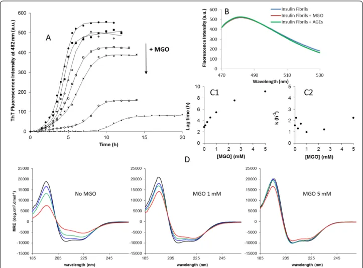
To investigate the effects of methylglyoxal on insulin fibril formation, insulin was incubated with methyl-glyoxal at different concentrations in the appropriate aggregation conditions described in the“Methods” sec-tion. The insulin fibrillation process as a function of time and methylglyoxal concentration was monitored by ThT fluorescence and circular dichroism (Figure 2). Methylglyoxal glycation of insulin resulted in a substan-tial dose-dependent decrease in ThT fluorescence inten-sity at the end of the fibrillation which is consistent with a reduced insulin fibril formation (Figure 2A). These differences were probed not to occur by ThT quenching caused by methylglyoxal or AGEs (Figure 2B). To further explore the biochemical mechanism on
the inhibition of fibril formation by methylglyoxal glyca-tion, a kinetic analysis was performed. The fibrillation kinetics represented in Figure 2A exhibit characteristic sigmoidal curves with an initial lag phase, a subsequent growth phase and a final equilibrium phase. Such curves are consistent with a nucleation-dependent polymeriza-tion model, in which the lag corresponds to the nuclea-tion phase and the exponential part to fibril growth (elongation) [36-39]. Equation 1 was fitted to the experi-mental data and yielded values for the fibrillation lag time and for the apparent first-order rate constant (kapp)
of fibrillation [40,41]. The dependence of the kinetic parameters of fibrillation on methylglyoxal concentra-tion is represented in Figure 2C1 and 2C2. Clearly, the lag time increases as a function of methylglyoxal con-centration, changing from 2.8 h in unmodified insulin to 9.1 h upon methylglyoxal glycation. By contrast, no sig-nificant changes in the apparent rate constant of fibrilla-tion were observed. These results show a longer nucleation phase which indicates that methylglyoxal gly-cation blocks the formation of the seeding nuclei, with-out changing the fibril elongation rate.
To detect changes in protein conformation during the fibrillation process, insulin fibril formation was moni-tored by circular dichroism (Figure 2D). Insulin pre-sented a mainly a-helical secondary structure with spectral local minima at 222 and 208 nm and a positive band below 200 nm, which are characteristics of a-heli-cal conformations (Figure 2 - time 0 h). CD spectra col-lected at several time points along the fibrillation pathway, showed that fibril formation is accompanied by a conformational transition, suggesting loss of a-helix and gain of b-sheet. This shift was most extensive when methylglyoxal was absent and decreases with methyl-glyoxal in a concentration-dependent manner. These results show that glycation preserves insulin native con-formation, blocking the a-helix to b-sheet transition characteristic of amyloid fibril formation. This is in agreement with the reduction of fibril formation observed in ThT kinetic measurements and suggests that there is a structural inertia to conformational changes in glycated insulin that is responsible for block-ing the seedblock-ing nuclei formation, leading to a reduced fibril formation.
Methylglyoxal induces protein oligomerization
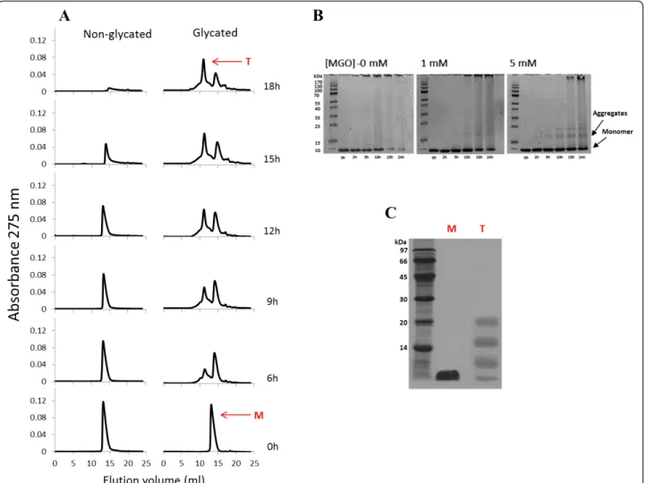
To investigate the early steps of protein aggregation, samples were collected at indicated incubation times and analyzed by size exclusion chromatography and PAGE (Figure 3). Non-glycated insulin appears as a sin-gle molecular species (elution volume of 14.04 ml) cor-responding to the insulin monomer mass. No hexameric insulin species were detected confirming that the insulin sample preparation produced monomeric solution. The
same feature was observed for glycated insulin at time 0 (elution volume of 13.68 ml), as it can be observed either by SEC or gel electrophoresis. The difference in the elution volumes is explained by an increased hydro-dynamic radius of glycated insulin, which may be caused by a less compact structure formed upon glycation. Dur-ing incubation time, the unmodified insulin monomer changes into amyloid fibrils. This can be observed from the native-PAGE (Figure 3B) where a reduction of insu-lin monomer (only species present at time 0 h) conco-mitant with the appearance of high molecular mass fibrils, unable to enter the separation gel, is clearly
detected. Likewise, the insulin amyloid fibrils are unable to pass through the SEC column’s filter and enter the stationary phase and thus a reduction of the SEC insulin monomer peak intensity with time is observed (Figure 3A). Interestingly, intermediate oligomeric species are apparently absent or in undetectable concentration. This may be due to the nature of soluble oligomers: they are intermediates of the aggregation process, and are there-fore an extremely transient and labile species [42]. As soon as their concentration reaches a few percent, the oligomers are rapidly converted into amyloid fibrils with an organized b-structure. A very different scenario
Figure 2 Effect of methylglyoxal concentration on the kinetics of fibril formation of human insulin. (A) Kinetics of fibrillation at different MGO concentrations monitored by ThT fluorescence. The symbols represent the average of ThT fluorescence intensities determined in three experiments, and the lines represent the best fit using the equation 1. Methylglyoxal concentrations used were 0 (•), 0.1 (■), 0.25 (◇), 0.5 (□), 1 (×), 2.5 (○) and 5 (+) mM. The decreasing in fluorescence intensities of the curves plateau are correlated with increasing methylglyoxal
concentrations. (B) Evaluation of ThT quenching by methylglyoxal and AGEs. Non-glycated insulin fibrils were probed by ThT fluorescence after 8 h incubation (blue). Subsequently insulin fibrils were mixed with methylglyoxal (red) and glycated insulin containing AGEs (green) and probed again by ThT fluorescence. Fluorescence spectra show no quenching of ThT fluorescence induced by either methylglyoxal (red) or AGEs (green). (C) Dependence of the kinetic parameters lag time (C1) and apparent rate constant (C2) as a function of methylglyoxal concentration. Lag time is
taken as x0-2τ and the k is given by 1/τ. (D) a- to b- transition of insulin at the indicated methylglyoxal concentrations during the fibrillation
process followed by circular dichroism. CD spectra were collected at time 0 h (black), 3 h (blue), 5 h (green) and 7 h (red) incubation. Measurements were all performed at 37°C with agitation of the reaction mixture.
emerged when methylglyoxal is added. In this case, SEC peak intensity also becomes reduced, but other species are clearly detected on the chromatogram, correspond-ing to insulin soluble aggregates (Figure 3A). These aggregates are also observed in gel electrophoresis and show apparent molecular masses consistent with tri-meric and tetratri-meric forms of insulin (Figure 3B). Moreover, high molecular mass species are only detected in the later incubation times compared to the control (without methylglyoxal). Taken together, these results show that methylglyoxal-induced glycation reduces insulin fibril formation and promotes the popu-lation of oligomeric states.
Protein glycation has been referred to induce protein aggregation due to cross-link formation [43,44]. How-ever, when using methylglyoxal, only the lysine-lysine dimer MOLD is formed [45], which is a minor advanced
glycation end-product compared to other AGE [46]. The fact that only a single arginine residue is glycated and that significant amounts of glycated insulin are in aggre-gated forms suggest that major non-covalent interac-tions are likely to be involved. The nature of the interactions in glycated insulin aggregates was evaluated by PAGE. The denaturing conditions of the SDS-PAGE induced significant dissociation of the glycated insulin tetramer (Figure 3C) showing that mainly non-covalent interactions are present in the insulin aggregates.
Methylglyoxal effects on insulin structure and stability
Our final set of experiments was aimed to investigate the structural changes imposed by methylglyoxal-derived glycation that might be associated to fibril inhibition and stabilization of oligomeric species. In these
Figure 3 Effects of methylglyoxal on the early steps of insulin aggregation. Insulin (3 mg.ml-1) was incubated in the absence of
methylglyoxal and in the presence of 5 mM of the glycation agent with stirring. Samples were collected at specific incubation times and immediately analysed by size exclusion chromatography (A) and PAGE (B). Sample buffer in PAGE did not contain SDS andb-mercaptoethanol in order to preserve the insulin oligomerization. To investigate the nature of insulin aggregates, the monomeric form of glycated insulin collected at incubation time 0 h (M) and the tetrameric form of glycated insulin collected at time 18 h (T) were analysed by a standard SDS-PAGE (C).
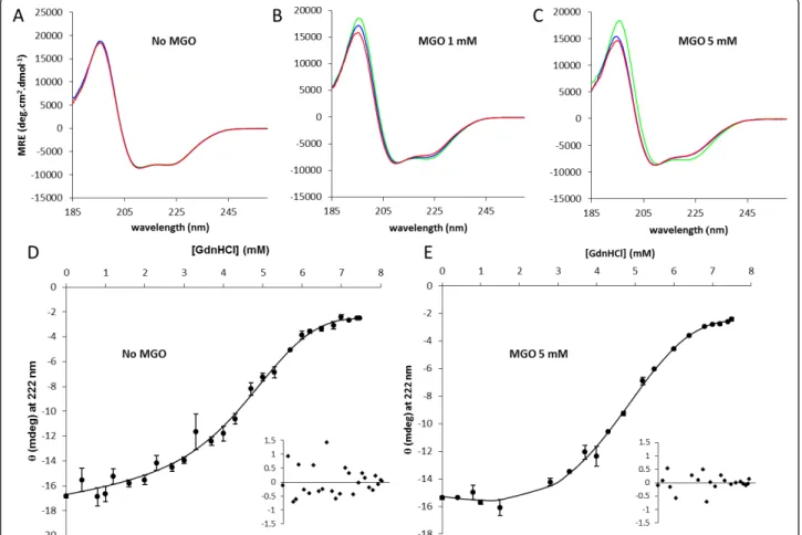
experiments insulin was incubated without agitation, a condition that does not promote aggregation, as observed by SEC experiments (see Additional File 1: Fig-ure S1). In these conditions, insulin is glycated but remains almost entirely in monomeric form. In contrast with the results obtained when insulin was incubated in aggregation conditions, the CD spectra of non-glycated insulin remains unchanged during the incubation period (Figure 4A), while glycated insulin undergoes slight spectral changes (Figure 4B and 4C). Spectra deconvolu-tion shows a redistribudeconvolu-tion of secondary structure ele-ments in glycated insulin with a respective increase in b-sheet content, an increase in unordered structure and a reduction in the relativea-helical content (Table 2). We then assess the conformational stability of glycated and native insulin (Figure 4D and 4E). GdnHCl-induced denaturation was found to be reversible, as judged by
CD experiments after dialysis of GdnHCl-denatured insulin (data not shown). Fits were made using the lin-ear extrapolation method [47] in a non-linlin-ear least squares fitting procedure and yielded values for ΔGo (H2O), the conformational stability, and m, the
depen-dence of ΔGoon denaturant concentration. Table 3 shows the values obtained from the curves in Figure 4D and 4E for ΔGo(H2O), m, and Cm, the denaturant con-centration at the midpoint of the unfolding transition. Glycated insulin has a smaller conformational stability with ΔGo(H2O) of 2.66 ± 0,27 kcal.mol-1against 3,34 ± 0,33 kcal.mol-1for unmodified insulin. This decrease in conformational stability is also supported by the smaller Cm value of glycated insulin. In addition, glycation resulted in a weaker GdnHCl-dependence of unfolding (smallerm-value). The m-value has been correlated with the difference between accessible surface areas in the
Figure 4 Effects of methylglyoxal on insulin structure and stability. Insulin (3 mg/ml) was incubated with 1 and 5 mM of methylglyoxal at 37°C without stirring for 48 h and compared with non-glycated insulin. Insulin secondary structure was monitored far-UV CD. Circular dichroism spectra were recorded as a function of time at different methylglyoxal concentrations (A - 0 mM; B - 1 mM; C - 5 mM). Spectra were collected at time zero (blue) and after 24 h (red) and 48 h (green) incubation. Deconvolution of the CD spectra are present in Table 2. Protein
conformational stability was evaluated for native insulin (D) and glycated insulin (E) by guanidinium hydrochloride equilibrium denaturation curves at pH 7.4 and 37°C monitored by circular dichroism at 222 nm. The curves are non-linear least squares fits to a two-state unfolding model equation [71,72] representing the entire denaturation curve and using a linear extrapolation method to the experimental circular dichroism data [47]. The insets are the residues plot.
unfolded and folded states:m ∝ ΔA, where ΔA = AU
-AN[48]. This weak dependence may reflect a less
com-pact folded structure or a more comcom-pact unfolded state. Putting these results together with the SEC experiments where glycated insulin has a small elution volume then the native insulin, suggest that the presence of a less compact structure is a more likely scenario, which may be the basis of a higher susceptibility to different unfold-ing and aggregation pathways.
Discussion
Insulin is a protein hormone that regulates glucose con-centration in blood. It is intimately related with glycae-mia and is vulnerable to glycation by glucose and other highly reactive carbonyls like methylglyoxal. Addition-ally, it has the ability to aggregate and form amyloid-like fibrils that are characteristic of a clinical condition called insulin injection amyloidosis [10]. In this work we have investigated the effects of methylglyoxal-modification of insulin on structural and fibril-forming properties. Mass spectrometry data showed that methylglyoxal specifically modifies a single arginine residue in the B-chain. This is in agreement with a previous study that observed a methylglyoxal-derived modification on the arginine resi-due of the B chain [30]. The glycation of insulin in our experimental conditions promoted the coexistence on insulin molecules with the arginine residue modified to a hydroimidazolone and to a tetrahydropirimidine
modification. This heterogeneity in in vitro glycation was already observed [32]. No modification on the lysine residues and N-terminal were detected by our experi-mental approach. Insulin glycation byD-glucose also led
to the coexistence of protein molecules glycated at dif-ferent residues [34]. In opposition to our results, the N-terminus of both chains and the lysine residue 29 were modified upon glucose glycation. This difference is not surprising since it is well documented that methyl-glyoxal preferentially reacts and modifies arginine resi-dues [35].
Previous reports showed that AGE modifications accelerated the fibrillation of several proteins and pep-tides including b-amyloid peptide, tau and albumin [49,50]. Additionally, AGE-modified proteins were detected in amyloid deposits from several amyloidosis such as Alzheimer’s [24,51], Parkinson’s [26,52] disease and FAP [27]. In contrast with those amyloidogenic pro-teins, modification ofb-2-microglobulin and a-synuclein by different glycation agents resulted in inhibitory effects on the formation and extension of fibrils [53,54]. Our data also showed that insulin fibril formation is substan-tially reduced upon methylglyoxal modification. The observed differences might be a consequence of the inherent properties of the native structure of each pro-tein, or differential structural changes induced by AGE modifications as result of different glycation agents. In most of the cases mentioned above, fibrillation enhance-ment is achieved by modifying amyloidogenic proteins with glycating sugars like glucose or fructose while small and highly reactive carbonyls like methylglyoxal are apparently more prone to reduce fibril formation. A good example comes froma-synuclein where glyoxal and methylglyoxal inhibit fibril formation [54] whileD
-ribose glycation does not [55]. This suggests that differ-ent glycation agdiffer-ents lead to specific structural con-straints that have a major role in protein fibrillation kinetics.
Table 2 Distribution of the structural element fractions for native and glycated insulin along time obtained by deconvolution of CD spectra using CDSSTR algorithm available on Dichroweb (Dichroweb; http://www.cryst.bbk.ac.uk/ cdweb/html/home.html) [69,70]
[MGO] (mM) Time (h) a-Helix b-Sheet b-Turns Unordered structure NRMSD
0 0 31 23 22 24 0.028 24 33 23 21 23 0.033 48 32 22 22 24 0.029 1 0 31 24 21 24 0.027 24 28 26 22 26 0.032 48 24 27 22 27 0.036 5 0 32 22 22 24 0.022 24 23 27 21 27 0.029 48 23 28 21 27 0.035
The NRMSD parameter represents the normalized root mean square deviance.
Table 3 Thermodynamic parameters from GdnHCl unfolding studies of native and glycated insulin
ΔGo(H 2O)
(kcal·mol-1) (kcal·molm-1.M-1) Cm(M)
Insulin 3.34 ± 0.33 0.63 ± 0.10 5.31 ± 0.98 Glycated Insulin 2.66 ± 0.27 0.52 ± 0.09 5.10 ± 0.98
Parameters were obtained by a direct fit of the model equations to experimental data in Figure 4 D and E.ΔGo(H2O) is the protein conformational stability;m is the dependence of ΔGo
on denaturant concentration;Cm is the denaturant concentration at the midpoint of the unfolding transition.
Insulin offers a structural simplicity of two short poly-peptide chains constrained by one intramolecular and two intermolecular disulphide bonds and has well-known molecular mechanisms of fibril formation [8,56]. The insulin B-chain segment with the sequence LVEA-LYL is the smallest segment in the basis of fibril assem-bly, being crucial to the cross-b spine of the insulin fibril [56]. In full-length insulin molecules, there must be conformational changes for the LVEALYL side chains of the segment to be exposed and to interact with each other [56]. However, insulin glycation leads to native-like aggregation, as showed by CD experiments. This suggests that glycation impairs insulin conformational alterations, causing the inhibitory effects observed in the fibrillation process. Moreover our kinetic analysis of insulin aggregation showed an increase in fibrillation lag time. The lag time can be used to monitor the nuclea-tion phase prior to the exponential stage of fibril elonga-tion. Increasing lag time indicates that methylglyoxal glycation inhibits the fibrillation process by blocking the formation of the seedingnuclei. Accordingly fibril for-mation is reduced due to lack of a critical concentration of seeds.
Despite the inhibition of fibril formation, size exclu-sion chromatography experiments showed that glycation induces insulin aggregation. However these aggregates are small, soluble, non-fibrillar and native-like in struc-ture, and apparently are not a consequence of a covalent crosslinking of insulin monomers. This implies that aggregation of modified insulin is not a merely result of a chemical reaction, but an outcome of complex folding interactions that are established and populates an off-pathway to fibril formation. A subject of intense investi-gation is whether the amyloid fibril deposits or the pre-fibrillar aggregates, called protofibrils, are the most potent mediators of cell damage, cytotoxicity and neuro-toxicity. The finding that the severity of cognitive impairment in protein misfolding diseases correlates with the levels of small oligomeric species and not with the large fibrillar species has led researchers to the con-clusion that the soluble small aggregates are the primary cause of the pathological symptoms [57-60]. Moreover, accumulation of AGE-modified proteins has been related to cellular responses including oxidative stress and the release of pro-inflammatory cytokines mediated by AGE:RAGE interaction [61,62]. Therefore it will be interesting to evaluate the cytotoxicity of the insulin gly-cated aggregates.
In order to understand what structural restrictions could cause this behavior, we investigated the effects of methylglyoxal glycation on the structure and stability of insulin. Circular dichroism experiments showed that modified insulin has a small conformational stability and a slight increase in b-sheet content when compared to
the unmodified protein. This lower conformational sta-bility is accompanied by a weaker dependence ofΔGoon denaturant concentration which is related to a less com-pact native structure or a more comcom-pact unfolded state [48]. Size exclusion chromatograms of glycated insulin showed a slight decrease in retention time of the insulin monomer, supporting the idea of a less compact native structure. Although most of the proteins have well-defined structures, they are not static molecules. Pro-teins are dynamic entities and possess an inherent flex-ibility. Having a lower contribution of van der Waals interactions, it is likely to expect that a less compact structure may result in a more dynamic one. The term dynamics is used for intrinsic protein molecular motions, while the term flexibility is used for the ability of a protein to adapt its structure to external stimuli. Accordingly, proteins are flexible as a consequence of their dynamics, yet their dynamics do not automatically result in flexibility. We propose that higher dynamics in glycated insulin could lead to impairment of the forma-tion of the rigid cross-b core structure found in amyloid fibrils, resulting in a higher susceptibility to different unfolding and aggregation pathways. In this case other aggregation pathways that preserve native-like structure and comparable dynamics, like the small and soluble aggregates of glycated insulin observed in size exclusion chromatography, could be more likely populated.
Conclusions
Insulin is a nearly all-alpha protein playing a central role in blood glucose homeostasis and is associated with a medical condition termed insulin injection amyloidosis, characterized by the formation and deposition of amy-loid fibrils from insulin. Due to its main physiological role, insulin is a target for glycation by methylglyoxal. Protein glycation mostly impairs protein functionality by changing protein structure and stability, and AGE-modi-fied proteins have been related to cellular responses including oxidative stress and the release of pro-inflam-matory cytokines. Glycation has been associated with human conformational diseases, such as Alzheimer’s dis-ease, Parkinson’s disease and Familiar Amyloidotic Poly-neuropathy, which are associated to the formation of amyloid fibrils. Our results show that glycation of insu-lin by methylglyoxal reduce insuinsu-lin fibril formation and leads to the formation of insulin native-like aggregates. In addition they suggest that modification of insulin leads to a less compact and less stable structure that may be associated to an increased dynamics, preventing the formation of the rigid cross-b core structure found in amyloid fibrils. Overall the present study points that methylglyoxal adducts can trigger a drifting from an amyloid aggregation to a native-like aggregation path-way, a mechanism that might be important in the
context of the amyloidogenicity of AGE-modified pro-teins involved in conformational diseases.
Methods
Insulin preparation and glycation
Insulin exists in solution as an equilibrium mixture of monomers, dimers, tetramers and hexamers, and possi-bly higher associated states, depending on concentration, pH, metal ions, ionic strength and solvent composition [63]. A solution containing only insulin in the mono-meric form was prepared taking into account the fluc-tuation of its association states in different milieu conditions as described [64]. Briefly, human zinc-free insulin (Sigma) was dissolved in ultra-pure miliQ water to a final concentration of 6 mg.ml-1and acidified with H3PO4to a pH of 5 in order to obtain monomeric
insu-lin. Insulin at pH 5 was then incubated for 15 min at room temperature and protein concentration was deter-mined by absorbance at 275 nm (ε275= 4560 M-1cm-1)
in a UV-Visible spectrophotometer Jasco V-530. Finally, insulin was neutralized to pH 7 with NaOH 0.1 M and diluted to a final concentration of 3 mg.ml-1. Insulin preparation was proven to be in the monomeric form after pH neutralization as evaluated from size exclusion chromatography and native-PAGE experiments as described below. Also circular dichroism experiments showed that no structural changes or unfolding occurred with pH variations. In all assay, monomeric insulin was prepared in exactly the same way.
For the methylglyoxal-derived glycation of insulin, the protein preparation (3 mg.ml-1) was incubated with methylglyoxal (at several concentrations ranging from 0.1 to 5 mM) (a kind gift from Dr. Carlos Cordeiro, Centro de Química e Bioquímica, FCUL, Lisbon, Portu-gal) in 50 mM potassium phosphate buffer, pH 7.4, sup-plemented with 150 mM of NaF, at 37°C in sterile conditions. Samples were collected at different incuba-tion times for analysis with the maximum incubaincuba-tion time of 48 hours. Control samples were treated in the same way but without methylglyoxal addition. To evalu-ate the effects of methylglyoxal on insulin stability and secondary structure changes, samples were incubated without stirring, a condition that avoid fibril formation, producing only glycated insulin in the monomeric state. In contrast, for the oligomerization and fibrillation kinetic studies, samples were incubated with vigorous agitation. Aliquots were collected in sterile conditions at defined incubation times from 0 to 4 hours and immedi-ately analyzed.
Characterization of insulin glycation by methylglyoxal using mass spectrometry and dot-blot analysis
Dot-blot assay was performed using a specific monoclo-nal antibody towards methylglyoxal-derived glycation (a
kind gift from Dr. Ram Nagaraj, Case Western Univer-sity, Cleveland, OH, USA), using a 1:2000 dilution. Washes, secondary antibody and detection procedures were performed using the BM Chemiluminescence Wes-tern Blotting Kit (Pierce) following the manufacturer’s instructions.
To characterize the protein modification and assign the amino acid residues modified by methylglyoxal, a chymotrypsin digestion of insulin was performed. Pro-tein samples were reduced with 10 mM dithiothreitol in 100 mM NH4HCO3buffer (pH 8.0) at 55°C for 1 h and
alkylated with 55 mM of iodoacetamide in 100 mM NH4HCO3 buffer (pH 8.0) in the dark for 30 min. In
solution digestion were performed with chymotrypsin (Promega) using 50:1 ratio of protein:protease in 100 mM Tris-HCl buffer (pH 7.8) containing 10 mM CaCl2
for 16 h. Protein digestion was stopped by the addition of formic acid [(final concentration of 1% (v/v)]. The obtained peptide mixture was purified and concentrated by solid-phase extraction using home-made R2 Pore microcolumns (Applied Biosystems) as previously described [65]. Peptide mixture were eluted directly onto the MALDI target plate with 0.5 μl of a-CHCA matrix (5 mg.ml-1) prepared in 50% (v/v) acetonitrile with 0.1% (v/v) formic acid. The mixture was allowed to air dry (dried droplet method). Sample peptides were analysed in a MALDI-TOF-TOF mass spectrometer 4800 plus (Applied Biosystems) in positive reflectron mode for peptide mass determination. The mass spec-trometer was externally calibrated using 4700 Calibra-tion Mix (Applied Biosystems). Mass spectra were collected in a result-independent acquisition mode, typi-cally using 1000 laser shots per spectrum and a fixed laser intensity of 3500 V. The peptides of interest (i.e., having a mass consistent with the mass increment of the modifications by methylglyoxal) were selected for MS/MS experiments using Collision Induced Dissocia-tion (CID), with 1 kV collision energy and an air pres-sure of 106 torr. Two thousand laser shots were collected for each MS/MS spectrum using a fixed laser intensity of 4500 V. Raw data were generated by the 4000 Series Explorer Software v3.0 RC1 (Applied Biosys-tems). The identification of MAGE-modified peptide and amino acid residues was further validated using Peaks Studio 4.5 software (Bioinformatic Solutions Inc.), combined with manual inspection of the assigned sequence.
Analysis of insulin-fibril formation and fibrillation kinetics
To investigate the effects of MGO in insulin fibril for-mation, solutions of monomeric insulin (prepared as described above) were incubated with stirring at 37°C in the presence of methylglyoxal at 0, 0.1, 0.25, 0.5, 1.0, 2.5 and 5.0 mM. Fibril formation was monitored with
thioflavin T (ThT) binding assay as previously described [65,66]. Briefly, aliquots of 5 μl were removed and added to 0.5 ml of 10μM ThT in 50 mM sodium phos-phate buffer (pH 7.4) at room temperature and immedi-ately analyzed. Fluorescence measurements were performed using a Perkin Elmer LS50B spectrofluori-meter, in quartz cuvettes with 1 cm excitation light path. ThT fluorescence was recorded immediately after ThT binding from 470 to 530 nm with excitation at 450 nm, an increment of 0.5 nm, an integration time of 1 s and 5 nm slits for both excitation and emission. For each sample, the signal was obtained as the ThT inten-sity at 482 nm from which was subtracted a blank mea-surement recorded prior to addition of insulin to the ThT solution. To test if methylglyoxal alone or the derived insulin AGEs interfere with ThT fluorescence of insulin fibrils, non-glycated insulin fibrils were produced in vigorous agitation by incubating monomeric insulin preparation (3 mg.ml-1) in the absence of methylglyoxal for 8 h. ThT fluorescence was then determined for insu-lin fibrils alone, in the presence of methylglyoxal (5 mM), and also in the presence of methylglyoxal-glycated insulin (3 mg.ml-1) prepared with vigorous agitation as described above.
ThT fluorescence measurements were plotted as a function of time and equation 1 was fitted to the experi-mental data [40,41]. Y = (yi+ mix) + (yf + mfx) 1 + e− x−x0 τ (1)
where Y is the fluorescence intensity and x0is the time
to 50% of maximal fluorescence. The initial base line dur-ing the lag phase is described by yi+ mix. The final base
line after the growth phase had ended is described by yf+
mfx. The apparent first-order rate constant (kapp) for the
growth of fibrils is calculated as 1/τ, and the lag time is calculated as x0-2τ. This expression is unrelated to the
underlying molecular events, but provides a convenient method for comparison of the fibrillation kinetics.
Size-exclusion and PAGE experiments
Aggregation of human insulin upon methylglyoxal glyca-tion was monitored by size exclusion chromatography (SEC) and Native-PAGE. Solutions of monomeric insu-lin were incubated and stirred at 37°C in the presence of methylglyoxal at 0, 1 and 5 mM. Samples were ana-lyzed by SEC at defined incubation times, after filtration with a 0.2μm Whatman filter. SEC was performed with HPLC Jasco PU-2080 Plus isocratic pump with an UV detector JASCO 2075. The mobile phase was 50 mM sodium phosphate buffer pH 7.4 with 150 mM NaF. Separation was achieved on a molecular exclusion analy-tical column (Amersham-Pharmacia Superdex™ 75 10/
300 GL) at a flow rate of 0.4 ml/min. Eluting peaks were monitored at 275 nm. Insulin samples were also separated by Native and SDS-PAGE on a Bio-Rad Mini-Protean 3 system, using a 12% separation gel and a 4% stacking gel. On Native-PAGE all buffers were prepared without SDS addition. Proteins were stained with Comassie Brilliant Blue [67].
Circular dichroism and conformational stability measurements
Secondary structure analysis was performed by far-UV (185-260 nm) CD in a Jasco J810 spectropolarimeter equipped with a temperature control unit Julabo F25 using an insulin concentration of 3 mg.ml-1. Far UV CD spectra were recorded with 0.01 cm (linear) path length quartz cuvette at 37°C in 50 mM sodium phosphate buf-fer pH 7.4 with 150 mM NaF. For each spectrum, three scans were averaged and protein concentration was determined by absorbance at 275 nm using the above mentioned insulin extinction coefficient in a UV-Visible spectrophotometer Jasco V-530. For protein secondary structure estimation, CD spectra were deconvoluted using the CDSSTR [68] deconvolution algorithm on Dichroweb [69,70]. CD spectra of the appropriate buffers were recorded and subtracted from the protein spectra.
CD denaturation curves for non-glycated and glycated insulin monomer were constructed using the ellipticity at 222 nm, monitored at 37°C after 24 h incubation with guanidinium hydrochloride (GdnHCl) at various concentrations. The denaturation of glycated and non-glycated insulin could be described as sigmoidal curves and were analyzed according to a two-state unfolding model M ↔ U using the linear extrapolation method [47] in a non-linear least squares fitting procedure and yielded values forΔGo(H2O), the conformational
stabi-lity, and m, the dependence ofΔGoon denaturant con-centration. Cm, the denaturant concentration at the midpoint of the unfolding transition was calculated as Cm= Go(H2O)/m. Denaturation curves for monomeric
species were analyzed considering the equation devel-oped by Santoro & Bolen [71,72].
Additional material
Additional file 1: Figure S1. Evaluation of insulin aggregation in non-stirring conditions. Insulin incubation in 50 mM potassium phosphate buffer, pH 7.4 supplemented with 150 mM of NaF, at 37°C in sterile conditions without stirring. Gel filtration experiments show that insulin does not aggregate in this incubation conditions, remaining in the monomeric form.
Abbreviations
α-CHCA: α-cyano-4-hydroxicinamic acid; Aβ: β-amyloid peptide; AGE: Advanced glycation end-products; CD: Circular dichroism; CID: Collision
induced dissociation; FAP: Familial amyloidotic polyneuropathy; GdnHCl: Guanidinium hydrochloride; MALDI: Matrix-assisted laser-desorption ionization; MAGE: Methylglyoxal-derived advanced glycation end-products; MGH: Hydroimidazolone; MGO: Methylglyoxal; MOLD: Methylglyoxal lysine dimer; RAGE: Receptor for advanced glycation end-products; SEC: Size exclusion chromatography; THP: tetrahydropyrimidine; ThT: Thioflavin T; TOF: Time of flight.
Acknowledgements
We thank Dr. Carlos Cordeiro for the gift of the methylglyoxal and Dr. Ram Nagaraj for the gift of the anti-MAGE antibody. We wish to acknowledge Dr. Carlos Cordeiro, Professor Ana Ponces Freire and Dr. Tiago F. Outeiro for helpful discussions and all the assistance provided. This work was supported by grants (PTDC/QUI/73430/2006), SFRH/BD/23604/2005 (L.M.A.O) and SFRH/ BPD/41037/2007 (R.A.G.) from the Fundação para a Ciência e a Tecnologia, Ministério da Ciência e Tecnologia, Portugal.
Author details
1Centro de Investigação Interdisciplinar Egas Moniz, Instituto Superior das
Ciências da Saúde Egas Moniz, Campus Universitário, Monte da Caparica 2829-511 Caparica, Portugal.2Centro de Química e Bioquímica,
Departamento de Química e Bioquímica, Faculdade de Ciências da Universidade de Lisboa, Edifício C8, 1749-016 Lisboa, Portugal.
3Departamento de Análises Clínicas e Saúde Pública, Escola Superior de
Saúde Dr. Lopes Dias, Instituto Politécnico de Castelo Branco, Campus da Talagueira 6000-767 Castelo Branco, Portugal.4Instituto de Tecnologia
Química e Biológica. Universidade Nova de Lisboa, 2780-901 Oeiras, Portugal.
Authors’ contributions
LMAO conceived the part of the study related with the fibrillation kinetics of insulin, carried out part of the experimental procedures and was mainly responsible for the experimental setup and data analysis as well as the initial drafting of the manuscript. AL is responsible for executing most part of the experiments. RAG conceived, executed and interpreted the experiments related with mass spectrometry and participated in the writing of the manuscript. HN executed the experiments associated to the thioflavin T curves. CF participated in the experiments to prove the nature of insulin aggregates. AVC contributed to this work with her expertise concerning mass spectrometry. AQ designed and coordinated the study and was mainly involved in the preparation and reviewing of the manuscript. All authors read and approved the final manuscript.
Received: 14 April 2011 Accepted: 5 August 2011 Published: 5 August 2011
References
1. Shepherd PR, Kahn BB: Glucose transporters and insulin action– implications for insulin resistance and diabetes mellitus. N Engl J Med 1999, 341(4):248-257.
2. Taylor R: Insulin action 1991. Clin Endocrinol (Oxf) 1991, 34(2):159-171. 3. Zierath JR, Krook A, Wallberg-Henriksson H: Insulin action and insulin resistance in human skeletal muscle. Diabetologia 2000, 43(7):821-835. 4. Baker EN, Blundell TL, Cutfield JF, Cutfield SM, Dodson EJ, Dodson GG,
Hodgkin DM, Hubbard RE, Isaacs NW, Reynolds CD, Sakabe K, Sakabe N, Vijayan NM: The structure of 2Zn pig insulin crystals at 1.5 A resolution. Philos Trans R Soc Lond B Biol Sci 1988, 319(1195):369-456.
5. Blundell TL, Cutfield JF, Cutfield SM, Dodson EJ, Dodson GG, Hodgkin DC, Mercola DA: Three-dimensional atomic structure of insulin and its relationship to activity. Diabetes 1972, 21(2 Suppl):492-505. 6. Nystrom FH, Quon MJ: Insulin signalling: metabolic pathways and
mechanisms for specificity. Cell Signal 1999, 11(8):563-574. 7. Ottensmeyer FP, Beniac DR, Luo RZ, Yip CC: Mechanism of
transmembrane signaling: insulin binding and the insulin receptor. Biochemistry 2000, 39(40):12103-12112.
8. Ahmad A, Uversky VN, Hong D, Fink AL: Early events in the fibrillation of monomeric insulin. J Biol Chem 2005, 280(52):42669-42675.
9. Brange J, Andersen L, Laursen ED, Meyn G, Rasmussen E: Toward understanding insulin fibrillation. J Pharm Sci 1997, 86(5):517-525. 10. Westermark P, Benson MD, Buxbaum JN, Cohen AS, Frangione B, Ikeda S,
Masters CL, Merlini G, Saraiva MJ, Sipe JD: Amyloid: toward terminology
clarification. Report from the Nomenclature Committee of the International Society of Amyloidosis. Amyloid 2005, 12(1):1-4. 11. Dische FE, Wernstedt C, Westermark GT, Westermark P, Pepys MB,
Rennie JA, Gilbey SG, Watkins PJ: Insulin as an amyloid-fibril protein at sites of repeated insulin injections in a diabetic patient. Diabetologia 1988, 31(3):158-161.
12. Storkel S, Schneider HM, Muntefering H, Kashiwagi S: Iatrogenic, insulin-dependent, local amyloidosis. Lab Invest 1983, 48(1):108-111. 13. Wilhelm KR, Yanamandra K, Gruden MA, Zamotin V, Malisauskas M,
Casaite V, Darinskas A, Forsgren L, Morozova-Roche LA: Immune reactivity towards insulin, its amyloid and protein S100B in blood sera of Parkinson’s disease patients. Eur J Neurol 2007, 14(3):327-334.
14. Jimenez JL, Nettleton EJ, Bouchard M, Robinson CV, Dobson CM, Saibil HR: The protofilament structure of insulin amyloid fibrils. Proc Natl Acad Sci USA 2002, 99(14):9196-9201.
15. Vestergaard B, Groenning M, Roessle M, Kastrup JS, van de Weert M, Flink JM, Frokjaer S, Gajhede M, Svergun DI: A helical structural nucleus is the primary elongating unit of insulin amyloid fibrils. PLoS Biol 2007, 5(5): e134.
16. Brownlee M: Biochemistry and molecular cell biology of diabetic complications. Nature 2001, 414(6865):813-820.
17. Abdel-Wahab YH, O’Harte FP, Ratcliff H, McClenaghan NH, Barnett CR, Flatt PR: Glycation of insulin in the islets of Langerhans of normal and diabetic animals. Diabetes 1996, 45(11):1489-1496.
18. Brownlee M: Advanced protein glycosylation in diabetes and aging. Annu Rev Med 1995, 46:223-234.
19. Lyons TJ, Silvestri G, Dunn JA, Dyer DG, Baynes JW: Role of glycation in modification of lens crystallins in diabetic and nondiabetic senile cataracts. Diabetes 1991, 40(8):1010-1015.
20. Miyata T, Ueda Y, Saito A, Kurokawa K:’Carbonyl stress’ and dialysis-related amyloidosis. Nephrol Dial Transplant 2000, 15(Suppl 1):25-28. 21. Kume S, Takeya M, Mori T, Araki N, Suzuki H, Horiuchi S, Kodama T,
Miyauchi Y, Takahashi K: Immunohistochemical and ultrastructural detection of advanced glycation end products in atherosclerotic lesions of human aorta with a novel specific monoclonal antibody. Am J Pathol 1995, 147(3):654-667.
22. Bucala R, Cerami A: Advanced glycosylation: chemistry, biology, and implications for diabetes and aging. Adv Pharmacol 1992, 23:1-34. 23. Vitek MP, Bhattacharya K, Glendening JM, Stopa E, Vlassara H, Bucala R,
Manogue K, Cerami A: Advanced glycation end products contribute to amyloidosis in Alzheimer disease. Proc Natl Acad Sci USA 1994, 91(11):4766-4770.
24. Yan SD, Chen X, Schmidt AM, Brett J, Godman G, Zou YS, Scott CW, Caputo C, Frappier T, Smith MA, Perry G, Yen SH, Stern D: Glycated tau protein in Alzheimer’s disease: a mechanism for induction of oxidant stress. Proc Natl Acad Sci USA 1994, 91(16):7787-7791.
25. Chen F, Wollmer MA, Hoerndli F, Munch G, Kuhla B, Rogaev EI, Tsolaki M, Papassotiropoulos A, Gotz J: Role for glyoxalase I in Alzheimer’s disease. Proc Natl Acad Sci USA 2004, 101(20):7687-7692.
26. Castellani R, Smith MA, Richey PL, Perry G: Glycoxidation and oxidative stress in Parkinson disease and diffuse Lewy body disease. Brain Res 1996, 737(1-2):195-200.
27. Gomes R, Sousa Silva M, Quintas A, Cordeiro C, Freire A, Pereira P, Martins A, Monteiro E, Barroso E, Ponces Freire A: Argpyrimidine, a methylglyoxal-derived advanced glycation end-product in familial amyloidotic polyneuropathy. Biochem J 2005, 385(Pt 2):339-345. 28. Richard JP: Mechanism for the formation of methylglyoxal from
triosephosphates. Biochem Soc Trans 1993, 21(2):549-553.
29. Chen K, Maley J, Yu PH: Potential inplications of endogenous aldehydes in beta-amyloid misfolding, oligomerization and fibrillogenesis. J Neurochem 2006, 99(5):1413-1424.
30. Jia X, Olson DJ, Ross AR, Wu L: Structural and functional changes in human insulin induced by methylglyoxal. FASEB J 2006, 20(9):1555-1557. 31. Gomes RA, Sousa Silva M, Vicente Miranda H, Ferreira AE, Cordeiro CA,
Freire AP: Protein glycation in Saccharomyces cerevisiae. Argpyrimidine formation and methylglyoxal catabolism. FEBS J 2005, 272(17):4521-4531. 32. Gomes RA, Oliveira LM, Silva M, Ascenso C, Quintas A, Costa G, Coelho AV, Sousa Silva M, Ferreira AE, Ponces Freire A, Cordeiro C: Protein glycation in vivo: functional and structural effects on yeast enolase. Biochem J 2008, 416(3):317-326.
33. O’Harte FP, Hojrup P, Barnett CR, Flatt PR: Identification of the site of glycation of human insulin. Peptides 1996, 17(8):1323-1330. 34. Guedes S, Vitorino R, Domingues MR, Amado F, Domingues P: Mass
spectrometry characterization of the glycation sites of bovine insulin by tandem mass spectrometry. J Am Soc Mass Spectrom 2009,
20(7):1319-1326.
35. Lo TW, Westwood ME, McLellan AC, Selwood T, Thornalley PJ: Binding and modification of proteins by methylglyoxal under physiological conditions. A kinetic and mechanistic study with N alpha-acetylarginine, N alpha-acetylcysteine, and N alpha-acetyllysine, and bovine serum albumin. J Biol Chem 1994, 269(51):32299-32305.
36. Jarrett JT, Lansbury PT Jr: Amyloid fibril formation requires a chemically discriminating nucleation event: studies of an amyloidogenic sequence from the bacterial protein OsmB. Biochemistry 1992, 31(49):12345-12352. 37. Jarrett JT, Lansbury PT Jr: Seeding“one-dimensional crystallization” of
amyloid: a pathogenic mechanism in Alzheimer’s disease and scrapie? Cell 1993, 73(6):1055-1058.
38. Lomakin A, Teplow DB, Kirschner DA, Benedek GB: Kinetic theory of fibrillogenesis of amyloid beta-protein. Proc Natl Acad Sci USA 1997, 94(15):7942-7947.
39. Wood SJ, Wypych J, Steavenson S, Louis JC, Citron M, Biere AL: alpha-synuclein fibrillogenesis is nucleation-dependent. Implications for the pathogenesis of Parkinson’s disease. J Biol Chem 1999,
274(28):19509-19512.
40. Nielsen L, Frokjaer S, Brange J, Uversky VN, Fink AL: Probing the mechanism of insulin fibril formation with insulin mutants. Biochemistry 2001, 40(28):8397-8409.
41. Munishkina LA, Ahmad A, Fink AL, Uversky VN: Guiding protein aggregation with macromolecular crowding. Biochemistry 2008, 47(34):8993-9006.
42. Lashuel HA, Lansbury PT Jr: Are amyloid diseases caused by protein aggregates that mimic bacterial pore-forming toxins? Q Rev Biophys 2006, 39(2):167-201.
43. Chellan P, Nagaraj RH: Protein crosslinking by the Maillard reaction: dicarbonyl-derived imidazolium crosslinks in aging and diabetes. Arch Biochem Biophys 1999, 368(1):98-104.
44. Verzijl N, DeGroot J, Ben ZC, Brau-Benjamin O, Maroudas A, Bank RA, Mizrahi J, Schalkwijk CG, Thorpe SR, Baynes JW, Bijlsma JW, Lafeber FP, Tekoppel JM: Crosslinking by advanced glycation end products increases the stiffness of the collagen network in human articular cartilage: a possible mechanism through which age is a risk factor for osteoarthritis. Arthritis Rheum 2002, 46(1):114-123.
45. Nagaraj RH, Shipanova IN, Faust FM: Protein cross-linking by the Maillard reaction. Isolation, characterization, and in vivo detection of a lysine-lysine cross-link derived from methylglyoxal. J Biol Chem 1996, 271(32):19338-19345.
46. Ahmed N, Thornalley PJ: Peptide mapping of human serum albumin modified minimally by methylglyoxal in vitro and in vivo. Ann N Y Acad Sci 2005, 1043:260-266.
47. Pace CN: Determination and analysis of urea and guanidine hydrochloride denaturation curves. Methods in enzymology 1986, 131:266-280.
48. Myers JK, Pace CN, Scholtz JM: Denaturant m values and heat capacity changes: relation to changes in accessible surface areas of protein unfolding. Protein Sci 1995, 4(10):2138-2148.
49. Ledesma MD, Bonay P, Colaco C, Avila J: Analysis of microtubule-associated protein tau glycation in paired helical filaments. J Biol Chem 1994, 269(34):21614-21619.
50. Loske C, Gerdemann A, Schepl W, Wycislo M, Schinzel R, Palm D, Riederer P, Munch G: Transition metal-mediated glycoxidation accelerates cross-linking of beta-amyloid peptide. Eur J Biochem 2000,
267(13):4171-4178.
51. Smith MA, Taneda S, Richey PL, Miyata S, Yan SD, Stern D, Sayre LM, Monnier VM, Perry G: Advanced Maillard reaction end products are associated with Alzheimer disease pathology. Proc Natl Acad Sci USA 1994, 91(12):5710-5714.
52. Munch G, Luth HJ, Wong A, Arendt T, Hirsch E, Ravid R, Riederer P: Crosslinking of alpha-synuclein by advanced glycation endproducts–an early pathophysiological step in Lewy body formation? J Chem Neuroanat 2000, 20(3-4):253-257.
53. Hashimoto N, Naiki H, Gejyo F: Modification of beta 2-microglobulin with D-glucose or 3-deoxyglucosone inhibits A beta 2M amyloid fibril extension in vitro. Amyloid 1999, 6(4):256-264.
54. Lee D, Park CW, Paik SR, Choi KY: The modification of alpha-synuclein by dicarbonyl compounds inhibits its fibril-forming process. Biochim Biophys Acta 2009, 1794(3):421-430.
55. Chen L, Wei Y, Wang X, He R: Ribosylation rapidly induces alpha-synuclein to form highly cytotoxic molten globules of advanced glycation end products. PLoS One 2010, 5(2):e9052..
56. Ivanova MI, Sievers SA, Sawaya MR, Wall JS, Eisenberg D: Molecular basis for insulin fibril assembly. Proc Natl Acad Sci USA 2009,
106(45):18990-18995.
57. Caughey B, Lansbury PT: Protofibrils, pores, fibrils, and
neurodegeneration: separating the responsible protein aggregates from the innocent bystanders. Annu Rev Neurosci 2003, 26:267-298.
58. Crowther DC, Kinghorn KJ, Miranda E, Page R, Curry JA, Duthie FA, Gubb DC, Lomas DA: Intraneuronal Abeta, non-amyloid aggregates and neurodegeneration in a Drosophila model of Alzheimer’s disease. Neuroscience 2005, 132(1):123-135.
59. Danzer KM, Haasen D, Karow AR, Moussaud S, Habeck M, Giese A, Kretzschmar H, Hengerer B, Kostka M: Different species of alpha-synuclein oligomers induce calcium influx and seeding. J Neurosci 2007, 27(34):9220-9232.
60. Lauren J, Gimbel DA, Nygaard HB, Gilbert JW, Strittmatter SM: Cellular prion protein mediates impairment of synaptic plasticity by amyloid-beta oligomers. Nature 2009, 457(7233):1128-1132.
61. Bierhaus A, Stern DM, Nawroth PP: RAGE in inflammation: a new therapeutic target? Curr Opin Investig Drugs 2006, 7(11):985-991. 62. Maczurek A, Shanmugam K, Munch G: Inflammation and the
redox-sensitive AGE-RAGE pathway as a therapeutic target in Alzheimer’s disease. Ann N Y Acad Sci 2008, 1126:147-151.
63. Brange J: Galenics of insulin. The physicochemical and pharmaceutical aspects of insulin and insulin preparations. Berlin: Springer-Verlag; 1987. 64. Groenning M, Frokjaer S, Vestergaard B: Formation mechanism of insulin
fibrils and structural aspects of the insulin fibrillation process. Curr Protein Pept Sci 2009, 10(5):509-528.
65. Naiki H, Higuchi K, Hosokawa M, Takeda T: Fluorometric determination of amyloid fibrils in vitro using the fluorescent dye, thioflavin T1. Analytical biochemistry 1989, 177(2):244-249.
66. Naiki H, Higuchi K, Matsushima K, Shimada A, Chen WH, Hosokawa M, Takeda T: Fluorometric examination of tissue amyloid fibrils in murine senile amyloidosis: use of the fluorescent indicator, thioflavine T. Lab Invest 1990, 62(6):768-773.
67. Wilson CM: Studies and critique of Amido Black 10B, Coomassie Blue R, and Fast Green FCF as stains for proteins after polyacrylamide gel electrophoresis. Analytical biochemistry 1979, 96(2):263-278.
68. Johnson WC: Analyzing protein circular dichroism spectra for accurate secondary structures. Proteins 1999, 35(3):307-312.
69. Lobley A, Whitmore L, Wallace BA: DICHROWEB: an interactive website for the analysis of protein secondary structure from circular dichroism spectra. Bioinformatics (Oxford, England) 2002, 18(1):211-212. 70. Whitmore L, Wallace BA: DICHROWEB, an online server for protein
secondary structure analyses from circular dichroism spectroscopic data. Nucleic acids research 2004, , 32 Web Server: W668-673.
71. Bolen DW, Santoro MM: Unfolding free energy changes determined by the linear extrapolation method. 2. Incorporation of delta G degrees N-U values in a thermodynamic cycle. Biochemistry 1988, 27(21):8069-8074. 72. Santoro MM, Bolen DW: Unfolding free energy changes determined by
the linear extrapolation method. 1. Unfolding of phenylmethanesulfonyl alpha-chymotrypsin using different denaturants. Biochemistry 1988, 27(21):8063-8068.
doi:10.1186/1471-2091-12-41
Cite this article as: Oliveira et al.: Insulin glycation by methylglyoxal results in native-like aggregation and inhibition of fibril formation. BMC Biochemistry 2011 12:41.