UNIVERSIDADE DE LISBOA
FACULDADE DE CIÊNCIAS
DEPARTAMENTO DE BIOLOGIA VEGETAL
Studying mechanisms of antifungal resistance in Candida
gla-brata clinical isolates: emphasis on the role of the CgPdr1
tran-scription factor
Danielle de Sotti Novais
Mestrado em Microbiologia Aplicada
Dissertação orientada por:
Prof. Doutor Nuno P. Mira
Prof. Doutora Margarida Barata
I
Acknowledgements
Firstly, I would like to express my gratitude to Professor Nuno Mira for having accepted to be my supervisor at the Institute for Bioengineering and Biosciences (iBB), Instituto Superior Técnico (IST), University of Lisbon, where my work was conducted. In particular, I would like to thank him for his guidance, encouragement, suggestions and mainly for his knowledge and scientific enthusiasm that were of the most valuable for my scientific formation. I would also like to thank Professor Margarida Barata for always being available to help any time it was needed.
Furthermore, I would like to acknowledge Professor Isabel Sá-Correia, for allowing me to do my pro-ject in Biological Sciences Research Group and for providing the necessary conditions to realize this thesis. My acknowledgements are also going to Professor Fernanda Carvalho from the Centro de Química Estrutural, Instituto Superior Técnico, for kindly providing me the compounds used in this thesis. I also would like to acknowledge the funding received by iBB-Institute for Bioengineering and Biosciences from FCT-Portuguese Foundation for Science and Technology (UID/BIO/04565/2013), from Programa Operacional Regional de Lisboa 2020 (Project N. 007317).
A special thanks goes to Sara Salazar for her patience in guiding me in the lab, even when she some-times had to interrupt her on work in order to help me with mine. It would have been truly difficult to advance with my work without her help so I really appreciate all the time she took to support me. I also want to express my gratitude to Pedro Pais, Ana Vila-Santa and Mafalda Cavalheiro for being ready to listen me and for helping me during my journey in the lab. In conclusion, I am most grateful for all my colleagues of the BSRG group for the given support and especially for all the laughs (and sense of humor) that motivated me to continue my project.
Moreover, I want to say thank to my friends outside the lab, specially, Ruth, Iara, Íris, Lígia and Filipa who helped me to keep my mental health during this year and for being always ready to listen to me even in more difficult times. Also, I want to say thank to my colleagues from work.
At last, I want to express my sincere gratitude to my mom and Fernando for all the support during my master degree. I know how difficult it was sometimes, particularly for my mom. I will never be able to thank her enough for the effort she made so that I could finish my studies.
II
Studying mechanisms of antifungal resistance in Candida glabrata
clinical isolates: emphasis on the role of the CgPdr1 transcription
factor
Danielle de Sotti Novais
2017This thesis was fully performed at Biological Sciences Research Group (BSRG),
Institute for Biotechnology and Bioengineering (IBB), Instituto Superior
Téc-nico, University of Lisbon under the direct supervision of Prof. Dr. Nuno P.
Mi-ra. Prof.ª DMi-ra. Margarida Barata was the internal designated supervisor in the
scope of the Master in Applied Microbiology of the Faculty of Sciences of the
University of Lisbon.
III
Resumo
Nos últimos anos tem-se verificado um aumento significativo na incidência de infeções causadas por espécies do género Candida, entre outras razões, devido ao aumento significativo da população imu-nocomprometida 1 2 3. C. albicans é a principal espécie causadora de infeções fúngicas, que variam
desde infeções superficiais nas mucosas até infeções sistémicas, potencialmente letais. No entanto, nos últimos tempos tem-se verificado um aumento significativo nas infeções originadas por outras espé-cies do género Candida, geralmente conhecidas por NCAC (Non-Candida albicans Candida speespé-cies)
1. Entre estas, C. glabrata tem emergido de forma particular sendo a segunda principal espécie
causa-dora de infeções fúngicas invasivas 8 9, com uma taxa de letalidade a rondar os 40 % 10 11. A
emergên-cia de estirpes NCAC (e, particularmente, de C. glabrata) resistentes a azóis, os principais antifúngi-cos usados na prática clínica 1, tem contribuído de forma significativa para a mudança da etiologia
observada nas infecções causadas por Candida. Os azóis têm como alvo a enzima citocromo P45014DM
envolvida na biossíntese de ergosterol 22 23. Em C. glabrata esta enzima é codificada pelo gene ERG11
e a inibição desse gene pelos azóis sabe-se levar à diminuição da síntese do ergosterol o que resulta na concomitante acumulação de esteróis metilados na posição 14α, altamente tóxicos para célula por le-varem a alterações significativas na integridade da membrana plasmática 22 23 25. Um dos principais
mecanismos de resistência aos azóis observado em estirpes clínicas de C. glabrata é a sobre-expressão de genes que codificam transportadores de múltiplas drogas pertencentes à Superfamília ABC
(ATP-binding cassette) como sejam Cdr1 ou Cdr2 29. Um dos mecanismos moleculares subjacentes à
sobre-expressão dessas bombas de efluxo em isolados clínicos resistentes está relacionada com aparecimento de mutações ganho de função no gene que codifica o factor de transcrição Pdr1 68. Vários estudos têm
reportado diferentes mutações de ganho de função no gene CgPDR1 47 68 70 73 80, mutações estas que
conduzem a uma ativação constante do regulador mesmo na ausência de qualquer droga. Estas muta-ções localizam-se em vários domínios da proteína incluindo o domínio de regulação (e.g. R376W e L328F) ou o domínio de ativação (e.g. D1082Q e E1083Q). A identificação destas mutações de ganho de função reveste-se de especial importância para tornar mais rápida a identificação de pacientes que possam estar colonizados com estirpes que serão intrinsecamente resistentes a azóis e que, portanto, vão necessitar de ser tratados com outro tipo de antifúngicos aos quais o regulador CgPdr1 não res-ponde. Neste trabalho foram comparadas as sequências codificantes do gene CgPDR1 de um conjunto de isolados clínicos de C. glabrata resistentes a fluconazol e voriconazol, com a sequência da estirpe de referência CBS138 que se sabe ser sensível a estes mesmos azóis. Na sequência dessa análise foram identificadas duas mutações, I392M e R35K, como possíveis candidatas a corresponderem a novas mutações de ganho de função de CgPDR1. Tal hipótese é suportada pelo fenótipo de resistência dos isolados clínicos e também pelo facto de se observar uma sobre-expressão dos genes CgCDR1 e
CgPUP1 nesses mesmos isolados, por comparação com os níveis de expressão observados na estirpe
CBS138. A substituição de aminoácido I392M está localizada no domínio regulatório inibitório (loca-lizado entre os resíduos 322-465), pelo que é possível que a alteração da isoleucina para a metionina resulte, de uma alguma forma, numa redução da inibição deste domínio permitindo assim aumentar a atividade de CgPdr1. Este domínio regulatório verificou-se também conter o domínio de ligação a xenobióticos, uma região responsável pela ativação de CgPdr1 por ligação a drogas, incluindo azois 77.
Assim, outra hipótese para uma eventual híper-ativação de CgPDR1 no mutante I392M pode resultar por uma alteração nesse domínio de ligação a drogas da proteína, fazendo um bypass à necessidade de haver ligação da droga à proteína para que ocorra ativação da mesma. A variante R35K está localizada no domínio de ligação ao DNA do CgPDR1 (localizado entre os resíduos 26 e 59) e está responsável pela ligação do regulador ao motivo de DNA PDRE (Pleiotropic drug resistance element) encontrada na região promotora dos genes alvos 75. Até ao momento não foram descritas variantes de CgPdr1
hiper-ativas com alterações no DBD de CgPDR1, pelo que serão necessários mais estudos para avaliar de que forma a mutação R35K contribui para uma híper-activação de CgPdr1.
IV
A constante emergência de estirpes resistentes a azóis torna essencial o desenvolvimento de novas moléculas com potencial antifúngico mas que tenham alvos de acção diferentes dos azóis, para ser assim possível sensitizar estirpes resistentes. Nesse sentido, neste trabalho foi aprofundado o estudo de um conjunto de complexos organometálicos derivados de Ag e de cânfora, recentemente demonstra-dos como tendo potencial antimicrobiano 87, como agentes anti-Candida e, em particular, como
agen-tes capazes de sensitizar isolados de C. glabrata resistenagen-tes a azois. Os compostos foram obtidos usando uma combinação de nitrato de prata e derivados de cânfora, que são dois tipos de compostos com propriedades farmacêuticas reconhecidas 89 90. Os 3 compostos Ag(I)-camphorimine usados neste
estudo apresentaram uma elevada atividade contra a estirpe de referência C. glabrata CBS138 (MIC na ordem de 15,63 mg/L para os compostos B e C, e 31,25 mg/L para o composto A), sendo esta ativi-dade particularmente notória em condições de pH neutro. Os valores de MIC obtidos para os isolados clínicos resistentes a fluconazol e voriconazol foram numa gama semelhante aos dos obtidos para a estirpe de referência (na gama entre os 15,63 mg/L e os 62,5 mg/L) indicando assim uma elevada ca-pacidade dos compostos Ag(I)-camphorimine em sensitizar as estirpes resistentes. Foi também obser-vado que a presença dos compostos Ag(I)-camphorimine aumenta o efeito inibitório do fluconazol, incluindo nas estirpes resistentes, indicando assim um efeito sinérgico significativo. Estes dois resulta-dos e também o facto de CgPDR1 e o seu gene alvo CgCDR1 se terem verificado ser dispensáveis para a tolerância de C. glabrata contra os compostos Ag(I)-camphorimine, indica que o mecanismo de ação destes compostos e dos azóis são diferentes. Por fim, foi observado que o efeito sinérgico obser-vado não resulta de um aumento da concentração intracelular de fluconazol nas células potenciado pela presença dos compostos Ag(I)-camphorimine.
Até ao momento, o efeito inibitório de Ag(I) ou de químicos derivados de Ag(I) tem sido pouco estu-dado em Candida glabrata e a maior parte desses estudos tem aborestu-dado somente o uso de nanopartí-culas. Os estudos que mostram a atividade desses compostos indicam que existe uma inibição signifi-cativa das células sendo os principais motivos, a inibição de formação de hifas, alteração da superfície celular levando a formação de poros na célula e também a alteração da parede celular dessas estirpes 87
104 105. Por este motivo no futuro, a fim de promover a implementação dos compostos
Ag(I)-camphorimine aqui estudados, como um possível tratamento da candidíase, estudos adicionais serão necessários de modo a elucidar quais os mecanismos de ação desses compostos em células de C.
gla-brata. Também é requerido obter informação quanto toxicidade desses químicos contra células
mamí-feras, tanto a nível local e sistémica.
Concluindo, considerando que o atraso no diagnóstico de estirpes resistentes aos antifúngicos e poste-rior ajuste dos tratamentos utilizados é um dos fatores com maior impacto na determinação da taxa de mortalidade de pacientes infetados com candidíase, o conhecimento obtido neste trabalho constitui uma contribuição significativa para melhorar o sucesso da atualidade estratégias utilizadas no trata-mento da candidíase.
Palavras-Chaves: CgPdr1, Resistência aos azóis, Candida Glabrata, Complexos
V
Abstract
Candida glabrata is emerging as a human pathogen, being the second major cause of invasive fungal
infections. Part of the success of C. glabrata as a pathogen rely on its rapid acquisition of resistance to azoles, the front line therapy used in prophylactic and active treatment of candidiasis. Acquisition of azole resistance in C. glabrata results, among other mechanisms, from alterations in the coding se-quence of the transcriptional regulator CgPdr1 that result in gain-of-function (GOF) variants of this protein that are constitutively active, even in the absence of azoles. In the present work the sequence of CgPDR1 alleles encoded by eight C. glabrata isolates found to be resistant to fluconazole and voriconazole was determined and compared with the sequence of the reference strain CBS138, suscep-tible to azoles. Two mutations, I392M and R35K, were identified and hypothesized to represent new gain-of-function CgPDR1 variants. CgCDR1 and CgPUP1, two known CgPdr1-targets, were found to be over-expressed in isolates FFUL412, FFUL443 and FFUL866 during growth in drug-free medium, reinforcing the idea that the identified mutations lead to constitutive activation of CgPDR1.
The efficacy of a set of Ag(I)-camphorimine complexes against C. glabrata, including against azole resistant clinical isolates (including those harboring CgPDR1 GOF variants), was also examined in this work. The results obtained showed a potent effect of Ag(I)-camphorimine complexes in inhibiting growth of C. glabrata, more marked at a near neutral pH. Remarkable, the level of inhibition of the azole resistant isolates was similar to the one registered for azole susceptible strains suggesting that the mechanism of action of azoles and of Ag(I)-camphorimine differs. A synergistic effect between Ag(I)-camphorimine complexes and fluconazole in inhibiting growth of C. glabrata, including of the azole-resistant isolates was also demonstrated, a strategy that could be used to improve the efficacy of azole-based therapies in the treatment of candidiasis.
Keywords: CgPdr1, CgPDR1 GOF variants, azole resistance, Candida glabrata,
VI
Table of contents
Acknowledgements ... I Resumo ... III Abstract ... V Table of contents ... VI List of figures ... VIII List of tables ... XIII Abbreviations ... XIV1. Introduction ... 1
1.1. Overview ... 1
1.2. A general overview on the molecular mechanisms of azole functioning in Candida cells and underlying resistance mechanisms ... 2
1.2.1. The CgPdr1-dependent regulatory network and its role in azole-resistance in C. glabrata ... 5
1.3. Introduction to the theme of thesis ... 8
2. Materials and Methods ... 10
2.1. Strains and growth media ... 10
2.2. Preparation of antifungal drugs and of camphorimine-derived chemicals ... 10
2.3. Genomic DNA extraction ... 11
2.4. PCR reaction and DNA sequencing by Sanger method of CgPDR1 gene ... 11
2.5. Cloning of CgPDR1 gene ... 12
2.6. Assessment of gene expression based on real time RT-PCR ... 14
2.7. Quantification of MIC50 using the microdilution method ... 15
2.8. Growth curves in the presence of camphorimine complexes ... 16
2.9. Synergy assays between the camphorimine compound and fluconazole ... 16
2.10. [1-3H]- Fluconazole accumulation assay ... 16
3. Results ... 17
3.1. Unveiling new gain-of-function mutations in CgPDR1 ... 17
3.1.1. Cloning of CgPDR1 gene of the reference strain and of the resistant clinical isolates FFUL412, FFUL443 and FFUL866 ... 19
3.2. Study on the antimicrobial effect against C. glabrata of Ag(I) camphorimine complexes ... 21
3.2.1. Assessment of the activity of compound A, B and C against C. glabrata azole-resistant clinical isolates ... 23
VII 3.2.3. The synergistic effect between Ag(I)-camphorimine complexes and fluconazole does not result from increased accumulation of the azole inside C.
glabrata cells ... 29
4. Discussion and Future Perspectives ... 30
References ... 33 Annex A ... 40 Annex B ... 41 Annex C ... 42 Annex D ... 43 Annex E ... 44 Annex F ... 45 Annex G ... 46 Annex H ... 47 Annex I ... 48 Annex J ... 49 Annex K ... 50 Annex L ... 51 Annex M ... 53 Annex N ... 54 Annex O ... 55
VIII
List of figures
Figure 1.1: Targets of antifungal drugs not present in mammalian cells. A. Azoles inhibits fungal
cytochrome P45014DM (14α-demethylase) which is encoded by ERG11 and catalyzes a late step in the
biosynthesis of ergosterol blocking the production of ergosterol and causing the accumulation of a toxic sterol intermediate 18. B. Polyenes intercalates into ergosterol-containing fungal membranes,
thereby forming membrane-spanning channels that lead to the leakage of cellular components and cell death. D. Echinocandins Interferes with fungal cell-wall biosynthesis by inhibiting β-(1,3)-d-glucan synthase 19. The image was altered from Kathiravan et al. 2012 20.
Figure 1.2: Chemical structural of fluconazole, voriconazole and miconazole. Image was modified
from Mast et al. 2013 26 and Snell et al. 2012 27.
Figure 1.3: Ergosterol biosynthesis pathway. The enzyme Erg11 targeted by azoles is highlighted in
the red box. ERG1, ERG4 and ERG6 genes sites are shown. Deletions of these genes increased suscep-tibility to azoles in C. glabrata showing that function of these enzymes in the ergosterol biosynthetic pathway is required for maximal tolerance of this yeast species to azoles 32 34.Image was modified
from Akins, Robert A., 2005 33.
Figure 1.4: Domain arrangement of the ABC transporters and a schematic representation of the predicted topology of the ABC transporter Cdr1p. Image was modified from Cannon D., Richard
et al., 2009 48.
Figure 1.5: Domain arrangement of MFS transporters and a schematic representation of the predicted topology of the MFS transporter Mdr1p. Image was modified from Cannon D., Richard
et al., 2009 48.
Figure 1.6: Example of a functional model of a new compound (iKIX1) that show to sensitize azole-resistant strains by hampering the Pdr1-Med interaction. The iKIX1 molecule block the
azole-induce recruitment of Gal11/Med15-Mediator to Pdr1 target genes upon azole treatment. This blocking prevent the upregulation of Pdr1 target genes, including those which encode drug efflux pumps which result to a restoration of the sensitivity to the azole antifungal. Image was retrieved from Nishikawa, J et al., 2016 78.
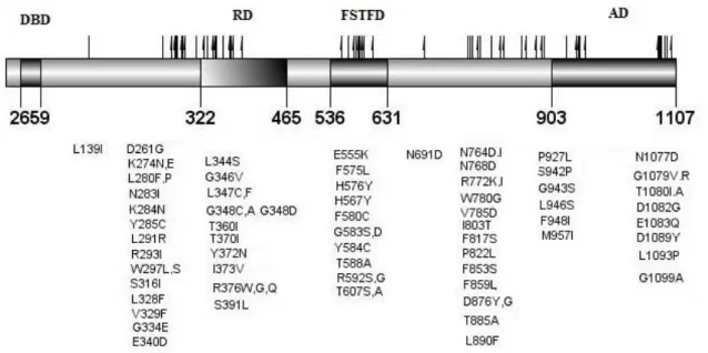
Figure 1.7: Described C. glabrata PDR1 gain-of-function mutations. The domains shown were
based on the homology between S. cerevisiae Pdr1p and CgPdr1p retrieved from Tsai, H., et al., 2010
70. DBD – DNA-binding domain; RD – Regulatory Domain; FSTFD – Fungus-specific Transcription
factor Domain; AD – Activation Domain. Mutations described according from the information availa-ble in Tsai H., et al.,2006,68 Torelli, B., et al.,2008,47 Ferrari, S., et al., 2009,80 Tsai, H., et al., 2010,70
Berila, N. and Subik, J., 2010,81 Caudle K., et al.,2011, 73 Vale-Silva, L., et al.,2015,82 Garnaud, C., et
al,2015,83 Katiyar, S., et al.,2016,84 Healey, K., et al.,201655.
Figure 1.8: Measured MIC values for the antifungals fluconazole, voriconazole of the cohort clinical isolates used in the previous work (Salazar, S., 2015) 86 and the reference strains CBS138
(White). Resistant isolates are market in black, while isolates classified as being susceptible or
inter-mediately resistant are marked in grey.
Figure 2.1: Schematic representation of the procedure used to prepare the 96-multiwell plates used to determine MIC of 3 different compounds for the different C. glabrata isolates. B: blank;
com-IX
pounds the same methodology was applied with the difference that the initial stock concentration was of 1000 mg/L.
Figure 3.1: Example of the result obtained after amplification of CgPDR1 gene using primer FW1 and REV1. Lane 1 represents the ladder (1 kb plus DNA Ladder) - The black arrows show the 3
kb fragment and the 4 kb fragment.
Figure 3.2: Comparison of the transcript levels of CgCDR1 and CgPUP1 genes in CBS138, FFUL29, FFUL412, FFUL443, FFUL830, FFUL866 and FFU878. Cells of the different isolates
were cultivated in RPMI growth medium until mid-exponential phase after which the expression of
CgCDR1 and CgPUP1 genes was compared by qRT-PCR. The values represented for the isolates
strains are relative to the value obtained for the CBS138 strain, which was considered to be equal to 1. Gene expression was calculated using RDN25 as an internal control. For the statistical analysis the results obtained for the resistant isolates strains were compared with those gathered for CBS138. * p-value below 0.05, ** p-p-value below 0,001, *** p-p-value below 0.0001.
Figure 3.3: Genetic engineering strategy scheme used to clone CgPDR1 gene encoded by the CBS138 and FFUL887. 1 – Amplification of CgPDR1 by PCR; 2 – Digestion of plasmid with SalI; 3
– Homologous recombination in S. cerevisiae (representation of linearized plasmid after insertion of the CgPdr1: black rectangle refers to CgPdr1)
Figure 3.4: PCR products fragments expected after amplification of CgPDR1 and separated by an agarose gel electrophoresis. The amplification of CgPDR1 gene was done using the genomic
DNA of the CBS138 (lane 3), FFUL412 (lane 4), FFUL443 (lane 5), FFUL866 (lane 8) and FFUL887 (lane10). Lane 1 represents the ladder (Nzy Ladder III) – The black arrows show the 3 kb fragment and the 4 kb fragment.
Figure 3.5: PCR colony of potential candidates of CBS138 and FFUL887. Lane 1 and 13
repre-sents the ladder (Nzy Ladder III). Lanes 2 to 6 reprerepre-sents CBS138 candidates and lanes 8 to 12 FFUL887 candidates. The expect fragment should be approximately 4500 bp. Black arrows show the 4 kb fragment and the 5 kb fragment.
Figure 3.6: Microdilution assay performed in several condition of the growth medium RPMI supplemented with compound A against the reference strain CBS138. a) Growth medium RPMI
(pH 7) prepared as recommended by EUCAST (for more information see materials and methods), b) RPMI (pH 5.38) prepared as recommended by EUCAST, c) RPMI (pH 7) with TrisBase instead of MOPS, d) RPMI (pH 5,38) with TrisBase instead of MOPS, e) RPMI (pH 7) without MOPS buffer, f) RPMI (pH 5.38) without MOPS buffer. The MIC50 was determined comparing the values of the
differ-ent isolates in the supplemdiffer-ented medium with chemical and the one registered in control conditions after 24 h of cultivation at 37 ºC in RPMI growth medium (pH 7), as described in materials and meth-ods. The MIC50 value in indicated by a black arrows and the 50 % reduction of the growth registered
in the absence of the chemicals is indicated by the red line.
Figure 3.7: Growth curves of KUE100 (●), ∆pdr1 ( ) and ∆cdr1 ( ) in growth medium RPMI (pH7), or with the same medium supplemented with compound A (7.81 mg/L and 31.25mg/L), B, and C (7.81 mg/L and 15.63 mg/L). The growth was followed during approximately 40 h and the
X
Figure 3.8: Heatmaps obtained in the synergism assay used to assess the potential of the syner-gistic effect of fluconazole and camphorimine compound A, B and C. Darker green means a larger
growth and red a greater inhibition.
Figure 3.9: Graphic representation of the ODs obtained in the assays to assess the effects of the potential synergistic combination of compound A and fluconazole against C. glabrata strains. In
the graphic is represented a control column (w/o FLC or A) with the growth medium without stress, or the medium supplemented only with compound A (only A), or with the concentration of the break-point value (32 mg/L for CBS138 (only 32) and 64 mg/L for the resistant isolates (only 64)), and one concentration below (16 mg/L for CBS138 (only 16) and 32 mg/L for the resistant isolates (only 32)) and at last a combination of the compound plus each one of the concentrations of fluconazole. Three non-inhibitory concentrations of the compound was used, 1.95 mg/L, 3.91 mg/L and 7.81 mg/L.
Figure 3.10: Graphic representation of the ODs obtained in the assays to assess the effects of the potential synergistic combination of compound B and fluconazole against C. glabrata strains. In
the graphic is represented a control column (w/o FLC or B) with the growth medium without stress, or the medium supplemented only with compound B (only B), or with the concentration of the breakpoint value (32 mg/L for CBS138 (only 32) and 64 mg/L for the resistant isolate (only 64)), and one concen-tration below (16 mg/L for CBS138 (only 16) and 32 mg/L for the resistant isolate (only 32)) and at last a combination of the compound plus each one of the concentrations of fluconazole. Three non-inhibitory concentrations of the compound was used, 7.81 mg/L, 3.91 mg/L and 1.95 mg/L.
Figure 3.11: Graphic representation of the ODs obtained in the assays to assess the effects of the potential synergistic combination of compound C and fluconazole against C. glabrata strains. In
the graphic is represented a control column (control) with the growth medium without stress, or the medium supplemented only with compound C (only C), or with the concentration of the breakpoint value (32 mg/L for CBS138 (only 32) and 64 mg/L for the resistant isolate (only 64)), and one concen-tration below (16 mg/L for CBS138 (only 16) and 32 mg/L for the resistant isolate (only 32)) and at last a combination of the compound plus each one of the concentrations of fluconazole. Three non-inhibitory concentrations of the compound was used, 7.81 mg/L, 3.91 mg/L and 1.95 mg/L.
Figure 3.12: Intracellular concentration of fluconazole inside the Candida glabrata cells potenti-ated by the Ag(I)-based drugs. Candida glabrata cells don’t accumulate more fluconazole
intracellu-lar with the RPMI supplemented with camphorimine compound than cells without the supplementa-tion. The results obtained were representative of, at least, three independent experiments.
Figure 4.1: Described GOF mutations and two aminoacid substitutions candidates to be new GOF variants of CgPdr1. A - Location of the described C. glabrata PDR1 GOF mutations B –
Loca-tion of the R35K and I392M replacement candidates to be new GOF variants of CgPdr1. The domains shown were based on the homology between S. cerevisiae Pdr1p and CgPdr1p retrieved from Tsai, H., et al., 2010 70. DBD – DNA-binding domain; RD – Regulatory Domain; FSTFD – Fungus-specific
Transcription factor Domain; AD – Activation Domain.
Annex A: Alignment of the sequence of CgPDR1 from isolate FFUL29 obtained with the sequence of
the CBS138 reference strain. The sequencing reaction was performed by STABVIDA as a paid service and Clustal Omega was used for sequence alignment. Aminoacids substitutions are represented by a black rectangle.
Annex B: Alignment of the sequence of CgPDR1 from isolate FFUL412 obtained with the sequence
XI
service and Clustal Omega was used for sequence alignment. Aminoacids substitutions are represented by a black rectangle.
Annex C: Alignment of the sequence of CgPDR1 from isolate FFUL443 obtained with the sequence
of the CBS138 reference strain. The sequencing reaction was performed by STABVIDA as a paid service and Clustal Omega was used for sequence alignment. Aminoacids substitutions are represented by a black rectangle.
Annex D: Alignment of the sequence of CgPDR1 from isolate FFUL674 obtained with the sequence
of the CBS138 reference strain. The sequencing reaction was performed by STABVIDA as a paid service and Clustal Omega was used for sequence alignment. Aminoacids substitutions are represented by a black rectangle.
Annex E: Alignment of the sequence of CgPDR1 from isolate FFUL677 obtained with the sequence
of the CBS138 reference strain. The sequencing reaction was performed by STABVIDA as a paid service and Clustal Omega was used for sequence alignment. Aminoacids substitutions are represented by a black rectangle.
Annex F: Alignment of the sequence of CgPDR1 from isolate FFUL830 obtained with the sequence
of the CBS138 reference strain. The sequencing reaction was performed by STABVIDA as a paid service and Clustal Omega was used for sequence alignment. Aminoacids substitutions are represented by a black rectangle.
Annex G: Alignment of the sequence of CgPDR1 from isolate FFUL866 obtained with the sequence
of the CBS138 reference strain. The sequencing reaction was performed by STABVIDA as a paid service and Clustal Omega was used for sequence alignment. Aminoacids substitutions are represented by a black rectangle.
Annex H: Alignment of the sequence of CgPDR1 from isolate FFUL878 obtained with the sequence
of the CBS138 reference strain. The sequencing reaction was performed by STABVIDA as a paid service and Clustal Omega was used for sequence alignment. Aminoacids substitutions are represented by a black rectangle.
Annex I: Antifungal activity of compound A against the resistant clinical isolates, FFUL29,
FFUL674, FFUL830, FFUL866, FFUL878, FFLU887 and the reference strain CBS138. The MIC50
was determined comparing the values of the different isolates in the supplemented medium with chem-ical and the one registered in control conditions after 24 h of cultivation at 37 ºC in RPMI growth me-dium (pH 7), as described in materials and methods. The MIC50 value in indicated by a black arrows
and the 50% reduction of the growth registered in the absence of the chemicals is indicated by the red line.
Annex J: Antifungal activity of compound B against the resistant clinical isolates, FFUL29,
FFUL674, FFUL830, FFUL866, FFUL878, FFLU887 and the reference strain CBS138. The MIC50
was determined comparing the values of the different isolates in the supplemented medium with chem-ical and the one registered in control conditions after 24 h of cultivation at 37 ºC in RPMI growth me-dium (pH 7), as described in materials and methods. The MIC50 value in indicated by a black arrows
and the 50% reduction of the growth registered in the absence of the chemicals is indicated by the red line.
XII
Annex K: Antifungal activity of compound C against the resistant clinical isolates, FFUL29,
FFUL674, FFUL830, FFUL866, FFUL878, FFLU887 and the reference strain CBS138. The MIC50
was determined comparing the values of the different isolates in the supplemented medium with chem-ical and the one registered in control conditions after 24 h of cultivation at 37 ºC in RPMI growth me-dium (pH 7), as described in materials and methods. The MIC50 value in indicated by a black arrows
and the 50% reduction of the growth registered in the absence of the chemicals is indicated by the red line.
Annex L: Heatmaps obtained in the synergism assay used to assess the potential of the synergistic
effect of fluconazole and camphorimine compound A. Darker green means a larger growth and red a greater inhibition.
Annex M: Graphic representation of the ODs obtained in the assays to assess the effects of the
poten-tial synergistic combination of compound A and fluconazole against C. glabrata clinical isolates. In the graphic is represented a control column (w/o FLC or A) with the growth medium without stress, or the medium supplemented only with compound A (only A), or with the concentration of the break-point value (32 mg/L for CBS138 (only 32) and 64 mg/L for the resistant isolate (only 64)), and one concentration below (16 mg/L for CBS138 (only 16) and 32 mg/L for the resistant isolate (only 32)) and at last a combination of the compound plus each one of the concentrations of fluconazole. Three non-inhibitory concentrations of the compound was used, 1.95 mg/L, 3.91 mg/L and 7.81 mg/L.
Annex N: Graphic representation of the ODs obtained in the assays to assess the effects of the
poten-tial synergistic combination of compound B and fluconazole against C. glabrata clinical isolates. In the graphic is represented a control column (w/o FLC or B) with the growth medium without stress, or the medium supplemented only with compound B (only B), or with the concentration of the breakpoint value (32 mg/L for CBS138 (only 32) and 64 mg/L for the resistant isolate (only 64)), and one concen-tration below (16 mg/L for CBS138 (only 16) and 32 mg/L for the resistant isolate (only 32)) and at last a combination of the compound plus each one of the concentrations of fluconazole. Three non-inhibitory concentrations of the compound was used, 1.95 mg/L, 3.91 mg/L and 7.81 mg/L.
Annex O: Graphic representation of the ODs obtained in the assays to assess the effects of the
poten-tial synergistic combination of compound C and fluconazole against C. glabrata clinical isolates. In the graphic is represented a control column (w/o FLC or C) with the growth medium without stress, or the medium supplemented only with compound C (only C), or with the concentration of the breakpoint value (32 mg/L for CBS138 (only 32) and 64 mg/L for the resistant isolate (only 64)), and one concen-tration below (16 mg/L for CBS138 (only 16) and 32 mg/L for the resistant isolate (only 32)) and at last a combination of the compound plus each one of the concentrations of fluconazole. Three non-inhibitory concentrations of the compound was used, 1.95 mg/L, 3.91 mg/L and 7.81 mg/L.
XIII
List of tables
Table 1.1: Biological class of genes regulated by CgPdr1. Information was retrieved from Claude,
K., et al., 2011 73, Tsai, H., et al., 201070, Vermitsky, J., et al., 2006 69 and Ferrari, S., et al., 2011 74.
Table 2.1: Description of the group of strains used in this work
Table 2.2: Primers sequences used to amplify the selected regions of CgPDR1 gene of the C. gla-brata resistant isolates.
Table 2.3: Reaction mixture used for the amplification of the selected regions of CgPDR1 gene. Table 2.4: Conditions of the PCR cycles for amplification of the selected regions of CgPDR1 gene.
Table 2.5: Primers used for the amplification of the CgPDR1 gene.
Table 2.6: Reaction mixture used for the amplification of the CgPDR1 gene. Table 2.7: Conditions of the PCR cycles for amplification of the CgPDR1 gene.
Table 2.8: Primer sequences used to measure expression of CgPDR1 gene using real time RT-PCR.
Table 3.1: Alterations in the amino acid sequence of CgPdr1 transcription factor encoded by FFUL29, FFUL412, FFUL443, FFUL674, FFUL677, FFUL830, FFUL866, FFUL878, FFUL887, FFUL4012, when compared to its counter-partners encoded by the CBS138 strain. In green the
susceptible mutations and in blue and lighter red the non-described mutations. * Represent the candi-dates to be new GOF variants of CgPdr1. In red is shown the GOF mutations found in other studies. The described mutations was retrieved from Ferrari, S., et al., 2009 80 Caudle, K., et al., 2011 73
Kati-yar, S., et al., 2016 84 and Salazar, S., et al., 2015 86.
Table 3.2: MIC50 values (mg/L) for Ag(I) camphor complexes A, B and C against the azoles
re-sistant isolates FFUL29, FFUL412, FFUL674, FFUL830, FFUL866, FFUL878 FFUL887, FFUL4012 and reference strain CBS138 based on the EUCAST microdilution method.
XIV
Abbreviations
ABC ATP-binding Cassette
AD Activation Domain
C. glabrata Candida glabrata
C. albicans Candida albicans
cDNA Complementary Deoxyribonucleic Acid
CDR Candida drug resistance
CFU Colony Forming Units
ddH2O Double Distilled Water
DMSO Dimethyl Sulfoxide
DNA Deoxyribonucleic Acid
DBD DNA-binding Domain
FSTFD Fungus-specific Transcription factor Domain
GOF Gain of function
MDR Multi-drug Resistance
MFS Major Facilitator (MF) Superfamily
MIC Minimum Inhibitory Concentration
NCAC Non-Candida albicans Candida Species
OD Optical Density
ORF Open Reading Frame
PDH Pleiomorphic Drug Resistance Homolog
PDR Pleiotropic Drug Resistance
PDRE Pleiotropic Drug Response Elements
PUP PDR1 Upregulated and Encoding a Mitochondrial Protein
RD Regulatory Domain
RNA Ribonucleic Acid
RPMI Roswell Park Memorial Institute Medium
RT-PCR Reverse Transcription Polymerase Chain Reaction S. cerevisiae Saccharomyces cerevisiae
spp. Species
TRIS Tris (Hydroxymethyl) Aminomethane
1
1. Introduction
1.1. Overview
The incidence of infections caused by Fungi has risen in the past decades emerging now as one of the main concerns in health. This increase has several origins out of which the increase in the size of the population of risk (e.g. immunocompromised patients undergoing aggressive therapeutic treatments that prolong patient’s stays in hospitals, sometimes in a fragile condition, or the elderly) stands out 12
3.
Fungal infections range from superficial rashes affecting the mucosas to life-threatening systemic in-fections in which the cells cross the bloodstream and may colonize any major internal organ 1. Almost
10 % of the bloodstream infections reported today are attributed to fungi, these having associated high rates of mortality and contributing to increase hospital stays, which together with the high costs asso-ciated with antifungal therapies, leads to a massive economic burden for public healthcare systems 4 5 6. Invasive infections caused by species of the Candida genus, generally known as candidemia or
inva-sive candidiasis, are responsible for more fatalities than any other systemic mycosis having an associ-ated lethality rate of around 40 % 4. Around 2/3 of the invasive candidemias reported are thought to
result from healthcare associated-infections, the main risk factors for this being the use of central ve-nous catheters, long term parenteral nutrition, surgery and transfer of the yeasts via nursing staff han-dling 1 2 3 7. Epidemiological surveys show that C. albicans is the more relevant causative agent of
invasive and superficial fungal infections; however, in the recent years the number of infections caused by non albicans Candida species (NCAC) has been raising significantly 1. In specific, C.
gla-brata has been emerging as a major pathogen being the second major cause of invasive fungal
infec-tions 89. The mortality of infections caused by C. glabrata is around 40 %, these rates being
compara-ble with those reported for C. albicans 10 11. However, in cases of delayed diagnosis the outcomes of
patients with candidemia caused by C. glabrata are more severe than those observed for patients colo-nized with C. albicans 10. One of the reasons underlying this emergence of infections caused by
NCAC and, in particular, by C. glabrata, relates with the high resistance of this yeast species to flu-conazole, the frontline drug used for both active and prophylactic treatments of candidiasis. In this context, the massive use of fluconazole in the clinical practice led to the selection of the more tolerant species, C. glabrata among them 1. More recently, the widespread use of agricultural fungicides
struc-turally similar to azoles used in the clinical practice was also demonstrated to be on the basis of the emergence of resistant strains, including those belonging to the C. glabrata species 12. C. glabrata was
also found to acquire resistance to antifungals at a higher rate than any other Candida spp. 13 14. To
overcome this problem of azole-resistance, drugs alternative to fluconazole have been developed in-cluding new azoles (e.g. voriconazole) and echinocandins 15. Nevertheless, the number of strains
re-sistant to these drugs is increasing 16 thereby rendering clear that a thorough understanding of the
mechanisms underlying the development of acquired resistance to azoles is urgent so that appropriate responses could be developed. Furthermore it is also recognized the need of developing new antifun-gal drugs targeting proteins other than those that are affected by azole drugs. The overall goal of this thesis is to contribute for both these two issues, either by deepening the study on the role played by the transcriptional regulator CgPdr1, a key player in mediating azole resistance in C. glabrata, and also to assess the efficacy of Ag-based chemicals as putative anti-Candida agents.
2
1.2.
A general overview on the molecular mechanisms of azole functioning
in Candida cells and underlying resistance mechanisms
Being eukaryotic cells, fungal cells share many proteins with those found in mammalian cells which poses a great challenge in the design of antifungal drugs. For that reason, most of the currently used antifungal drugs target the cell wall and/or the properties of the plasma membrane that are not present in mammalian cells, as shown in Figure 1.1 17.
Figure 1.1: Targets of antifungal drugs not present in mammalian cells. A. Azoles inhibits fungal cytochrome P45014DM
(14α-demethylase) which is encoded by ERG11 and catalyzes a late step in the biosynthesis of ergosterol blocking the pro-duction of ergosterol and causing the accumulation of a toxic sterol intermediate 18. B. Polyenes intercalates into
ergosterol-containing fungal membranes, thereby forming membrane-spanning channels that lead to the leakage of cellular components and cell death. D. Echinocandins Interferes with fungal cell-wall biosynthesis by inhibiting β-(1,3)-d-glucan synthase 19. The
image was altered from Kathiravan et al. 2012 20.
Azoles arrived in late 1960s and until now they are still the most used antifungals in the clinic practice
21. The target of azoles is ergosterol biosynthesis, in specific the cytochrome P450
14DM
14α-demethylase 22 23. In C. glabrata this enzyme is encoded by the ERG11 gene. Ergosterol is a major
component of fungal cytoplasmic membrane being responsible for the maintenance of membrane flu-idity, asymmetry and integrity 24. The inhibition of ERG11 prompted by azoles decrease ergosterol
synthesis and lead to a concomitant accumulation of 14α-methylated sterols that are often highly toxic for the yeast cells by causing significant alterations in the integrity of the plasma membrane structure and perturbing its function as a selective barrier and a matrix for membrane proteins 22 23 25 (Figure
1.3). There are two types of azoles: i) imidazoles (e.g. ketoconazole and miconazole), which contain an imidazole in the ring system, and that are mainly applied topically because of poor water solubility and limited oral bioavailability; and ii) triazoles (including, among others, fluconazole, itraconazole, and voriconazole), which have a triazole ring and that exhibit more amenable properties from the pharmacological point of view.
In Figure 1.2 it is shown the chemical structure of voriconazole and fluconazole, the two azoles more focused on this thesis, as well as of miconazole (an imidazole).
3
Figure 1.2: Chemical structural of fluconazole, voriconazole and miconazole. Image was modified from Mast et al. 2013 26 and Snell et al 2012 27.
In several species of Candida, alteration of the azole binding site and increased expression due to point-mutations in ERG11 gene or in its promoter has been highlighted as a key resistance mechanism, however in C. glabrata this is not case 28 2930. One of the reasons underlying the higher resilience of
C. glabrata to azoles results from these cells not accumulating in the plasma membrane the toxic ster-ol 14α-methyl 3,6-diol upon azole exposure, but rather accumulates 14-α-methyl sterols, which are non-toxic 3132. Thus, there is no selective pressure for the selection of Erg11 variants.
Figure1.3: Ergosterol biosynthesis pathway. The enzyme Erg11 targeted by azoles is highlighted in the red box. ERG1,
4
that function of these enzymes in the ergosterol biosynthetic pathway is required for maximal tolerance of this yeast species to azoles 3233. Image was modified from Akins, Robert A., 2005 34.
The ability of C. glabrata to promote the uptake of sterols (cholesterol or ergosterol) from the growth medium is also believed to contribute for the increased resilience of this species to azole stress 313235.
It is thought that these sterols taken from the growth medium can complement the defect of ergosterol biosynthesis in C. glabrata that occurs upon azole stress 32. To support this, it has been shown that
CgAUS1 and the mannoprotein CgTIR3 are highly up-regulated when cellular sterol synthesis is
blocked by fluconazole 36 37 38. In a different study it was also shown that CgUPC2A and CgUPC2B
genes, encoding two transcriptional regulators, are required for full induction of CgAUS1 in response to serum and for the growth restoration of fluconazole-inhibited C. glabrata cells in response to se-rum, suggesting a possible cross-regulation between the ergosterol biosynthesis and ergosterol trans-porters 39.
The activity of drug-efflux pumps has also been implicated in the development of the azole-resistance phenotype in C. glabrata, as well as in other Candida species 40 41 42 43. In specific, transporters
be-longing to the ATP-binding cassette (ABC) Superfamily Cdr1, Cdr2, Phd1 and Snq2 have been found to be over-expressed in azole-resistant C. glabrata isolates 18293040414244454647 (Figure 1.4).
Figure 1.4: Domain arrangement of the ABC transporters and a schematic representation of the predicted topology of the ABC transporter Cdr1p. Image was modified from Cannon D., Richard et al., 2009 48.
Besides these ABC-MDR transporters, more recently transporters belonging to the Major Facilitator Superfamily have also been found to contribute for azole resistance in C. glabrata including, CgAqr1, CgQdr2, CgTpo1_1, CgTpo1_2 and CgTpo3 49 5051 52 53 (Figure 1.5). Consistently, the up-regulation
of some of these MFS-MDR transporters in azole-resistant clinical isolates has been described to occur
5
Figure 1.5: Domain arrangement of MFS transporters and a schematic representation of the predicted topology of the MFS transporter Mdr1p. Image was modified from Cannon D., Richard et al., 2009 48.
Recent studies have also shown that defects in DNA repair account for accelerated emergence of vari-ous genetic changes responsible for drug resistance. In specific, it has been shown that C. glabrata clinical isolates carrying loss-of-function mutations in the mismatch repair (MMR) gene MSH2, led to a hyper-mutator phenotype, resulting in an increase in the emergence of antifungal drug resistance 55.
Interestingly, the MSH2 partial loss of function genotypes found in the study appear to be geograph-ically dependent, being the difference probably explained by different medical care habits and recom-mendations 5657.
1.2.1.
The CgPdr1-dependent regulatory network and its role in
azole-resistance in C. glabrata
The regulation of drug-resistance genes has been well studied in a multitude of organisms, however, in Fungi most of the knowledge has been gathered in S. cerevisiae 58 59. Two orthologous transcription
factors, ScPdr1 and ScPdr3, were found to mediate pleiotropic drug resistance (PDR) in S. cerevisiae by controlling the expression of a panoply of drug-efflux pumps belonging to the ABC (e.g. Pdr5p, Snq2p and Yor1p) or MFS Superfamily (e.g. Tpo1, Tpo2 and Tpo3) 60 61 62 63 64. A complementary
roles for this two transcription factors has been described, since the double mutant ∆pdr1∆pdr3 in-crease multidrug sensitivity of S. cerevisiae 65 66. Consistently, studies have shown that ScPdr1 and
ScPdr3 can act as homo- and heterodimers and can positively or negatively regulate expression of target genes, indicating that additional factor modulate their activity 67.
In C. glabrata, a homolog of Saccharomyces cerevisiae Pdr1/Pdr3 was found to be conserved, desig-nated as CgPdr1. This regulator was demonstrated to play a key regulator in the regulation of the ex-pression of the drug-efflux pumps CgCDR1, CgCDR2 and CgSNQ2, among others 4247 68 69. Besides
regulating the expression of drug-efflux pumps, CgPdr1 has also been found to regulate the expression a multitude of genes with other functions including transcription, stress response, adhesion, metabo-lism of lipids, sterols, and fatty acids 69 70 71 72 , as briefly shown in Table 1.1. The involvement of
CgPdr1 in the regulations of these genes has been demonstrated in laboratory strains but also in azole-resistant clinical isolates 69707374.
6
Table 1.1: Biological class of genes regulated by CgPdr1. Information was retrieved from Claude, K., et al., 2011 73 , Tsai,
H., et al., 2010 70, Vermitsky, J., et al., 2006 69 and Ferrari, S., et al., 2011 74 .
In S. cerevisiae Pdr1 and Pdr3 promote the up-regulation of their target genes by binding to the DNA motif, 5’-TCCGCGGA-3’, generally known as PDRE (pleiotropic drug resistance element), present in the promoter region of target genes 75. In C. glabrata, studies found functional PDRE sequences in the
promoters of several genes including the ATP binding cassete (ABC)-transporters CgCDR1, CgCDR2 and CgSNQ2 70 726876. Significant contributions for the understanding of the mechanism of activation
of ScPDR1 and CgPDR1 have been given recently in the field 77 78. In a study was shown that Pdr1p
orthologues in S. cerevisiae and C. glabrata can directly bind to structurally diverse drugs and xenobi-otics, resulting in stimulated expression of drug efflux pumps and induction of MDR. This means that drugs can directly trigger PDR1 activation. It was also shown that Pdr1p interacts physically and func-tionally with the Gal11p/MED15 subunit of the mediator, a key component of transcriptional machin-ery. This interaction was interesting as it opened the door to the development of new compounds that could sensitize azole-resistant strains by hampering the Pdr1-Med interaction 78.
Figure 1.6: Example of a functional model of a new compound (iKIX1) that show to sensitize azole-resistant strains by hampering the Pdr1-Med interaction. The iKIX1 molecule block the azole-induce recruitment of Gal11/Med15-Mediator
to Pdr1 target genes upon azole treatment. This blocking prevent the upregulation of Pdr1 target genes, including those which encode drug efflux pumps which result to a restoration of the sensitivity to the azole antifungal. Image was retrieved from Nishikawa, J et al., 2016 78.
ABC transporter CDR1, PDH1, YOR1, SNQ2, YBT1
MFS-transporter QDR2, TPO3
Mitochondrial porin CAGL0J09900g
Transcription PDR1, SUN4, RPN4, MEC3
Response to stress YIM1, HSP12, RTA1 , CAGL0H08844g, CAGL0I05874g, CAGL0M09713g
Lipid, fatty acid and sterol metabolism ATF2, HFD1, RTA1, ERG4 Biological fuction Unkown YJL163C, YIL077C, YNL134C
Virulence PWP6, EPA1 , CAGL0E06600g, EPA8, PUP1
Uncharacterized CAGL0G01122g, CAGL0M14091g
Class of genes regulated by PDR1
7
It has been shown that the azole-resistance phenotype in clinical isolates often arises from mutations in CgPDR1 which lead to constitutive activation of the transcription factor and, consequently, to the over-expression of its target genes out of which the drug efflux pumps Cdr1 and Cdr2 stand out for their prominent role in azole resistance 47687980. In Figure 1.7 are shown the multitude of point
muta-tions that had already been identified in CgPDR1 and that have been found to result in hyper-activation of this regulator.
Figure 1.7: Described C. glabrata PDR1 gain-of-function mutations. The domains shown were based on the homology
between S. cerevisiae Pdr1p and CgPdr1p retrieved from Tsai, H., et al., 2010 70. DBD – DNA-binding domain; RD –
Regu-latory Domain; FSTFD – Fungus-specific Transcription factor Domain; AD – Activation Domain. Mutations described ac-cording from the information available in Tsai H., et al.,2006,68 Torelli, B., et al.,2008,47 Ferrari, S., et al., 2009,80 Tsai, H., et
al., 2010,70 Berila, N. and Subik, J., 2010,81 Caudle K., et al.,2011, 73Vale-Silva, L., et al.,2015,82 Garnaud, C., et al,2015,83
Katiyar, S., et al.,2016,84 Healey, K., et al.,2016 55.
These substitutions are distributed throughout the various domains of the protein being found near the inhibitory domain, in the MHR (Middle homology region) and also in the activation domain 80. The
localization of these mutations along the protein is similar to GOF mutations described in the S.
cere-visiae homologue Pdr1p 66. The comparison of the set of genes regulated by different gain-of-function
mutations in CgPDR1 shows an overlap of only two genes, CgCDR1 and PUP1, encoding a mito-chondrial protein 74. These observations suggest that the mechanism by which CgPdr1 becomes
acti-vated could be different depending on the gain-of-function mutation that occurs 7380.
Besides enhancing azole resistance, GOF mutation in CgPDR1 are also shown to prompt an increase in adherence and virulence in host cells 85. In specific, Ferrari et al. showed that mutations L280F,
R376W, Y584C, T588A and D1082G on CgPDR1 enhance virulence of C. glabrata, this being in part at the result of the up-regulation of CgCDR1 and CgPUP1 74. It was also shown that gain of function
mutation L280F in CgPDR1 modulate the expression of the adhesin EPA1 increasing adherence to host tissues 71. It thus seems that the origin of CgPDR1 GOF mutations results not only in increased
resistance to azoles, but also has other outcomes with impact in the context of infections prompted by
8
1.3. Introduction to the theme of thesis
The emergence of resistance among C. glabrata clinical isolates poses a serious challenge to control infections prompted by this pathogenic yeast species. A better understanding of the molecular mecha-nisms underlying this phenotypic trait is thought to help in the design of more effective antifungal drugs as well as to the development of more rapid strategies for the detection of resistant strains, thereby fastening the application of appropriate treatments to patients. It has to be highlighted that the delay in diagnosis of Candida infections as well as of the choice of the more suited antifungal drugs has been found to significantly increase risk of death in infected patients. The present work follows a previous one in which a collection of C. glabrata clinical isolates has been phenotyped to search for azole-resistant strains 86. In that study a collection of 8 strains were found to be identified as being
resistant to fluconazole and voriconazole, as shown by the results presented in Figure 1.8. One of the resistant strains, FFUL887, was found to encode a CgPdr1 gain-of-function mutant exhibiting a point mutation K274Q that had not been previously described 86.
Figure 1.8: Measured MIC values for the antifungals fluconazole, voriconazole of the cohort clinical isolates used in the previous work (Salazar, S., 2015) 86 and the reference strains CBS138 (White). Resistant isolates are market in black,
9
In this thesis it were used the isolates FFUL29, FFUL412, FFUL443, FFUL674, FFUL830, FFUL866 and FFUL878 resistant to fluconazole and voriconazole and FFUL677 resistant only to voriconazole. The first task that was performed was the sequencing of the CgPDR1 in all these strains to search for eventual point mutations. Afterwards several strategies were designed in order to determine on wheth-er or not the CgPDR1 genes encoded by the above-mentioned clinical isolates indeed encoded gain-of-function mutations. Simultaneously it was examined the efficacy of a set of Ag(I)-camphorimine com-plexes, recently described to have a good antimicrobial effect against all non-albicans Candida species
87 88, in inhibiting growth of C. glabrata including the above-mentioned azole resistant strains (in
col-laboration with the group of Professor Fernanda Carvalho from CQE at IST). The compounds were obtained using a combination of silver nitrate and camphor derivatives which are two types of com-pounds with recognized pharmaceutical properties. The usefulness of camphor and silver has long been known in pharmacological uses. (e.g. anesthetic, burns, muscular relaxant, nasal decongestant etc
89 90 91 92). In recent years, camphor derivative have been explored to inhibit influenza virus 93 94 95.
Silver nitrate is known to have a potential antimicrobial effect, however, its toxicity severely limits its usefulness 96 97. The compounds used in this thesis have the general formula {Ag(NO3)(YL)}n, in
which YL are the camphorimine compound having the bicyclic structure of camphor (camphor
dia-mine) and used as ligands. To obtain the compounds a reaction was performed using AgNO3 and
cam-phorquinone (designated compound A), mono-camphor (designated compound B) and camphor sul-phonilimine (designated compound C) (Figure 1.9).
Figure 1.9: Camphorimine compounds used as ligand. LIII was used in complex A, LV in complex B and LVIII in complex
C.
Further insights into the mechanisms by which the Ag(I)-camphorimine drugs could interact with C.
glabrata cells were also further scrutinized in this study, with emphasis on an eventual involvement of
10
2. Materials and Methods
2.1. Strains and growth media
A cohort of C. glabrata clinical isolates previously described to be resistant to azoles named FFUL29, FFUL412, FFUL443, FFUL674, FFUL830, FFUL866 and FFUL878 resistant to fluconazole and voriconazole and FFUL677 resistant only to voriconazole 86 was used. These clinical isolates were
recovered from patients attending three major hospitals of the Lisbon area through the years 2000 and 2008 and were kindly provided by Prof. Maria Manuel Lopes, Faculdade de Farmácia da Universidade de Lisboa and Dr. Rosa Barros, Head of the Microbiology Laboratory of “Centro Hospitalar de Lisboa Central”. Besides these clinical isolates, a set of laboratory strains were also used as described in Table 2.1. The deletion strains were kindly provided by Dr. Hiroji Chibana from Chiba University.
Saccha-romyces cerevisiae strain BY4741 (MATa his3Δ1 leu2Δ0 met15Δ0 ura3Δ0; acquired from Euroscarf)
and E. coli XL1-Blue strain were used as host for molecular biology procedures.
Table 2.1: Description of the group of strains used in this work
Strain
Genotype/Description
Reference
C. glabrata
CBS138 C. glabrata laboratory standard strain CBS-KNAW fungal biodiversity Centre98
C. glabrata
KUE100
Parent strain, histidine auxotroph, the recipient ena-ble high efficient gene targeting which yku80 is
repressed with SAT1flipper Ueno et al., 2007 99
C. glabrata
∆cdr1 KUE100_∆cdr1
Prof. Hiroji Chibana, Medical Mycology Research Center, Chiba University, Chipa, Japan
C. glabrata
∆pdr1 KUE100_∆pdr1
Prof. Hiroji Chibana, Medical Mycology Research Center, Chiba University, Chipa, Japan
C. glabrata cells were batch-cultured at 30 ºC, with orbital agitation (250 rpm) in rich growth medium
Yeast Peptone Dextrose (YPD) or RPMI (from Roswell Park Memorial Institute Medium). YPD con-tains, per liter, 20 g glucose (Merck Millipore), 10 g yeast extract (HiMedia Laboratories, Mumbai, India) and 20 g Peptone (HiMedia Laboratories). RPMI, contains, per liter 20.8 g RPMI-1640 synthet-ic medium (Sigma), 36 g glucose (Merck Millipore), 0.3 g of L-glutamine (Sigma) and 0.165 mol/L of MOPS (3-(N-morpholino) propanesulfonic acid, Sigma). The different C. glabrata strains were main-tained at -80 ºC in YPD medium supplemented with 30 % glycerol (v/v) (Merck). All media were prepared in deionized water. YPD medium was sterilized by autoclave for 15 min at 121 ºC and 1 atm. RPMI medium was filtered with a 0.22 μm pore size filter and preserved at 4 ºC until further use. Un-less otherwise specified the pH of the RPMI growth medium was adjusted to 7 using NaOH as the alkalinizing agent. E. coli XL1-Blue strain was maintained and cultivated in LB medium (Sigma) or in this same medium supplemented with 150 mg/L of ampicillin. LB medium contains, per liter, 10 g tryptone, 10 g NaCl and 5 g Yeast extract.
2.2.
Preparation of antifungal drugs and of camphorimine-derived
chemi-cals
The stock solution of fluconazole, voriconazole and of camphorimine compounds A, B and C were all prepared from the powder and using DMSO (Dimethyl sulfoxide, Sigma) as the solvent. The concen-trations tested ranged from 0.125 mg/L to 64 mg/L for Fluconazole and 0.015 mg/L to 8 mg/L for Voriconazole. The range of concentrations used for each compound in this study were from 1.953 mg/L to 1000 mg/L.
11
2.3. Genomic DNA extraction
Genomic DNA was extracted from different C. glabrata/S. cerevisiae colonies by harvesting three loops of biomass that were afterwards inserted in a 1.5 mL Eppendorf tube. To this mixture it were added approximately 100 µL of glass beads (0.5 mm) and 200 µL lysis buffer (Tris 50 mM, EDTA 50 mM, NaCl 250 mM, SDS 0.3 %). The tubes were vortexed for 2 min at maximum speed and then in-cubated at 65 ºC for 1 h. Afterwards, the tubes were put on ice for 2 min and then a second round of vortexing was performed. The obtained disrupted cell suspension was centrifuged at 13000 rpm for 15 min at 4 ºC and the supernatant transferred to a clean Eppendorf. After, were added 20 µL of NaAC 3 M (pH 4.8) and 400 µL of cold Ethanol to the suspensions to induce DNA precipitation. The samples were left at -20 ºC for, at least, 30 min and then centrifuged at 13000 rpm, during 20 min at 4 ºC. The pellet obtained was washed with 500 µL of ethanol 70 %, and then centrifuged at 13000 rpm during 8 min at 4 ºC. After, supernatant was discarded and the pellet was dried in speed vacuum and finally resuspended in 30 µL deionized water.
2.4. PCR reaction and DNA sequencing by Sanger method of CgPDR1
gene
The sequence of CgPDR1 gene of the different C. glabrata strains was obtained by primer walking. For this 6 rounds of PCR were performed for each strain using specifically primers whose sequence is indicated in Table 2.2.
Table 2.2: Primers sequences used to amplify the selected regions of CgPDR1 gene of the C. glabrata resistant isolates.
Name Sequence 5' 3' Primer hybridization position
FW1 CTT CCA TTA CTT CGT ACC C 5'- 120 to 101 -3' - before the beginning of the gene
FW2 GCC TAG TAC AAG AAG AAC AAA AGT TG 5'- 56 to 82 -3'
FW3 TCC ATT GAC GCC ATT GAG TTA CAA C 5'- 721 to 746 -3'
REV3 CAG AGT GCC AAA GTA TGC AGC CTT 5'- 2581 to 2604 -3'
REV2 CGG CGA GGG TAA ATT CAA CTG ATA C 5'- 3023 to 3047 -3'
REV1 GAC AGT GTG CAT AGC CTG 5'- 14 to 32 -3' - after the end of the gene
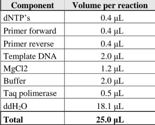
The PCR amplification was performed using the reaction mixture and the experimental conditions described in Tables 2.3 and 2.4. The sequencing reaction was performed by STABVIDA as a paid service. Clustal Omega was used for sequence alignment establishing as a reference for the compara-tive analysis the sequence of CgPDR1 from the reference strain C. glabrata CBS138, susceptible to azoles (Figure 1.8.).
12 Table 2.3:Reaction mixture used for the amplification of the selected regions of CgPDR1 gene.
Component Volume per reaction
dNTP’s 0.4 μL Primer forward 0.4 μL Primer reverse 0.4 μL Template DNA 2.0 μL MgCl2 1.2 μL Buffer 2.0 μL Taq polimerase 0.5 μL ddH2O 18.1 μL Total 25.0 μL
Table 2.4: Conditions of the PCR cycles for amplification of the selected regions of CgPDR1 gene.
Time Temperature 1 min 94 ºC 30 s 94 ºC 1 min 56 ºC x32 1 min 30 s 72 ºC 10 min 72 ºC ∞ 8 ºC
2.5. Cloning of CgPDR1 gene
To clone gene CgPDR1 from C. glabrata CBS138 or from the resistant clinical isolates FFUL412, FFUL443, FFUL866 and FFUL887 in the pGREG526 plasmid 100 a strategy based on homologous
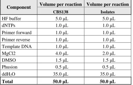
recombination was used. The pGREG526 plasmid was digested with 15 U of SalI (Takara Bio USA, Inc.) at 37 ºC during 3 h then the digested product was incubated with 1 U of Calf Intestinal Alkaline Phosphatase (CIAP) (Invitrogen) at 37 ºC for 1 h to prevent re-circularization. The CgPDR1 gene was amplified by PCR using as a template the genomic DNA of the strains with the specific primers listed in Table 2.5. The reaction mixture and the experimental conditions used are also described in Tables 2.6 and 2.7.
Table 2.5: Primers used for the amplification of the CgPDR1 gene.
Name Sequence 5’ 3’
CgPdr1_rev GCGTGACATAACTAATTACATGACTCGAGGTCGACGCATCACAAGTAAACATCAG CgPdr1_fw GAATTCGATATCAAGCTTATCGATACCGTCGACAATGCAAACATTAGAAACTACATC