Diagnosis and management of systemic
hypertension due to renovascular and aortic stenosis
in patients with Williams-Beuren syndrome
Erika Arai Furusawa, MD, PhD1 Camila Sanches Lanetzki Esposito, MD1 Rachel Sayuri Honjo, MD, PhD2 Lisa Suzuki, MD, PhD3 Gabriela Nunes Leal, PhD3
Chong Ae Kim, MD, PhD2
Benita Galassi Soares Schvartsman, MD, PhD1
1. Pediatric Nephrology Unit, Institute of Children, Hospital das Clínicas, Faculty of Medicine, University of São Paulo, São Paulo, Brasil. 2. Genetics Unit, Institute of Children, Hospital das Clínicas, Faculty of Medicine, University of São Paulo, São Paulo, Brasil. 3. Radiology Unit, Institute of Children, Hospital das Clínicas, Faculty of Medicine, University of São Paulo, São Paulo, Brasil.
http://dx.doi.org/10.1590/1806-9282.64.08.723
SUMMARY
AIM: To describe the incidence, diagnosis, and management of systemic arterial hypertension related to renal artery stenosis in patients with Williams-Beuren syndrome.
METHODS: Sixty-five patients with Williams-Beuren syndrome were evaluated for hypertension. Enrolled patients underwent Doppler sonography of the renal arteries and Doppler echocardiography. Those with Doppler sonography-detected lesions or with normal Doppler sonography but severe hypertension underwent computed tomography or gadolinium-enhanced magnetic resonance an-giography of the aorta and renal vessels. Patients needing vascular therapeutic intervention underwent conventional anan-giography. RESULTS: Systemic arterial hypertension was diagnosed in 21/65 patients with Williams-Beuren syndrome (32%; 13 male) with a mean age of 13.9 years (5mo-20yrs). In 8/21 patients renovascular hypertension was detected. Angioplasty was unsuccessful in five patients with renal artery stenosis, requiring additional treatment. Doppler echocardiography showed cardiac abnormalities in 16/21 (76%) hypertensive patients.
CONCLUSION: Cardiac abnormalities and hypertension in patients with Williams-Beuren syndrome are common. Thus, thorough eval-uation and follow-up are necessary to reduce cardiovascular risks and mortality of these patients
KEYWORDS: children, hypertension, renal artery stenosis, Williams-Beuren syndrome
DATE OF SUBMISSION: 18-Nov-2016
DATE OF ACCEPTANCE: 20-Nov-2016
CORRESPONDING AUTHOR: Erika Furusawa
Hospital das Clínicas, Av. Dr. Eneas de Carvalho Aguiar, 647 05403-000, Cerqueira Cesar, São Paulo, Brasil
E-mail: [email protected]
INTRODUCTION
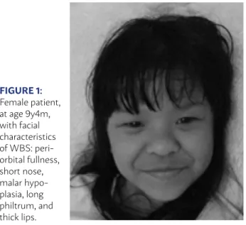
Williams-Beuren syndrome (WBS) is a genetic disorder characterized by facial dysmorphisms, con-genital heart defects, growth retardation, infantile hypercalcemia, renal and vascular abnormalities,
and intellectual disability1. Clinical diagnosis is
usu-ally performed during childhood when the typical facial changes and cognitive profile become more
apparent1 (Figure 1). Genetic confirmation can be
carried out using FISH2 (fluorescence in situ
hybrid-ization) or MLPA3 (multiplex ligation-dependent probe
amplification), or microarray tests for identification of the causal microdeletion at 7q11.23. Urinary tract system abnormalities in WBS have been described in
ectopia, hydronephrosis, renal agenesis or hypopla-sia, vesicoureteral reflux, and voiding dysfunction. Nephrocalcinosis, proteinuria, and chronic renal
fail-ure have also been reported in some cases series4,5,6
Cardiovascular abnormalities are also quite com-mon in patients with WBS and have been observed in
more than 80% of cases7,8. Supravalvular aortic
steno-sis (SVAS) is the most frequent abnormality, with an
estimated incidence of 64%(9,10). Systemic arterial
ab-normalities include localized or diffuse narrowing of the thoracic or abdominal aorta, coronary, renal and
other visceral arteries11,12. According to Lacolley et al.13
vascular injury in patients with WBS may be associat-ed with rassociat-educassociat-ed elastin synthesis and increasassociat-ed prolif-eration of vascular smooth muscle cells.
Arterial hypertension arterial (SAH) is also
ob-served with high prevalence in WBS14. In a minority
of patients, renal artery stenosis, diffuse narrowing of the aorta, aortic coarctation or a combination of
these abnormalities have been implicated4,5. Renal
artery stenosis is usually found at the origin of the
renal arteries7 (Figure 2). Nonetheless, there are few
reports about the origin and management of SAH in WBS, and the diagnosis is often not made.
This study aimed to describe the incidence of hy-pertension among 65 patients with WBS, as well as the diagnosis and management of hypertension due to renovascular or aortic stenosis.
METHODS
Sixty-five patients who were being treated from 1993 to 2010 at the Pediatric Nephrology and Genet-ics Units at the Institute of Children, Hospital das
Clínicas of the Faculty of Medicine of the University of São Paulo were included in this study. All patients were diagnosed with WBS based on clinical findings and had the presence of the 7q11.23 microdeletion
confirmed by the FISH(2) or MLPA test (with a
spe-cific kit for WBS) (3).
Patients with blood pressure (BP) values at or above the 95th percentile for age, gender, and height
confirmed on 3 different occasions15 were
includ-ed in the study and followinclud-ed prospectively. Clinical and laboratory parameters such as the age of onset of hypertension, associated symptoms, baseline BP,
fundus examination, microalbuminuria/creatinine16
and calcium/creatinine ratio17 in spot urine samples,
estimated creatinine clearance18, and serum ionized
calcium were evaluated.
All enrolled patients were initially investigated by Doppler echocardiography(DE) and renal ultrasound (RU) with color-flow Doppler sonography of the re-nal arteries (DS). Those with findings of rere-nal artery
stenosis19 or hypertension stage II15 with a normal
DS underwent computed tomographic angiography
(CTA)(20) and/or gadolinium-enhanced magnetic
reso-nance angiography (MRA) of the aorta and renal
ves-sels21. Patients with unclear diagnosis by CTA and/or
MRA or who required vascular therapeutic interven-tion (angioplasty) underwent conveninterven-tional angiogra-phy (CA).
RESULTS
Of the 65 patients with WBS included in this study, 21 (32%; mean age of 13.9 years, range: 5 months to 20 years, 13 males) had hypertension and
FIGURE 2: Digital subtraction angiography demonstrates discreet stenosis of the abdominal aorta anda severe stenosis of the left and right renal arteries. Also note several collateral arteries from the aorta on the left side
FIGURE 1:
were submitted to further imaging studies. In this group, the mean age at WBS diagnosis was 5.2 years (ranging from 8 months to 12 years). All patients were asymptomatic, and hypertension was detected by active investigation during routine medical visits.
The evaluation of the renal arteries by DS was normal in 12/21 patients. Of these, five patients did not undergo a CTA or MRA scan because of a low clinical suspicion of hypertension due to renovascu-lar or aortic stenosis. In these five patients, BP was adequately controlled with one or two medications, and none had secondary involvement of target organs during follow-up. Of the 12 patients with normal DS results, seven had persistent severe hypertension and were therefore submitted to further testing. Two of them had findings consistent with SAH as-sociated with vascular stenosis, as detected by CTA and conventional arteriography (one had abdominal aorta narrowing, and the other had right renal artery stenosis), indicating that DS resulted in false nega-tive results for these patients. The remaining five pa-tients had normal CTA results.
Nine patients with DS suggesting renal artery ste-nosis were also submitted to CTA or MRA, and only six patients had vascular lesions confirmed by one of these methods. In three patients, the DS yielded false positive results. Of the 21 hypertensive patients, 16 underwent DS followed by complementary renal and aortic vascular investigation with CTA or MRA. The DS showed discordant results when compared with CTA or MRA in five of those patients (31%).
Hypertension associated with renal or aortic le-sions was confirmed in 8/21 patients (Table 1), cor-responding to 12% of the cases of WBS and 38% of patients with arterial hypertension. Renal artery stenosis was detected in six patients (28.6%), aortic coarctation in one patient, and diffuse narrowing of the aorta in another one. One patient with renal ar-tery stenosis also had aortic stenosis.
Percutaneous transluminal balloon angioplasty was performed in five patients with renal artery ste-nosis, and it was unilateral in two and bilateral in three patients. In one patient with bilateral steno-sis, a stent was placed in the renal arteries. All four patients who underwent angioplasty had treatment failure, and two of them required surgical inter-vention for stenosis correction. An aortorenal graft was performed in one patient. For another patient who had stenosis of both renal arteries and of the aorta, an iliac renal graft, as well as a graft of the descending aorta to the infrarenal abdominal aorta, were inserted. Two patients who did not undergo surgical intervention continued with conservative treatment with antihypertensive medications (am-lodipine, carvedilol, hydralazine, and diuretic). Two patients with lesions of the aorta underwent surgi-cal correction.
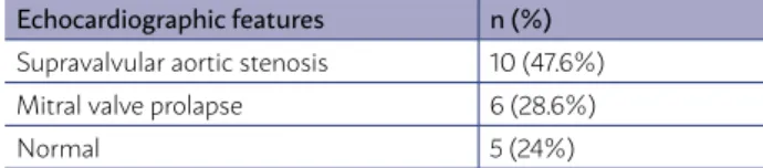
All patients underwent DE at the initial exam-ination, and the alterations found are described in Table 2. SVAS was the most prevalent malforma-tion (46%). Two patients underwent surgical cor-rection of SVAS and had mitral valve regurgitation, eccentric left ventricular hypertrophy, and left ven-tricular systolic dysfunction. Three patients with mitral valve prolapse also had eccentric left ven-tricular hypertrophy and left venven-tricular systolic dysfunction.
Hypercalciuria was found in four patients, and hypercalcemia was diagnosed in one patient. Esti-mated creatinine clearance and albuminuria at the beginning of the study are shown in Table 3. One pa-tient had hypertension after clinically presenting he-molytic uremic syndrome. Unilateral pyelocalyceal dilatation was observed in 2/21 patients submitted to renal sonography.
In this series, three patients died during follow-up due to heart failure: one patient with surgically cor-rected SVAS, one with both bilateral renal artery and aortic stenosis who had been submitted to angio-plasty and iliac-renal and aortic grafts, and one with SVAS who died following heart transplantation. TABLE 1: RENAL AND AORTIC VASCULAR LESIONS IN
8/21 PATIENTS WITH WILLIAMS-BEUREN SYNDROME AND SYSTEMIC ARTERIAL HYPERTENSION.
Patients Type of lesion
Renal artery stenosis 6 unilateral 2 / bilateral 4*
Aorta coarctation 1 thoracic
Diffuse aorta narrowing 1 descendent and
abdominal
*One patient also had aortic stenosis.
TABLE 2: ECHOCARDIOGRAPHIC FEATURES IN THE 21 WBS PATIENTS WITH SYSTEMIC ARTERIAL HYPERTENSION.
Echocardiographic features n (%)
Supravalvular aortic stenosis 10 (47.6%)
Mitral valve prolapse 6 (28.6%)
DISCUSSION
WBS is a congenital multisystem disease affect-ing the cardiovascular system, nervous system, and connective tissues that often involves hypertension, although the etiology of this symptom is not fully un-derstood. Possible explanations include disorganiza-tion of the elastic layer and hypertrophy of smooth muscle cells and collagen fibers. As a result, diffuse or localized progressive narrowing of the arterial wall leads to increased arterial stiffness and sympathetic
activity23. The prevalence of hypertension in WBS is
widely variable4,5,6,8,14 (5 to 70% in series including all
ages and 5 to 46% in children). It can appear as ear-ly as infancy, but the average age of diagnosis varies from 6.5 to 38 years. In the present study, the inci-dence of hypertension was 32%, with a mean age of diagnosis of 13.9 years (range: 5 months to 20 years).
As observed by other authors8, we confirmed that
patients with WBS are initially asymptomatic for hy-pertension, which highlights the need for health pro-fessionals to actively measure BP in these children in all routine visits.
In the present study, which included only chil-dren and adolescents, systemic arterial hyperten-sion associated with vascular stenosis was observed in 8/21 hypertensive patients (38%), which included renal artery stenosis (28.6%) and aortic lesions. In the literature; the frequency of arterial stenosis is variable across case series (e.g., 2 to 70%)5,8,12,14,22.
Renal DS is usually indicated as an initial diag-nostic method for screening of arterial stenosis. It is a non-invasive, relatively inexpensive test that is available in most centers but may have a high rate
of technical failure (approximately 30%)24 because it
is an operator-dependent method that also requires patients’ cooperation. In our study, we found a
simi-lar failure rate for DS (31%). The main difficulties are the detection of intra-renal and bilateral stenosis, the presence of obesity and the inadequate preparation of the patient. In WBS, psychomotor and behavioral disorders (agitation and anxiety) may represent
addi-tional complications1. The sensitivity and specificity
of DS in the detection of vascular stenosis have been shown to be between 60 and 98% and between 62 and
98%, respectively24;25. It should be emphasized that
the current sample was too small, since we conduct-ed further imaging tests only on severely hyperten-sive patients, hindering this type of analysis.
Computed tomographic angiography or MRA is commonly used in patients with WBS, but these techniques are not always available and require se-dation or general anesthesia in children. MRA has
high sensitivity (64-93%) and specificity (72-97%)25,26
but may overestimate renal arterial lesions or un-derdiagnose intra-renal lesions. On the other hand, CTA requires the use of iodinated contrast and ion-izing radiation. Most of our patients underwent CTA because of the limited availability of the MRA equipment at our unit. However, we were unable to establish the accuracy of these methods in our study, because only some of the patients who received MRA or CTA also underwent CA, which is considered the
gold standard in the diagnosis of arterial stenosis21.
Angioplasty was performed in five patients with stenosis of the renal artery but was unsuccessful, as
observed by other authors26,27. Patients with aortic
coarctation or stenosis were treated with surgical correction, with graft insertion in severe cases.
Other causes of hypertension such as hypercalce-mia, renal scarring secondary to recurrent urinary in-fections, obesity, and essential hypertension may also be involved in hypertension in WBS. Hypercalcemia
is frequently described in WBS 29 and occurred
tran-siently in one patient in this study. It may manifest early or later in life, and patients should, therefore, be periodically monitored for possible calcium
distur-bances1,6,9. Broder et al.30 found a higher incidence of
hypertension in patients with infantile hypercalcemia, but to date, no direct links between hypertension and hypercalcemia have been established.
Congenital cardiovascular abnormalities are prev-alent in patients with WBS and can occur in about
75% of cases7,8. SVAS is the most prevalent disease,
and is also present in up to 75% of cases9,14,30. Mitral
valve prolapse, bicuspid aortic valve, and coronary
abnormalities have also been described10,11,12. SVAS
TABLE 3: LABORATORY FEATURES (MEAN; RANGE) IN 21 PATIENTS WITH WBS AND ARTERIAL HYPERTENSION.
Laboratory features Mean; range n
Serum ionized calcium Hypercalcemiaa
1.23 (1.1-1.4) mmol/L 1
Urinary calcium Hypercalciuriab
0.15 (0-0.94) mg/mg creatinine 4
eGFRc
CKD class II
118.23 (68-183) ml/min/1.73m2
3 Albuminuria
Microalbuminuria d
13.31 (3.55-36.79) mg/g creatinine 1
a serum ionized calcium >1.4mmol/L ; b calcium /creatinine ratio of random spot urine > 0.21 mg/mg 17; c glomerular filtration rate estimated by serum creatinine18 , d
albu-min/creatinine ratio of random urine>30 mg/g creatinine16; CKD: chronic kidney
RESUMO
OBJETIVO: Descrever a incidência, o diagnóstico e o tratamento da hipertensão arterial sistêmica relacionada com estenose da artéria renal em pacientes com síndrome de Williams-Beuren.
MÉTODOS: Sessenta e cinco pacientes com síndrome de Williams-Beuren foram avaliados quanto à presença de hipertensão. Os pa-cientes foram submetidos à ultrassonografia com Doppler das artérias renais e ecocardiograma Doppler. Aqueles com suspeita de hipertensão renovascular foram submetidos à tomografia computadorizada ou angiografia por ressonância magnética da aorta e vasos renais ou angiografia convencional.
RESULTADOS: A hipertensão arterial sistêmica foi diagnosticada em 21/65 pacientes com síndrome de Williams-Beuren (32%, 13 do sexo masculino), com idade média de 13,9 anos (5 meses-20 anos). Em 8/21 pacientes foi detectada a hipertensão renovascular. Angio-plastia não teve sucesso em cinco pacientes com estenose da artéria renal, necessitando de tratamento adicional. O ecocardiograma Doppler mostrou anormalidades cardíacas em 16/21 (76%) pacientes hipertensos.
CONCLUSÃO: As anormalidades cardíacas e hipertensão arterial em pacientes com síndrome de Williams-Beuren são muito frequentes, sendo necessários uma avaliação minuciosa e seguimento para diminuir o risco cardiovascular e a morbimortalidade desses pacientes PALAVRAS-CHAVE: Criança. Hipertensão. Estenose de artéria renal. Síndrome de Williams-Beuren.
REFERENCES
1. American Academy of Pediatrics: Health care supervision for children with Williams syndrome. Pediatrics 2001; 107: 1192–204.
2. Borg I, Delhanty JD, Baraitser M. Detection of hemizygosity at the elastin locus by FISH analysis as a diagnostic test in both classical and atypical cases of Williams syndrome. J Med Genet 1995; 32: 692–696.
3. Dutra RL, Pieri Pde C, Teixeira AC, Honjo RS, Bertola DR, Kim CA. Detec-tion of deleDetec-tions at 7q11.23 in Williams– Beuren syndrome by polymorphic markers. Clinics (Sao Paulo) 2011; 66: 959–964.
4. Sugayama SM, Koch VH, Furusawa EA, Leone C, Kim CA. Renal and uri-nary findings in 20 patients with Williams–Beuren syndrome diagnosed by fluorescence in situ hybridization ( FISH). Rev Fac Med Hosp Clin Sao Paulo 2004; 59: 266–272.
5. Pankau R, Partsch CJ, Winter M, Gosch A, Wessel A. Incidence and spec-trum of renal abnormalities in Williams-Beurens syndrome. Am J Med Genet 1996; 63: 301–304.
6. Pober BR, Lacro RV, Rice C, Mandell V, Teele RL. Renal finding in 40 indi-viduals with Williams syndrome. Am J Med Genet 1993; 46: 271–274.
7. Donnai D, Karmiloff-Smith A. Williams syndrome. From genotype through to the cognitive phenoype. Am J Med Genet 2000; 97: 164–171.
8. Bouchired K, Boyer O, Bonnet D, Brunelle F, Decramer S, Landthaler G et al. Clinical features and management of arterial hypertension in chil-dren with Williams–Beuren syndrome. Nephrol Dial Transplant 2010; 25: 434–438
9. Morris CA. Williams Syndrome. 1999 Apr 9 [Updated 2013 Jun 13]. In: Pa-gon RA, Adam MP, Ardinger HH, et al., editors. GeneReviews® [Internet]. Seattle (WA): University of Washington, Seattle; 1993-2014. Available from: http://www.ncbi.nlm.nih.gov/books/NBK1249/
10. Vernant P, Corone P, Rossignol AM, Bielman C. 120 cases of the Williams and Beuren syndrome. Arch Mal Coer Vaiss 1980; 73: 661–666.
11. Bernand Y, Didier D, Bozio A, Champsaur G, Renaud JC, Maurat JP. Coro-nary anomalies associated with the Williams–Beuren syndrome. Apros of 2 cases. Arch Mal Coeur Vaiss 1985; 78: 791–795.
12. Zalzstein E, Moes CA, Musewe NN, Freedom RM. Spectrum of cardio-vascular anomalies Williams–Beuren syndrome. Pediatr Cardiol 1991; 12: 219–223.
13. Lacolley P, Boutouyrie P, Glukhova M, Daniel Lamaziere JM, Plouin PF, Bruneval P et al. Disruption of the elastin gene in adult Williams
syn-may be severe in up to 30% of patients30, and surgical
correction is necessary in such cases27. Consistent
with earlier reports10,12,23, the most common
car-diovascular abnormality observed in our study was SVAS, and it was severe in two patients who required surgical intervention.
CONCLUSION
Hypertension is a common finding in children with WBS and should be tested and investigated routinely as early as possible in this population. We recommend that CTA or MRA be used whenever pos-sible for cases of severe hypertension.
drome is accompanied by a paradoxical reduction in arterial stiffness. Clin Sci (Lond) 2002; 103: 21–29.
14. Honjo RS, Dutra RL, Furusawa EA, Zanardo EA, Costa LSA, Kulikows-ki LD, Bertola DR, Kim AE. Williams-beurens syndrome: A clinical study of 55 brazilian patients and the diagnosis use of MLPA. Biomed Res Int. 2015:903175
15. National High Blood Pressure Program Working Group on High Blood Pressure in Children and Adolescents. The fourth report on the diagnosis, evaluation and treatment of high blood pressure in children and adoles-cents. Pediatrics 2004; 114: 555–576.
16. Karalliedde J, Vibert G. Microalbuminuria and cardiovascular risk. Am J Hy-pertens 2004; 17(10): 989-93
17. Butani L, Kalia A. Idiopathic hypercalciuria in children-how valid are the existing diagnostic criteria? Pediatric Nephrol 2004; 19: 577-582.
18. Schwartz GJ, Haycock GB, Edelmann CM Jr, Spitzer A. A simple estimate of glomerular filtration rate in children derived from body length and plas-ma creatinine. Pediatrics 1976; 58:259-263.
19. Granata A, Fiorinin E, Andrulli S, Loggias F, Gallieni M, Sicurezza E et al. Doppler ultrasound and renal artery stenosis: An overview. J Ultrasound
2009; 12: 133–143.
20. Kurian J, Epelman M, Darge K, Meyers K, Nijs E, Hellinger JC. The role of CT angiography in the evaluation of pediatric renovascular hypertension.
Pediatr Radiol 2013; 43: 490–501.
21. O’Neill WC, Bardelli M, Yevzlin AS. Imaging for renovascular disease. Se-2min Nephrol 2011; 31(3): 272-282.
22. KDIGO 2012 Clinical practice guideline for the evaluation and manage-ment of chronic kidney disease. Kidney Int Suppl 2013; 3:1-150.
23. Collins RT 2nd, Kaplan P, Somes GW, Rome JJ. Long-term outcomes of
patients with cardiovascular abnormalities and Williams syndrome. Am J Cardiol 2010; 105: 874–878.
24. Wessel A, Pankau R, Kececioglu D, Ruschewski W, Bursch JH. Three de-cades of follow up of aortic and pulmonary vascular lesions in the Wil-liams–Beuren syndrome. Am J Med Genet 1994; 52: 297–301.
25. Conkbayir I, Yucesoy C, Edguer T, Yanik B, Yasar AU, Hekimoglu B. Dop-pler sonography in renal artery stenosis. An evaluation of intrarenal and extrarenal imaging parameters. Clin Imaging 2003; 27: 256–260.
et al. Imaging modalities for renal artery stenosis in suspected renovascu-lar hypertension: prospective intraindividual comparison of color Doppler US, CT angiography, GD–enhanced MR angiography, and digital subtrac-tion angiography. Ren Fail 2007; 29: 295–302.
27. Actis Dato GM, La Torre M, Caimmi P, Actios Dato A Jr., Centofanti P, Ot-tino GM et al. Williams-Beuren syndrome. Long-term results of surgical treatments in six patients. J Cardiovasc Surg (Torino) 1997; 38: 125–129.
28. Kumada Y, Yasuda H, Sasaki E, Murakawa S, Mori Y, Hirose H. Reoperation
for diffuse supravalvar aortic stenosis with Williams syndrome–extended path aortoplasty and extra–anatomic bypass from the ascending aorta to the descending aorta in a median sternotomy. J Thorac Cardiovasc Surg
1998; 46: 1061–1064.
29. Udwin O. A survey of adults with Williams syndrome and idiopathic infan-tile hypercalcaemia. Dev Med Child Neurol 1990; 32: 129–141.