Resumo
Introdução: O transtorno depressivo maior (TDM) é uma doença mental incapacitante caracterizada por episódios de pelo menos 2 semanas de mudanças claras no afeto, cognição e funções neurovegetativas. Vários estudos de neuroimagem, realizados através de imagem de ressonância magnética (IRM), examinaram mudanças morfométricas em pacientes com TDM, com resultados não conclusivos. Este estudo tem como objetivo revisar a literatura e realizar uma metanálise sobre o volume do hipocampo (VHc) em pacientes com TDM.
Métodos: Estudos de VHc em pacientes com TDM foram identificados a partir dos principais bancos de dados (MEDLINE, EMBASE, The Cochrane Library, Scopus, PsycINFO e SciELO) usando os seguintes termos: depression, major depressive disorder, MDD, unipolar, magnetic resonance imaging, MRI e hippocampus.
Resultados: Foi realizada uma metanálise de 29 estudos que preencheram os critérios específicos. A amostra foi composta por 1327 pacientes e 1004 indivíduos saudáveis. Os estudos foram altamente heterogêneos em relação a idade, gênero, idade do primeiro episódio e duração média da doença, mas o efeito combinado da depressão foi significativo em ambos os hipocampos. O TDM foi associado à atrofia do hipocampo à direita [-0,43; intervalo de confiança de 95% (IC95%) -0,66 a -0,21] e à esquerda (-0,40; IC95% -0,66 a -0,15).
Conclusões: O TDM parece estar associado à atrofia global do VHc. Estudos longitudinais com maior tempo de seguimento, projetados para analisar a influência dos fatores sociodemográficos nessa relação, são necessários para obter evidências mais robustas.
Descritores: Volume hipocampal, transtorno depressivo maior, RM, depressão.
Abstract
Introduction: Major depressive disorder (MDD), an incapacitating mental disorder, is characterized by episodes of at least 2 weeks of apparent changes in mood, cognition, and neurovegetative functions. Many neuroimaging studies using magnetic resonance imaging (MRI) have examined morphometric changes in patients with MDD, but the results are not conclusive. This study aims to review the literature and perform a meta-analysis on hippocampal volume (HcV) in patients with MDD.
Methods: Studies on HcV in patients with MDD diagnosis were identified from major databases (MEDLINE, EMBASE, The Cochrane Library, Scopus, PsycINFO, and SciELO) using the search terms depression, major depressive disorder, MDD, unipolar, magnetic resonance imaging, MRI, and hippocampus. Results: A meta-analysis of 29 studies fulfilling specific criteria was performed. The sample included 1327 patients and 1004 healthy participants. The studies were highly heterogeneous with respect to age, sex, age of onset, and average illness duration. However, the pooled effect size of depression was significant in both hippocampi. MDD was associated with right (-0.43; 95% confidence interval [95%CI] -0.66 to -0.21) and left (-0.40; 95%CI -0.66 to -0.15) hippocampal atrophy.
Conclusions: MDD seems to be associated with global HcV atrophy. Larger longitudinal follow-up studies designed to analyze the influence of sociodemographic variables on this relationship are required to yield better evidence about this topic.
Keywords: Hippocampal volume, major depressive disorder, MRI, depression.
1 Grupo de Pesquisa em Epidemiologia e Cardiologia, Universidade Federal de Pernambuco, Recife, PE, Brazil. 2 Centro Universitário Maurício de Nassau, Recife,
PE, Brazil. 3 Hospital da Restauração, Recife, PE, Brazil. 4 Pronto-Socorro Cardiológico Universitário de Pernambuco (PROCAPE), Universidade de Pernambuco
(UPE), Recife, PE, Brazil.
This study was conducted at Departamento de Medicina, Centro Universitário Maurício de Nassau, Recife, PE, Brazil. Submitted Oct 24 2017, accepted for publication May 06 2018.
Suggested citation: Santos MAO, Bezerra LS, Carvalho ARMR, Brainer-Lima AM. Global hippocampal atrophy in major depressive disorder: a meta-analysis of magnetic resonance imaging studies. Trends Psychiatry Psychother. 2018;40(4):369-378. Epub Sep 17 2018. http://dx.doi.org/10.1590/2237-6089-2017-0130
Global hippocampal atrophy in major depressive disorder:
a meta-analysis of magnetic resonance imaging studies
Atrofia global do hipocampo no transtorno depressivo maior:
uma metanálise de estudos com ressonância magnética
Marcelo Antônio Oliveira Santos,1,2 Lucas Soares Bezerra,1,2 Ana Rita Marinho Ribeiro Carvalho,2,3
Introduction
Major depressive disorder (MDD) is an incapacitating mental disorder characterized by episodes of at least 2 weeks of apparent changes in affection, cognition, and neurovegetative functions.1 Patients with MDD
present a lower quality of life and higher prevalence
of medical conditions. The global prevalence of MDD
is approximately 5%. According to the World Health
Organization, MDD is estimated to become the second most disabling condition by 2020.2
Several neurobiological models have been proposed to explain the pathogenesis of MDD. Because MDD is primarily related to lower affection and humor, many authors have studied and emphasized the role
of dysfunctional cortico-limbic networks in MDD.3
Postmortem and animal model studies have reported lower hippocampal volume (HcV) in participants with depressive disorders. The hippocampus is involved in episodic and declarative memory, as well as in learning, areas that often present deficits in patients with depression.4 The suggestion that HcV is lower because of depression has been influential in guiding neuroimaging studies on the analysis of the
hippocampus.5
Many neuroimaging studies using magnetic resonance imaging (MRI) have examined morphometric changes in patients with MDD, but the results are not conclusive. Some studies have reported bilateral,6 unilateral right,7 or unilateral left hippocampal hypotrophy in these patients compared with healthy participants,8,9 whereas others have reported no changes.10,11 Furthermore, some systematic reviews and meta-analyses have examined studies on HcV in the first depressive episode or in
specific groups, such as the elderly (aged ≥ 60 years)
with depression.7,12-14 However, to date, no reviews have systematically examined the macro influence of depression on HcV.
This study aims to examine whether patients with MDD present hippocampal atrophy compared with
non-depressive participants. We considered HcV reduction
as the primary outcome.
Methods
This systematic review was conducted in accordance with the Preferred Reporting Items for Systematic Reviews and Meta-Analysis (PRISMA) statement. Details of the study protocol were registered on the International Prospective Register of Systematic Reviews (PROSPERO) and can be accessed at www.crd.york.ac.uk/PROSPERO/ display_record.asp?ID=CRD42018086196.
The electronic databases MEDLINE, EMBASE, The Cochrane Library, Scopus, PsycINFO, and SciELO were
searched for papers published between January 1960 and October 2017. The search terminology included the terms depression, major depressive disorder, MDD, unipolar, magnetic resonance imaging, MRI, and hippocampus. Only studies written in English, Portuguese, or Spanish were reviewed. At least two of
the authors performed each search. Furthermore, all
reference lists of the obtained papers were checked for studies not indexed in electronic databases. The complete search strategy was as follows: Depression; Major depressive disorder; MAJOR DEPRESSIVE
DISORDER (MeSH); MDD; (unipolar) AND (depression); Magnetic resonance imaging; MAGNETIC RESONANCE IMAGING (MeSH); MRI; Hippocampus; HIPPOCAMPUS (MeSH); (hippocampal) AND (volume); (1 OR 2 OR 3 OR 4 OR 5) AND (6 OR 7 OR 8) AND (9 OR 10 OR 11).
All observational studies and clinical trials that evaluated the relationship between MDD and HcV measured through MRI were included in this review. Studies with sample groups in which patients presented any other associated neuropsychiatric or metabolic condition were excluded. Inclusion criteria were as follows: patients with a primary diagnosis of MDD assessed using international diagnostic criteria (Diagnostic and Statistical Manual of Mental Disorders,
Fourth Edition [DSM-IV] or International Statistical
Classification of Diseases and Related Health Problems, Tenth Revision [ICD-10]); a healthy comparison group; participants screened for neurological, psychiatric, and other medical disorders that could affect brain structure, including alcohol and substance abuse; MRI as the primary measurement tool; and a continuous measure of HcV as the dependent variable.
Two authors examined the abstracts of the articles retrieved against the defined inclusion criteria. All potentially relevant full-text articles were retrieved for
quality and satisfaction assessment of inclusion criteria. Figure 1 summarizes the study inclusion process.
For each data criterion, a data extraction table
was pilot-tested on five randomly-selected studies and adjusted accordingly. Two independent reviewers assessed the following data from the patient and control samples: number of participants, HcV means and standard deviations, and mean disease duration. A third author double-checked the extracted data. Disagreements were resolved by consensus between the authors; if no consensus could be reached, a third author made a decision. All volumes were converted to
mm3 before being entered into the meta-analysis.
To certify the quality and validity of the eligible
assessed each study using the Strengthening The Reporting of Observational Studies in Epidemiology (STROBE) checklist.16
Calculations were performed using the STATA software version 13.0 SE. The meta-analyses were performed using a random effects model weighting the studies by the inverse variance and calculating the Hedges’ g effect size. The random effects model using the DerSimonian-Laird method was selected over the
fixed effects approach because previous analyses have indicated considerable between-study heterogeneity.
To assess for between-study heterogeneity, the Cochran Q test was performed and I² statistic was recorded and further analyzed by meta-regression. Begg’s and Egger’s tests were used to determine publication bias.
Results
Twenty-nine studies comprising 1327 patients and 1004 healthy participants fulfilled both inclusion and exclusion criteria and were included in this meta-analysis.6-9,11-14,17-37 One study that found lower left HcV was excluded because volume measurements were unavailable.38 A report by Andreescu et al.39 was also excluded because only global HcV measurements were available. Pantel et al.40 reported a study in German that was excluded from the analysis because of language limitations.
All studies selected for this review were cross-sectional studies published in English, except one cohort study.30 The main inclusion criteria entailed adults (≥18 years). The Hamilton Depression Scale was the main
Records identified through
database searching (n = 187)
Identificatio
n
Screening
Eligibility
Included
Additional records identified
through other sources (n = 2)
Records after duplicates removed (n = 178)
Records screened (n = 178)
Records excluded (n = 102)
Records not meeting criteria (n = 44)
Full-text articles excluded with
reasons (n = 3):
• Language limitations (n = 1) • Hippocampal volume not
available (n = 2) Records assessed for
inclusion criteria (n = 76)
Articles meeting inclusion criteria
(n = 32)
Studies included in
qualitative synthesis
and meta-analysis (n = 29)
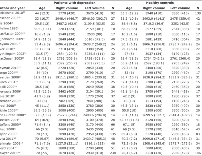
instrument used to assess the severity of depressive symptoms. Mean age of participants in the study groups varied from 16 to 74 years, and the percentage of male participants in each group varied from 0 to 63. The average HcV measured showed slight variations, and two of these measurements markedly deviated from the mean.9,14 Sample characteristics of the included studies are presented in Table 1. Some of the studies included patients with recurrent depression,6 first-episode depression,12,17,26 and late-onset depression.7,13,21 Eight studies evaluated illness duration, which varied from 7
to 57 months.6,12,18,25,27,28,33,35 Table 2 shows a summary
of the included studies.
Because the study performed by MacQueen et al.26
analyzed two samples (first-episode versus multiple episodes of depression) that did not overlap, it was considered as having two distinct samples for statistical
calculation. Therefore, the degrees of freedom (df), which is typically the number of studies minus 1, was 29 instead of 28.
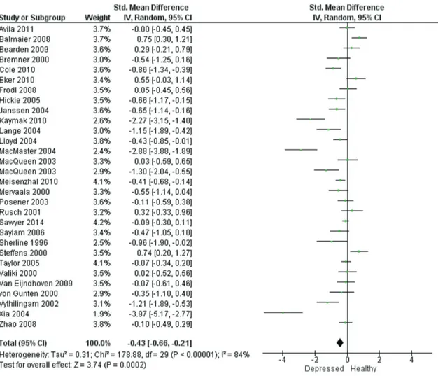
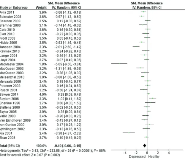
The Q test of heterogeneity (df = 29) was highly significant, as expected (both right and left hippocampus: p < 0.00001). The percentage of heterogeneity given
by the I² statistic was 84% (95% confidence interval [95%CI] 77.8 to 88.1, p < 0.00001) for the right hippocampus and 88% (95%CI 83.4 to 90.7, p <
0.00001) for the left hippocampus, which provides high evidence of between-study heterogeneity. Therefore, the effect size was calculated under the assumption of
a random effects model. The DerSimonian-Laird pooled
effect size revealed bilateral statistical significance:
-0.43 (95%CI -0.66 to -0.21) for the right hippocampus (Figure 2) and -0.40 (95%CI -0.66 to -0.15) for the left hippocampus (Figure 3).
Table 1 - Studies of hippocampal volume in patients with major depressive disorder
Author and year
Patients with depression Healthy controls
Age Right volume Left volume N Age Right volume Left volume N
Meisenzhal 201027 44 (12.3) 3770 (420) 3670 (410) 92 33.3 (12.2) 3940 (410) 3830 (410) 138
Posener 200328
33 (10.7) 2948.4 (446.7) 2546.00 (392.7) 27 33.2 (10.8) 2993.9 (414.2) 2475 (359.4) 42
Xia 200435 39.5 (12) 3487.2 (62.9) 3109.8 (83.5) 22 35.4 (8.8) 3710.3 (36.6) 3352 (43.5) 13
Sheline 19966
68.5 (10.4) 2283 (324) 2159 (301) 10 68.5 (9.5) 2577 (259) 2544 (333) 10
MacMaster 200425 16.6 (1.8) 2540 (120) 2530 (90) 17 16.2 (1.6) 2880 (110) 3050 (110) 17
Eijndhoven 200933 34.9 (11.5) 3848 (444) 3610 (372.5) 40 37.3 (12.7) 3881 (429) 3716 (364) 20 Saylam 200612 33.4 (9.3) 2696.4 (194.4) 2638.7 (249.2) 24 30.1 (6.1) 2806.3 (256.8) 2786.7 (249.2) 24
Eker 201018 32.1 (9.3) 3310 (420) 3380 (350) 25 29.7 (6.4) 3110 (260) 3330 (300) 22
Vythilingam 200234 33.5 (7) 2884 (120.5) 3235.5 (111) 32 27 (5) 3037 (134) 3179 (123) 14
MacQueen 200326 28.4 (11.8) 2793 (303.8) 2738 (301.1) 20 28.4 (11.5) 2784 (342.2) 2761 (368.4) 20
35.9 (11.1) 2392 (256.7) 2381 (273.5) 17 36.2 (11.9) 2692 (190.1) 2703 (249) 17
Kaymak 201022 32 (8.5) 2720 (320) 2850 (330) 20 29.3 (5.8) 3400 (250) 3520 (330) 15
Lange 200423 34 (10) 2670 (500) 2790 (410) 17 32 (6) 3190 (370) 2990 (460) 17 Bearden 200937 32.9 (11.9) 1911.1 (280.1) 1885.4 (230.8) 31 36.7 (10.7) 1828.9 (284.2) 1851.9 (326.8) 31
Rusch 200129 33.2 (9.5) 2290 (300) 2170 (260) 25 37.4 (14.4) 2200 (240) 2130 (270) 15
Vakili 200010 38.5 (10) 2610 (580) 2640 (550) 38 40.3 (10.4) 2600 (510) 2460 (380) 20
Mervaala 20008 42.2 (12.2) 3462 (405) 3104 (391) 34 42.1 (14.6) 3700 (467) 3441 (436) 17
Cole 201017 41.9 (8.9) 1860 (220) 1770 (260) 37 42.2 (9) 2080 (280) 1980 (290) 37
Bremner 20009 43 (8) 982 (269) 940 (208) 16 45 (10) 1113 (194) 1166 (248) 16
Frodl 200819 45 (11.1) 3850 (330) 3780 (380) 30 46.3 (13.1) 3830 (420) 3760 (400) 30
Hickie 200520 53.5 (13.5) 3000 (400) 2900 (400) 66 55.8 (10) 3300 (600) 3300 (500) 20 von Gunten 200011 57.6 (13.9) 2597.9 (244) 2498.6 (294.8) 14 58.1 (11.4) 2699.5 (312.7) 2644.4 (409.9) 14
Janssen 200421 64 (10.9) 2840 (390) 3100 (370) 28 62.37 (11.3) 3120 (450) 3200 (520) 41
Steffens 200013 71.4 (8.4) 3300 (440) 3170 (440) 66 67.1 (5) 2980 (390) 2920 (360) 18
Zhao 200813 66 (5.5) 3660 (560) 3435 (550) 61 69 (5.5) 3720 (590) 3510 (620) 43
Taylor 200531 70 (7.3) 3090 (420) 2950 (430) 135 69.4 (6.3) 3120 (440) 2960 (450) 83 Avila 201136 70 (6.67) 3099.3 (489.9) 3439.7 (405.3) 48 70.2 (7.2) 3099.8 (271.6) 3446.5 (334.2) 31 Ballmaier 200814 71.1 (7.6) 1127.5 (231.1) 1116.1 (222) 46 72.3 (6.9) 1308.4 (245.6) 1272.7 (275.6) 34
Lloyd 200424 74 (6.3) 2800 (500) 2700 (400) 51 73.1 (6.7) 3000 (400) 2800 (400) 39
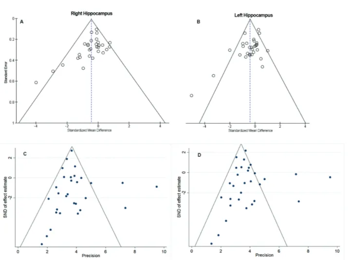
Begg’s and Egger’s tests were performed and
showed no evident signs of publication bias (Figure 4).
The meta-analysis was repeated, omitting one study at a time, to ensure that the result was not skewed by a single study. This analysis did not change the random-effects estimate, and the results continued to be statistically significant.
Because the studies had significant heterogeneity,
the data were analyzed using meta-regression. We
assumed that differences in age, sex, duration of illness, and year of publication could explain some of the variation. These variables were analyzed separately and together, but were not significantly correlated with the random-effects estimate in either the right or left hippocampus.
Discussion
The studies included in the analysis yielded highly
heterogeneous results. Notably, this heterogeneity
was expected because there were marked differences among the patient groups with respect to age and sex distribution, type of MDD (e.g., first episode, dysthymia, refractory, late onset), illness duration, age at the first episode, and factors related to treatment. Moreover, an increase in HcV variation was expected considering various protocols for the scanning and delineation of hippocampal structures.
However, a meta-analysis plays an important role in the analysis of scientific evidence because these possible confounding factors are diluted or neutralize
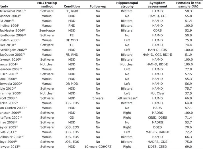
Table 2 - Summary of included studies evaluating hippocampal atrophy in MDD
Study
MRI tracing
method Condition Follow-up
Hippocampal atrophy
Symptom assessment
Females in the sample (%)
Meisenzhal 201027 Software FE, RMD No Bilateral HAM-D 58.3
Posener 200328 Manual MDD No No HAM-D, CGI 55.8
Xia 200435 Manual MDD No Bilateral HAM-D 51.4
Sheline 19966 Manual RMD No Bilateral HAM-D 100.0
MacMaster 200425 Semi-auto MDD No Bilateral CDRS 52.9
Eijndhoven 200933 Software FE No No HAM-D 50.0
Saylam 200612 Manual DF MDD No Left HAM-D 75.0
Eker 201018 Software FE No No HAM-D 74.4
Vythilingam 200234 Manual MDD No Left HAM-D, ZDS 100.0
MacQueen 200326 Manual FE, RMD No Bilateral HAM-D, CGI, BDI-II 51.9
Kaymak 201022 Software MDD No Bilateral HAM-D 100.0
Lange 200423 Not clear MDD No Not clear HAM-D, BDI-II 100.0
Bearden 200937 Manual MDD No Left HAM-D 77.0
Rusch 200129 Software MDD No No HAM-D 57.5
Vakili 200010 Manual MDD No No HAM-D 55.3
Mervaala 20008 Manual DR MDD No Left HAM-D 56.9
Cole 201017 Software MDD No Bilateral HAM-D 75.7
Bremner 20009 Not clear MDD No Left Not Clear 37.5
Frodl 200819 Software MDD 3 years Left increase* HAM-D 66.0
Hickie 200520 Manual LOS, EOS No Bilateral HAM-D 64.0
von Gunten 200011 Manual MDD No No HADS 57.1
Janssen 200421 Software MDD No Right MADRS 100.0
Steffens 200013 Software GD No Right CESD, DDES 71.4
Zhao 200813 Software MDD No No MADRS 53.7
Taylor 200531 Software LOS, EOS No Right DDES 70.6
Avila 201136 Manual LOS, EOS No Left MADRS, HAM-D 72.2
Ballmaier 200814 Manual LOS, EOS No Bilateral HAM-D 66.3
Lloyd 200424 Software LOS, EOS No Bilateral MADRS, GDS 75.0
Sawyer 201330 Software MDD 10 years COHORT Right DDES, CESD 69.0
BDI-II = Beck Depression Inventory II; CDRS = Childhood Depression Rating Scale; CESD = Center for Epidemiologic Studies Depression scale; DDES = Duke Depression Evaluation Schedule; DF = drug-free; DR = drug-resistant; EOS = early-onset depression; FE = first episode; GCI = Global Clinical Impression Scale; GD = geriatric depression; GDS = Geriatric Depression Scale; HADS = Hospital Anxiety and Depression Scale; HAM-D = Hamilton Depression Scale; LOS = late-onset depression; MADRS = Montgomery-Åsberg Depression Rating Scale; MDD = major depressive disorder; MRI = magnetic resonance imaging; RMD = recurrent major depression; ZDS = Zung Depression Scale.
each other based on the large number of participants analyzed. Meta-analyses are conducted to determine whether an effect is present, and summarize the data to determine if this effect is positive or negative. This process increases the external validity of the studies as well as the extendibility of results to the general population of patients with MDD.
It is established that HcV losses are expected as a natural process of aging41; however, Sheline et al.42 demonstrated that illness duration, not age, predicted hippocampal loss in women with recurrent MDD. Only eight studies included in our analysis presented information about depression duration. However, in the meta-regression, neither illness duration nor age were significantly correlated with the random-effects estimate.
Seven studies failed to find significant HcV differences in patients with depression compared with healthy participants.11,18,19,28,29,32,33 Six studies demonstrated significant reduction only in the right
hippocampus.7,20,21,30,31,35 Six other studies showed
similar findings in the lower left hippocampus.8,9,12,13,36,37 Bilateral hippocampal atrophy was reported in 10 studies.6,14,22-27,34
MacQueen et al.26 evaluated one group of patients during their first depressive episode and another during multiple episodes and compared them to healthy matched participants. After the first episode, there were no differences between the depression and comparison groups. However, in the group with multiple depressive episodes, there was bilateral hippocampal atrophy compared with healthy participants.
Vythilingam et al.34 theorize that HcV is bilaterally reduced in patients with depression who experienced sexual abuse in childhood compared with participants with depression but with no similar experience. Those
authors reported 18% smaller volumes for the left hippocampus and 15% smaller ones for the right
hippocampus.
According to the results of Vakili et al.,32 a smaller volume of the right hippocampus was associated with poor responses to antidepressants. This result is incipient, as investigating this association was not the primary objective of the study. However, if confirmed, that finding would be clinically interesting as a potential predictor of treatment response.
The pathophysiological pathways that explain HcV reduction in MDD remain unclear. Some authors theorize that HcV reduction is associated with disturbed hypothalamic pituitary adrenal axis function and adrenal hypersecretion of glucocorticoids, particularly cortisol. According to this hypothesis, cortisol leads to neural atrophy and the inhibition of neurogenesis in the hippocampus.43 Another important supporting
mechanism is the glutamate N-methyl-D-aspartate (NMDA) channel present in inhibitory neurons that
comprise this pathway. Subiculum neurons make a
synapse with a hypothalamic neuron, inhibiting it
through NMDA receptors. The hypothalamic neuron
modulates the corticotropic cell, stimulating it through gamma-aminobutyric acid (GAMA) liberation, which culminates in the production and release of the adrenocorticotropic hormone (ACTH). Serum cortisol concentration increases as a product of ACTH, released from the adrenal gland.44-46
It is important to address the potential protective properties of selective serotonin reuptake inhibitors (SSRIs) in patients with MDD. As demonstrated by
Frodl et al.,19 after a follow-up of 3 years, patients
with MDD who used SSRI antidepressants showed protective effects against hippocampal atrophy as well as an increase in the left HcV. Although this is the only study to demonstrate this association, many studies have indicated that SSRIs reduce functional deficits in
inflammatory and ischemic events.47
Study limitations
In principle, cross-sectional studies such as those included in the present analysis do not allow conclusions about causality to be drawn.
Further, socioeconomic and lifestyle characteristics
from specific population groups can act as confounders.
For example, the habit of regular exercise training is a
protective factor against hippocampal atrophy and may be responsible for hippocampus hypertrophy in healthy participants.48
Other factors can also act as confounders, such as the comorbidity of MDD and anxiety disorders,49 which was not assessed in any of the studies.
Longitudinal follow-up studies with large samples
are the best design to allow the drawing of conclusions about the association between hippocampal atrophy and MDD.
Conclusions
Although the studies available in the literature are
quite heterogeneous, MDD seems to be associated
with global HcV atrophy. Many confounding factors may have influenced the divergence between studies, in particular sociocultural variables, which are responsible for the construction of social identity and affect the way in which stressful and depressive situations may contribute to changes in hippocampal
neuronal circuits. Nevertheless, idiosyncratic biological
factors may influence neuronal circuitry development and plasticity and play a role in the fine adjustment of hippocampal dynamics.
Larger longitudinal follow-up studies that consider
these aspects are needed to yield better evidence about this topic.
Disclosure
No conflicts of interest declared concerning the
publication of this article.
References
1. American Psychiatric Association. Diagnostic and Statistical Manual of Mental Disorder. 5th ed. Washington: American Psychiatric Publishing; 2013.
2. Bromet E, Andrade LH, Hwang I, Sampson NA, Alonso J, de Girolamo G, et al. Cross-national epidemiology of DSM-IV major depressive episode. BMC Med. 2011;9:90.
3. Pizzagalli DA. Frontocingulate dysfunction in depression: toward biomarkers of treatment response. Neuropsychopharmacology. 2011;36:183-206.
4. Schwert C, Aschenbrenner S, Weisbrod M, Schröder A. Cognitive impairments in unipolar depression: the impact of rumination. Psychopathology. 2017;50:347-54.
5. Rajkowska G. Postmortem studies in mood disorders indicate altered numbers of neurons and glial cells. Biol Psychiatry. 2000;48:766-77.
6. Sheline YI, Wang PW, Gado MH, Csernansky JG, Vannier MW. Hippocampal atrophy in recurrent major depression. Proc Natl Acad Sci U S A. 1996;93:3908-13.
7. Steffens DC, Byrum CE, McQuoid DR, Greenberg DL, Payne ME, Blitchington TF, et al. Hippocampal volume in geriatric depression. Biol Psychiatry. 2017;48:301-9.
8. Mervaala E, Föhr J, Könönen M, Valkonen-Korhonen M, Vainio P, Partanen K, et al. Quantitative MRI of the hippocampus and amygdala in severe depression. Psychol Med. 2000;30:117-25. 9. Bremner JD, Narayan M, Anderson ER, Staib LH, Miller HL,
Charney DS. Hippocampal volume reduction in major depression. Am J Psychiatry. 2000;157:115-8.
10. Vakili K, Pillay SS, Lafer B, Fava M, Renshaw PF, Bonello-Cintron CM, et al. Hippocampal volume in primary unipolar major depression: a magnetic resonance imaging study. Biol Psychiatry. 2017;47:1087-90.
11. von Gunten A, Fox NC, Cipolotti L, Ron MA. A volumetric study of hippocampus and amygdala in depressed patients with subjective memory problems. J Neuropsychiatry Clin Neurosci. 2000;12:493-8.
12. Saylam C, Üçerler H, Kitiş Ö, Ozand E, Gönül AS. Reduced hippocampal volume in drug-free depressed patients. Surg Radiol Anat. 2006;28:82-7.
13. Zhao Z, Taylor WD, Styner M, Steffens DC, Krishnan KRR, MacFall JR. Hippocampus shape analysis and late-life depression. PLoS One. 2008;3:e1837.
14. Ballmaier M, Narr KL, Toga AW, Elderkin-Thompson V, Thompson PM, Hamilton L, et al. Hippocampal morphology and sistinguishing late-onset from early-onset elderly depression. Am J Psychiatry. 2008;165:229-37.
15. Moher D, Liberati A, Tetzlaff J, Altman DG, Altman D, Antes G, et al. Preferred reporting items for systematic reviews and meta-analyses: The PRISMA statement. PLoS Med. 2009;6:e1000097. 16. von Elm E, Altman DG, Egger M, Pocock SJ, Gøtzsche PC, Vandenbroucke JP, et al. The Strengthening the Reporting of Observational Studies in Epidemiology (STROBE) statement: guidelines for reporting observational studies. J Clin Epidemiol. 2008;61:344-9.
17. Cole J, Toga AW, Hojatkashani C, Thompson P, Costafreda SG, Cleare AJ, et al. Subregional hippocampal deformations in major depressive disorder. J Affect Disord. 2010;126:272-7.
18. Eker C, Kitis O, Taneli F, Eker OD, Ozan E, Yucel K, et al. Correlation of serum BDNF levels with hippocampal volumes in first episode, medication-free depressed patients. Eur Arch Psychiatry Clin Neurosci. 2010;260:527-33.
19. Frodl T, Jäger M, Smajstrlova I, Born C, Bottlender R, Palladino T, et al. Effect of hippocampal and amygdala volumes on clinical outcomes in major depression: a 3-year prospective magnetic resonance imaging study. J Psychiatry Neurosci. 2008;33:423-30. 20. Hickie I, Naismith S, Ward PB, Turner K, Scott E, Mitchell P, et al. Reduced hippocampal volumes and memory loss in patients with
early- and late-onset depression. Br J Psychiatry. 2005;186:197-202.
21. Janssen J, Pol HEH, de Leeuw F, Schnack HG, Lampe IK, Kok RM, et al. Hippocampal volume and subcortical white matter lesions in late life depression: comparison of early and late onset depression. J Neurol Neurosurg Psychiatry. 2007;78:638-40. 22. Kaymak SU, Demir B, Şentürk S, Tatar I, Aldur MM, Uluğ B.
Hippocampus, glucocorticoids and neurocognitive functions in patients with first-episode major depressive disorders. Eur Arch Psychiatry Clin Neurosci. 2010;260:217-23.
23. Lange C, Irle E. Enlarged amygdala volume and reduced hippocampal volume in young women with major depression. Psychol Med. 2004;34:1059-64.
24. Lloyd AJ, Ferrier IN, Barber R, Gholkar A, Young AH, O’Brien JT. Hippocampal volume change in depression: late- and early-onset illness compared. Br J Psychiatry. 2004;184:488-95.
25. MacMaster FP, Kusumakar V. Hippocampal volume in early onset depression. BMC Med. 2004;2:2.
26. MacQueen GM, Campbell S, McEwen BS, Macdonald K, Amano S, Joffe RT, et al. Course of illness, hippocampal function, and hippocampal volume in major depression. Proc Natl Acad Sci U S A. 2003;100:1387-92.
27. Meisenzahl EM, Seifert D, Bottlender R, Teipel S, Zetzsche T, Jäger M, et al. Differences in hippocampal volume between major depression and schizophrenia: a comparative neuroimaging study. Eur Arch Psychiatry Clin Neurosci. 2010;260:127-37. 28. Posener JA, Wang L, Price JL, Gado MH, Province MA, Miller MI, et
al. High-dimensional mapping of the hippocampus in depression. Am J Psychiatry. 2003;160:83-9.
29. Rusch BD, Abercrombie HC, Oakes TR, Schaefer SM, Davidson RJ. Hippocampal morphometry in depressed patients and control subjects: relations to anxiety symptoms. Biol Psychiatry. 2001;50:960-4.
30. Sawyer K, Corsentino E, Sachs-Ericsson N, Steffens DC. Depression, Hippocampal volume changes, and cognitive decline in a clinical sample of older depressed outpatients and non-depressed controls. Aging Ment Health. 2012;16:753-62. 31. Taylor WD, Steffens DC, Payne ME, MacFall JR, Marchuk DA,
Svenson IK, et al. Influence of serotonin transporter promoter region polymorphisms on hippocampal volumes in late-life depression. Arch Gen Psychiatry. 2005;62:537.
32. Vakili K, Pillay SS, Lafer B, Fava M, Renshaw PF, Bonello-Cintron CM, et al. Hippocampal volume in primary unipolar major depression: a magnetic resonance imaging study. Biol Psychiatry. 2000;47:1087-90.
33. van Eijndhoven P, van Wingen G, van Oijen K, Rijpkema M, Goraj B, Jan Verkes R, et al. Amygdala volume marks the acute state in the early course of depression. Biol Psychiatry. 2009;65:812-8. 34. Vythilingam M, Heim C, Newport J, Miller AH, Anderson E, Bronen
R, et al. Childhood trauma associated with smaller hippocampal volume in women with major depression. Am J Psychiatry. 2002;159:2072-80.
35. Xia J, Chen J, Zhou Y, Zhang J, Yang B, Xia L, et al. Volumetric MRI analysis of the amygdala and hippocampus in subjects with major depression. J Huazhong Univ Sci Technolog Med Sci. 2004;24:500-2, 506.
36. Avila R, Ribeiz S, Duran FLS, Arrais JPJ, Moscoso MAA, Bezerra DM, et al. Effect of temporal lobe structure volume on memory in elderly depressed patients. Neurobiol Aging. 2011;32:1857-67. 37. Bearden CE, Thompson PM, Avedissian C, Klunder AD, Nicoletti
M, Dierschke N, et al. Altered hippocampal morphology in unmedicated patients with major depressive illness. ASN Neuro. 2009;1:AN20090026.
38. Shah PJ, Ebmeier KP, Glabus MF, Goodwin GM. Cortical grey matter reductions associated with treatment-resistant chronic unipolar depression. Controlled magnetic resonance imaging study. Br J Psychiatry. 1998;172:527-32.
39. Andreescu C, Butters MA, Begley A, Rajji T, Wu M, Meltzer CC, et al. Gray matter changes in late life depression -- a structural MRI analysis. Neuropsychopharmacology. 2008;33:2566-72. 40. Pantel J, Schröder J, Essig M, Schad LR, Popp D, Eysenbach
K, et al. [Volumetric brain findings in late depression. A study with quantified magnetic resonance tomography]. Nervenarzt. 1998;69:968-74.
42. Sheline YI, Sanghavi M, Mintun MA, Gado MH. Depression duration but not age predicts hippocampal volume loss in medically healthy women with recurrent major depression. J Neurosci. 1999;19:5034-43.
43. Sapolsky RM. Depression, antidepressants, and the shrinking hippocampus. Proc Natl Acad Sci U S A. 2001;98:12320-2. 44. Radley JJ. Toward a limbic cortical inhibitory network: implications
for hypothalamic-pituitary-adrenal responses following chronic stress. Front Behav Neurosci. 2012;6:7.
45. Kang SJ, Kaang B-K. Metabotropic glutamate receptor dependent long-term depression in the cortex. Korean J Physiol Pharmacol. 2016;20:557-64.
46. Ardalan M, Wegener G, Rafati AH, Nyengaard JR. S-ketamine rapidly reverses synaptic and vascular deficits of hippocampus in genetic animal model of depression. Int J Neuropsychopharmacol. 2016;20:247-56.
47. Liechti FD, Grandgirard D, Leib SL. The antidepressant fluoxetine protects the hippocampus from brain damage in experimental pneumococcal meningitis. Neuroscience. 2015;297:89-94. 48. Erickson KI, Voss MW, Prakash RS, Basak C, Szabo A, Chaddock
L, et al. Exercise training increases size of hippocampus and improves memory. Proc Natl Acad Sci U S A. 2011;108:3017-22. 49. Hirschfeld RMA. The comorbidity of major depression and anxiety disorders: recognition and management in primary care. Prim Care Companion J Clin Psychiatry. 2001;3:244-54.
Correspondence:
Marcelo Antônio Oliveira Santos
Rua Jonathas de Vasconcelos, 316, Boa Viagem 51021-140 - Recife, PE - Brazil