Printed version ISSN 0001-3765 / Online version ISSN 1678-2690 www.scielo.br/aabc
Neuronal changes caused by
Trypanosoma cruzi
: an experimental model
NEIDE M. MOREIRA1, DÉBORA M.G. SANT’ANA2, EDUARDO J.A. ARAÚJO2, MAX J.O. TOLEDO3, MÔNICA L. GOMES3 and SILVANA M. DE ARAÚJO3
1Programa de Pós-graduação em Ciências da Saúde, Universidade Estadual de Maringá, Avenida Colombo, 5790, Zona 07, 08020-900 Maringá, PR, Brasil
2Universidade Paranaense, Praça Mascarenhas de Moraes, 4282, Centro, 87502-210 Umuarama, PR, Brasil 3Departamento de Ciências Básicas da Saúde – Parasitologia, Universidade Estadual de Maringá,
Avenida Colombo, 5790, Zona 07, 08020-900 Maringá, PR, Brasil
Manuscript received on June 26, 2010; accepted for publication on August 18, 2010
ABSTRACT
Define an experimental model by evaluating quantitative and morphometric changes in myenteric neurons of the colon of mice infected withTrypanosoma cruzi. Twenty-eightSwissmale mice were distributed into groups: control (CG, n=9) and inoculated with 100 (IG100, n=9) and 1000 (IG1000, n=10) blood trypomastigotes, Y strain-T. cruziII. Parasitemia was evaluated from 3-25 days post inoculation (dpi) with parasites peak of 7.7 ×10⁶and 8.4×10⁶
trypomastigotes/mL at 8th dpi (p>0.05) in IG
100and IG1000, respectively. Chronic phase of the infection was ob-tained with two doses of 100mg/Kg/weight and one dose of 250mg/Kg/weight of Benznidazole on 11, 16 and 18 dpi. Three animals from each group were euthanized at 18, 30 and 75 dpi. The colon was stained with Giemsa. The quantitative and morphometric analysis of neurons revealed that the infection caused a decrease of neuronal density on 30thdpi (p<0.05) and 75 dpi (p<0.05) in IG
100and IG1000. Infection caused death and neuronal hyper-trophy in the 75th dpi in IG
100 and IG1000 (p<0.05, p<0.01). The changes observed in myenteric neurons were directly related to the inoculate and the time of infection.
Key words:Chagas disease, colon, enteric nervous system, Giemsa,Trypanosoma cruzi.
INTRODUCTION
Chagas disease is caused by Trypanosoma cruzi,
af-fecting 8-9 million people in Latin America and the Caribbean (Hotez et al. 2008), having 50,000 new cases yearly (Senior 2007) and representing a huge socio-economic problem. Multinational efforts conducted in the past 15 years have reduced by 50% the number of new cases in the Southern Cone and Central American countries (Jannin and Vila 2007, Senior 2007).
However, Marin-Neto et al. (2007) showed that 5.4 million people will develop the chronic Chagas dis-ease, and 900,000 will develop megaesophagus and megacolon in the coming years (Pan American Health Organization – PAHO 2007). The manifestations in
Correspondence to: Neide Martins Moreira
E-mail: [email protected]/[email protected]
the digestive tube occur because of the non-uniform de-struction of the neuronal cells in the Enteric Nervous System – ENS (Adad et al. 2001, Silveira et al. 2007a). The denervation of the myenteric plexus becomes evid-ent in the Chagas disease and it is assumed to be the major causal factor of the malfunction of the mecha-nisms of motility and intestinal secretion observed in the chronic phase (Maifrino et al. 1999, 2005). Experimen-tal infections indicated that neuronal denervation plays a crucial role in the changes of the gastrointestinal func-tions (Maifrino 1996, Maifrino et al. 1999, 2005).
effective with respect to the total elimination of the parasite (Fabbro et al. 2007). Therefore, even patients undergoing etiologic treatment can present symptoms of constipation and other severe intestinal complica-tions (Santos and Monteiro 2002) which, turns the study of alternative ways to prevent the assisted ones from having extreme symptoms into a big challenge.
The Laboratório de Doença de Chagas da Uni-versidade Estadual de Maringá (LDCh/UEM) has the purpose of improving the assistance to patients infected with T. cruzi(Araújo et al. 2000). It has been invest-ing on basic and applied researchinvest-ing, as well as evalu-ating classic and alternative interventions (Aleixo et al. 2008, D’Arce Mota et al. 2008, Schebeleski-Soares et al. 2009). Experimental models are necessary for the conduction of basic research. Myenteric denervation has already been reported for murine models (Maifrino et al. 1999, 2005) and can be used as an effective marker for therapeutic interventions. However, it is necessary to evaluate the occurrence of such disorders and their properties concerning the conditions of each research laboratory.
TheT. cruziY strain is polyclonal and largely used in experimental studies. It is composed of a number of subpopulations of the parasite and has been recently
classified as belonging to theT. cruziII genetic lineage
(Araújo and Chiari 1988, Murta and Romanha 1998, Martins et al. 2008). Some factors, such as culture
me-dia and the schedule of in vitromaintenance, lineages
and age of the experimental mice used, inocula,
pas-sages by the vector andin vitromaintenance, can favor
the predominance of certain subpopulations, thus chang-ing the behavioral standard of the original strain.
This study had the purpose of establishing an ex-perimental model approaching the quantitative and mor-phometric changes in the myenteric neurons of the colon
of mice infected with the strain Y of T. cruziII in the
LDCh/UEM, Paraná, Brazil.
MATERIALS AND METHODS
ETHICALASPECTS
This study was approved by the Comitê de Conduta
Ética no uso de Animais em Experimentação (CEAE) of the UEM (Protocol 046/2009), and complies with the ethical principles involving animal experimentation
adopted by Sociedade Brasileira de Ciência em
Ani-mais de Laboratório(SBCAL).
ANIMALS ANDINFECTION
Twenty-eightSwissmale mice,Mus musculus, 60 days
of age, 38.26±4.27g, were divided into 3 groups:
Con-trol Group (CG, n=9), Group Inoculated with 100 (IG100,
n=9) (via i.p.) and 1,000 (IG1000, n=10) (via i.p.) blood
trypomastigotes from the strain Y ofT. cruzi.
The animals were housed in polypropylene rodent
cages (414×344×168mm) and allocated in a
temperat-ure-controlled vivarium (21-23◦C) with a 12h dark/light
cycle. They received water (chlorinated) and a
commer-cial rodent diet (Nuvilab Cr-1r, Nuvitalr)ad libitum.
ASSESSEMENT OF THEEVOLUTION OF THEINFECTION
Parasitemia was evaluated by using Brener’s technique (1962). The counting of the parasites was conducted daily, from 3 to 13 days post inoculation (dpi) and on alternate days from 14 to 25 dpi. The parasitemia curve was obtained by using the average parasitemia in each group. The following parameters were evaluated:
• Infectivity;
• Prepatent period: average time, in days, between
inoculation and the day positivity in the fresh blood examination for each group;
• Patent period: average time, in days, in each group
showed parasitemia detected in the fresh examina-tion;
• Peak of parasites: represented by the highest
num-ber of parasites observed in each group from the curve of average parasitemia;
• Total parasitemia: average of the sum of the
para-sitemia of each day in each group;
• Cumulative mortality: registered throughout the
whole experiment – 75 days;
• Weight of the animals: verified in the beginning of
the experiment, at 18, 30, and 75 dpi.
INDUCTION OF THECHRONICPHASE
EUTHANASIA OFANIMALS ANDCOLLECTION OFORGANS
At 18, 30, and 75 dpi, 3 animals from each group were euthanized by an overdose of Ether inhalation. After vertical laparotomy, they had their colon removed and washed in a 0.85% saline solution, filled and fixed in formalin-acetic acid solution for 48h, dissected, and the whole mounts of the serosa and the muscle tunic were stained with Giemsa (Barbosa 1978). The measuring of width and length of the organ was conducted on the colons removed at 75 dpi to calculate its area.
QUANTITATIVEANALYSIS
At 18, 30, and 75 dpi, the myenteric neurons in 120 microscopic fields equitably distributed all over the in-testinal circumference of the proximal, middle and dis-tal segments in the colon of each mice were quantified, totalizing 360 fields examined. A photonic microscope
(Olympus CBA) with a 40× objective was used. The
denervation (neuronal death) was evaluated by compar-ing the infected and control groups and expressed as a percentage. As there was no statistic differences among the population density of the neurons in the three seg-ments of the colon evaluated in the same group, the data are presented as the total density of the neuronal popu-lation (proximal + middle + distal colons) for the com-parisons amongst the groups.
MORPHOMETRICANALYSIS
The area of the body cell and nucleus of 300 neurons of the myenteric plexus equitably distributed all over the intestinal circumference of the colon of the animals at 75 dpi was measured. The images were captured by using a trinocular microscope (Motic B5) attached to a high definition digital camera (MOTICAM 2000). The area of the cytoplasm – the difference between the area of the body cell and the area of the nucleus (1) and the ratio between the area of the nucleus and the area of the body cell (2) – was calculated. The neurons were distributed into class intervals of area of cell body, and the ratio among the areas of the body cell, nucleus and cytoplasm was established. It was performed a correla-tion analysis among the areas of the cell body, nucleus and cytoplasm.
STATISTICALANALYSIS
Data with normal distribution were expressed as mean
±standard deviation. Data with non-specified
distribu-tion were expressed as median and percentiles 25 and 75 (P25 and P75), respectively. The ANOVA-Tukey tests (data with normal distribution), Kruskall-Wallis and test of Medians (data with non-specific distribution), with a significance level of 5%, were used for the comparison
of the data among the groups. BioEstat 5.0rwas used.
RESULTS
EVOLUTION OF THEINFECTION
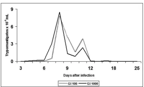
The curve of parasitemia presented the characteristic
profile of the strain Y with a peak of 7.72×10⁶ and
8.47×10⁶trypomastigotes/mL at 8 dpi in the animals
from IG100and IG1000. Likewise, there was no statistic
differences amongst infectivity, prepatent period, patent period and total parasitemia in the groups evaluated (Fig. 1, Table I). The body weight of the animals did not vary significantly amongst the groups from the beginning of the experiment to 18, 30 and 75 dpi (Table II).
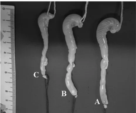
At 75 dpi, the colon of the mice infected with 100 and 1,000 blood trypomastigotes presented 9.3cm and 10.0cm length (p=0.0591) and 1.2cm and 1.5cm width
(p>0.05), respectively. Therefore, the total area of the
organ was larger in the proportion of 32.74% in IG1000, when compared to the CG, without presenting statistic
differences (p>0.05) (Table III and Figure 2).
QUANTITATIVEANALYSIS OF THEMYENTERICPLEXUS
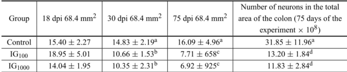
In Table IV, the neuronal density of the CG was observed to have remained constant throughout time
(p>0.05), whereas in the IG this parameter decreased
significantly (p<0.05). At 18 dpi, a decrease of 0% in
the neuronal density in the IG100and 9% in the IG1000
in relation to the CG was observed – without statistic
differences (p>0.05). At 30 dpi, the animals from the
IG100 and IG1000 presented a decrease of the neuronal
density of 28.1% and 35.1%, respectively, in relation
to the CG (p<0.05). At 75 dpi, a decrease of 52.0%
in the IG100 and 57.0% in the IG100 was observed in
relation to the CG (p<0.05). When the projection of
the number of neurons in the total area of the colon was
Fig. 1 – Curve of average parasitemia in 60-day-oldSwissmale mice infected by the strain Y ofT. cruzi. IG100– animals inoculated with 100 blood trypomastigotes, IG1000– animals inoculated with 1000 forms of blood trypomastigotes.
TABLE I
Parasitological parameters evaluated in 60-day-oldSwissmale mice experimentally infected byT. cruziY strain.
Group Infectivity Prepatent period Patent period Peak of parasites Total parasitemia (%) (days) (days) (trypomastigotes/mL×106) (trypomastigotes/mL×106)
IG100 100 4.89±0.93 12.67±1.94 7.72±2.96 18.23±6.70
IG1000 100 4.00±0.00 12.20±2.39 8.67±7.74 16.78±6.17
IG100, IG1000– animals inoculated with 100 or 1000 blood trypomastigotes.
TABLE II
Average body weight ofSwissmale mice infected with 100 (IG100) and 1000 (IG1000) forms of blood trypomastigotes fromT. cruziY strain
and the non-infected controls (Control Group – CG).
Groups Weight (g)
T0(n=28) T18(n=28) T30(n=19) T75(n=10) CG 40.16±3.96 44.27±4.09 45.72±6.30 37.03±26.73
IG100 37.22±4.32 41.06±4.15 45.24±5.36 36.32±27.09
IG1000 37.49±4.35 39.69±4.96 44.70±4.18 40.17±24.11
T0= beginning of the experiment, T18, 30and75= 18, 30 and 75 dpi – days post inoculation. Values expressed as mean±standard deviation. There was no statistic difference among the values observed in the different groups at the times evaluated.
63.4% in the IG1000(p<0.05) was verified. Statistic
dif-ferences in the same group were observed on different days of infection. At 18 and 75 dpi, there was a
signifi-cant difference (p<0.01) for both IG100and IG1000.
MORPHOMETRY OF THEMYENTERICNEURONS
The values for the area of the cell body, area of the nucleus, area of the cytoplasm and nucleus/body cell ratio for the groups studied are presented in Table V.
Hypertrophy was observed in all parameters evaluated, except for the nucleus/body cell ratio, as follows. The
increase of 154.0% 55.9µm2) and 176.0% (60.7µm2)
in the area of the nucleus in the IG100 and IG1000,
re-spectively, was observed when compared to the CG
(22.0µm2) (p<0.05). The area of the cytoplasm
pre-sented an increase of 181.0% (95.8µm2) and 199.0%
(101.8µm2) in the IG100 and IG1000, respectively,
TABLE III
Mean and standard deviation in length, width and area of the colon ofSwissmale mice infected by the strain Y ofT. cruzi
(IG100and IG1000) at 75 dpi – days post inoculation and the non-infected controls (CG).
Groups Length (cm) Width (cm) Area (cm2) CG 9.0±2.3 1.2±0.1 11.3±4.1
IG100 9.3±2.0 1.2±0.2 11.1±2.0
IG1000 10.0±1.1 1.5±0.1 15.0±2.3
Values expressed as mean±standard deviation. There was no statistic differ-ence among the values observed in the different groups at the times evaluated. IG100, IG1000– animals inoculated with 100 or 1000 blood trypomastigotes.
Fig. 2 – Colon ofSwissmale mice inoculated with 1000 (A) and 100 (B) blood trypomastigotes from the strain Y ofT. cruziat 75 dpi – days post inoculation and the non-infected controls (C).
also be observed that the area of the cell body
in-creased 173.0% (153.5µm2) in the IG100 and 198.0%
(167.5µm2) in the IG1000, in relation to the CG
(56.2µm2) (p<0.0001). A significant increase of the
area of the body cell, cytoplasm and nucleus was also
observed in the animals in the IG100 and IG1000. The
ratio for the area of the nucleus and the area of the body of the myenteric neurons in the animals in the
IG100 and IG1000 presented a significant decrease (p<
0.05) in relation to the CG; however, no statistic differ-ences were observed when comparing the IGs.
The degree of correlation among the area of the body, nucleus and cytoplasm of the neurons measured is presented in Table VI.
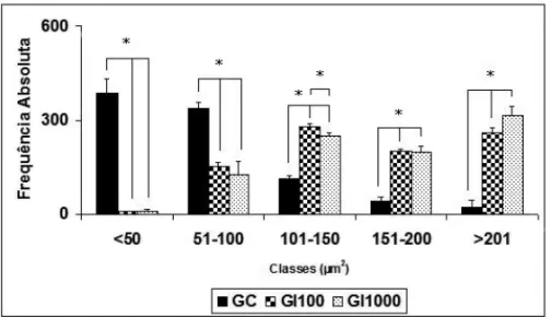
Statistic differences among the CG, GI100 and
GI1000for the classes of neurons according to the area
of the body cell in intervals of<50µm2; 51-100µm2;
101-150µm2; 151-200µm2 and >201µm2 (p<0.01)
and between GI100and GI1000in the class 101-150µm2
(p<0.0001) are presented in Figure 3.
Statistic differences among the CG and GI100 for
the class of neurons with nucleus/body ratio 0.21-30µm2
(p<0.05) are presented in Figure 4.
DISCUSSION
Most of the individuals infected with T. cruzi is
TABLE IV
Mean and standard deviation of the myenteric neuronal density* in the colon ofSwissmale mice infected by the strain Y with 100 (IG100) and 1000 (IG1000) forms of trypomastigotes
and the non-infected controls.
Number of neurons in the total Group 18 dpi 68.4 mm2 30 dpi 68.4 mm2 75 dpi 68.4 mm2 area of the colon (75 days of the
experiment×108)
Control 15.40±2.27 14.83±2.19a 16.09±4.96a 31.85±11.96a IG100 18.95±5.01 10.66±1.53b 7.71±658c 13.20±1.84d
IG1000 14.04±1.95 10.35±2.31b 6.92±925c 11.83±2.84d
*In 360 microscopic fields and area of 68.4 mm2. Data in the same column with different letters present significant differences (a×b: p<0.05; a×c: p<0.01; a×d: p<0.05) dpi – days post inoculation.
TABLE V
Median of the area of the nucleus, cytoplasm and body cell of the myenteric neurons in the colon ofSwissmale mice infected byT. cruziY strain with 100 (IG100) and 1000 (IG1000) forms
of trypomastigotes and the non-infected controls.
Parameter Median (percentile 25; 75)
CG IG100 IG1000
Area of the Nucleus (µm2) 22.0a(15.2; 32.8) 55.9b(42.0; 70.8) 60.7c(46.3; 79.7) Area of the Cytoplasm (µm2) 34.1a(21.0; 57.4) 95.8b(63.6; 147.7) 101.8c(65.4; 161.4)
Area of the Body Cell (µm2) 56.2a(38.1; 89.8) 153.5b(112.8; 213.0) 167.5c(119.8; 234.1) Nucleus/Body Cell ratio 0.38a(0.30; 0.47) 0.37b(0.28; 0.44) 0.37b(0.28; 0.46)
Values followed by different letters in the same line express statistically significant differences (p<0.05).
TABLE VI
Correlation index among the area of the nucleus, cytoplasm and body cell in the myenteric neurons of the colon ofSwissmale mice infected by 100 (IG100) and 1,000 (IG1000) trypomastigotes forms ofT. cruzi
Y strain and the non-infected controls. Relation between the Correlation index
considered areas CG IG100 IG1000 Body cell×Nucleus 0.84 0.68 0.70
Body cell×Cytoplasm 0.95 0.96 0.95
Nucleus×Cytoplasm 0.66 0.48 0.50
treatment (Jurberg 2009). Some of the major compli-cations of the Chagas disease are related to the com-promising of the functioning of the digestive tube and heart. Among them, megacolon stands out as a result of the destruction of the enteric neurons (Silveira et al. 2008). These neurons are located in the wall of the colon, organized in ganglions and interconnected by bundles of nerve fibers that constitute the myenteric and
sub-mucosal plexus (Furness and Costa 2006). Experimen-tal models that are capable of reproducing the changes observed in the Chagas disease and enable the evalua-tion of intervenevalua-tions are necessary and important.
This paper had the purpose of establishing an
ex-perimental model of the chronic infection byT. cruziby
for innovative approaches for therapeutic interventions in infected individuals.
The parasitological parameters evaluated in the strain Y showed high parasitemia according to the characteristics of such strain described in the literature (Brener 1962). Likewise, in a recent study, Schebeleski-Soares et al. (2009) showed the same behavior of the strain Y in BALB/c female mice, but with mortality for all the infected animals. In this study, the chronifica-tion of the infecchronifica-tion with low mortality was due to the discontinuous treatment with benznidazole. The use of two different inocula, despite of not reflecting the dif-ferentiation of parasitological parameters, caused differ-ences in the morphometry of the myenteric neurons.
According to the literature, the infection byT. cruzi
causes progressive weight loss and even cachexy in humans (Meyer et al. 2006). In this study, the body weight of the animals was not significantly different amongst the infected and control groups at 18, 30, 75 dpi. This was probably due to the treatment conducted with benznidazole for the chronification of the infec-tion. Although this drug did not present full efficacy in terms of cure, it restrains the evolution of the parasite-mia, which is, at last, one of the causes of morbidity of
the infection byT. cruzi(Fabbro et al. 2007).
Regarding the number of neurons amongst the dif-ferent segments of the colon (proximal, middle and dis-tal), no differences were observed for the mice inoc-ulated with 100 and 1,000 blood trypomastigotes and the control group. These data are in accordance with those of Maifrino (1996), who also observed no signif-icant difference, in the number of neurons among the three segments of the colon of mice at 50 days of
infec-tion with 1,000 blood form of the strain Y ofT. cruzi
and its controls.
At 75 dpi, the increase of 0% and 25% in the
width of the colon in the IG100and IG1000, respectively,
was observed in relation to the non-infected group. The increase of 3.3% (IG100) and 11.1% (IG1000) in length was verified in relation to the CG. Thus, the infection was observed to cause a tendency to the hypertrophy of the colon and, consequently, the total area of the
or-gan with 0.0% IG100and 32.7% IG1000, when compared
to the CG, although presenting no statistic differences
(p>0.05). Neither macroscopic changes nor fecal
im-paction were observed. In studies conducted with pa-tients with Chagas disease, the neuronal decrease was observed related to the megacolon (Adad et al. 2001, Iantorno et al. 2007, Silveira et al. 2007a, 2008). In this study, the decrease of the neuronal population was not related to the change in width and height of the organ. This certainly can be related to other factors such as the parasite-host relation, the inoculum and, mainly, the time of infection to which the animals were submitted.
Several studies had already showed that the de-struction of the myenteric neurons triggers an import-ant change of motility in the colon causing megacolon (Koberle 1968, 1970, Tafuri et al. 1971, Maifrino et al. 1999, 2005, Iantorno et al. 2007, Silveira et al. 2007a, b, 2008) – a phenomenon dependent on the degree of parasitism and time of infection.
The animals in the IG100 and IG1000 had
signifi-cant lower density of myenteric neurons than the ani-mals in the CG. The data showed a tendency to den-ervation what was dependent on the time of infection. At 18 dpi, no difference was verified among the neu-ronal density of the colon of the mice in the three groups. These data differ from those of Arantes et al. (2004) who observed neuronal decrease of 60% at 10 dpi in the colon of C57BL/6 mice inoculated with 100 blood trypomastigotes of the strain Y, highlighting the importance of the genetic characteristics of the host
in the determination of morbidity in the infection byT.
cruzi. Although C57BL/6 mice are more resistant than Swiss mice, the high variation on genetic background in nonisogenic models may be responsible for the high degree of intrinsic variation and conclusions must be regarded with caution.
In this experiment, at 30 dpi, the decrease of neu-ronal density was 28.1% and 35.1% with the inocula of 100 and 1,000 forms of trypomastigotes, respectively.
At 75 dpi, the decrease was 52.0% IG100 and 57.0%
IG1000(p<0.01) compared to control. These data agree
with those of other researchers (Maifrino 1996, Maifri-no et al. 1999, 2005) who observed neuronal decrease from 39.0% to 85.7% in the colon of mice infected with 100 and 1,000 forms of blood trypomastigotes of the strain Y, validating our findings for the establishment of
a model of denervation of the chronic infection by T.
Fig. 3 – Histogram of the classes of myenteric neurons in the colon ofSwissmale mice according to the area (µm2) of the body cell in the animals inoculated with 100 (IG100) and 1000 (IG1000) trypomastigotes ofT. cruziY strain at 75 dpi – days post inoculation and the non-infected controls (CG). (*) = (P<0.0001).
Fig. 4 – Histogram of the nucleus/body cell ratio in the myenteric neurons in the colon of
Swissmale mice inoculated with 100 (IG100) and 1,000 (IG1000) trypomastigotes ofT. cruzi Y strain at 75 dpi – days post inoculation and the non-infected controls (CG) (*)=(p<0.05).
Different authors showed that the colon of hu-mans with Chagas disease presents a neuronal decrease from 55.0% to 66.67% (Adad et al. 2001, Silveira et al. 2007a, 2008). The differences in the percentages obtained by different researchers are probably due to different neuronal classes investigated, strain of the parasite, inoculum and time of infection.
At 75 dpi, the projection of the number of neurons for the total area of the colon was evaluated. A
neu-ronal death index of 59.0% in the IG100 and 63.4% in
the IG1000(p<0.05) was verified when compared to the
CG. It was possible to notice that such evaluation was not conducted in former studies (Maifrino 1996,
Mai-frino et al. 1999, 2005). Statistic differences (p<0.01)
were observed within the same experimental group from
18 to 75 days of infection in both IG100and IG1000.
The data showed hypertrophy of the myenteric
neurons in the animals in the IG100 and IG1000 as the
nucleus increased 154.0% (IG100) and 176.0% (IG1000).
The cytoplasm increased 181.0% and 199.0% and,
consequently, the cell body increased significantly (p<
0.0001) 173.0% and 198.0%, respectively, in IG100and
IG1000. The increase of the body cell suggested to have been caused mainly by the increase of the cytoplasm as the degree of correlation among these parameters, is high despite the size of the inoculum (100: r=0.96/
p<0.0001 and 1.000: r=0.95/p<0.0001). It also
be-tween the cytoplasm and the nucleus of the neurons
in the animals in IG100 (r=0.48 and p<0.0001) and
IG1000 (r=0.50 and p<0.0001) has contributed for the
decrease of the area of the nucleus/cell body ratio in the IGs, indicating that the cytoplasm began to occupy more space in the cell. The neuronal hypertrophy
ob-served in the animals in IG100 and IG1000 explains the
progressive increase of the number of neurons when distributed in classes according to the area of the cell body. The inversely proportional distribution for this number of neurons when comparing the CG to the IGs stands out, and the class marking this inversion was the
one with neurons with a median from 101 to 150µm2.
Although a discrete decrease of the area of the nucleus/ area of the cell body ratio was observed, considering the infected and control groups, practically no change was observed in the distribution of the number of neurons in the intervals of class regarding this parameter, showing that this phenomenon occurred homogeneously among the neurons in the colon. Maifrino et al. (1999), while studying specific subpopulations of neurons (NADH-d
and AChE) in the colon ofSwiss male mice infected
with 1,000 forms of trypomastigotes of the strain Y of T. cruzi for 60 days, observed a significant death of medium and large neurons. Nevertheless, while using the inoculum with 100 forms of trypomastigotes of the same strain and time of infection, no significant morphometric change was observed in the neurons stained with NADH-d in the colon of BALB/c male mice (Maifrino et al. 2005).
This morphometric analysis of the myenteric neu-rons enabled to sustain the hypothesis that the physi-ology of the myenteric neurons is changed when the
host is parasited by T. cruzi. The hypertrophy of the
survival neuronal population was observed in the in-fected animals. Besides, in these animals, the neurons also had their nuclei increased, presenting a significant
change between IG100and IG1000, and this relation
de-monstrated to be proportionally different the inoculum. This finding suggests that there was an increase of the gene expression for a higher production of proteins with consequent increase of the cytoplasm. Chagas disease is characterized by the development of generalized
inflam-matory process (De Lana and TafuriapudNeves et al.
2005). Silveira et al. (2007b) observed, in the
myen-teric plexus in the colon of patients with Chagas dis-ease with megacolon, that the excitatory neurons that produce substance P had an increase in the frequency of neurochemical markers. Substance P, while secreted, contributes for the neurogenesis of the inflammation (Winter et al. 1995). Likewise, Silveira et al. (2008) found high levels of substance P while studying the ex-pression of this neuropeptide in patients with chagasic megacolon. In this study, hypertrophy in the neurons is suggested to be related to the neurochemical marker. Future studies involving the analysis of neurotransmit-ters and neuromodulators are necessary to understand the mechanism of action of the parasite in the myenteric denervation, considering the proposed model.
Considering that the Laboratório de Doença de
Chagas/UEM has the purpose of improving the
atten-tion provided to the subject infected by T. cruzi, even
when basic research is conducted, we sought in this study a direct relation between the findings and their practical application. Literature shows that capsaicin (an active component of black pepper) stimulates the release of substance P; however, the continuous use of capsaicin leads to the depletion of the substance P in-volved in the inflammatory process (Graton 2009, Win-ter et al. 1995). Therefore, we have evidences to support the hypothesis that the continuous use of capsaicin can result in benefits for chagasic patients in order to de-crease substance P and, consequently, reduce morbid-ity by decreasing the generalized inflammatory process. This approach is one of the perspectives for the continu-ation of this study. Another aspect to be discussed is the fact that substance P is related to the stimulation of the production of interleukins IL1, IL6, IL8, and TNF-alfa (Adad et al. 2001), which are involved in the resistance of the parasite. Once the treatment with capsaicin is able to deplete them, other perspective for the expansion of this study would be the investigation of the effect of the treatment with capsaicin on the production of different
interleukins bySwissmice infected byT. cruzi,
associ-ated or not to different interventions.
CONCLUSION
The model of the chronic infection inSwissmale mice
infected byT. cruziY strain and chronified by the
observing the changes in the myenteric neuronal pop-ulation (the decrease in the density/neuronal death and morphometric changes), as they are directly related to the inoculum and the time of infection with consequent plastic changes culminating in the hypertrophy of these neurons.
ACKNOWLEDGMENTS
This research was supported by grants from Coorde-nação de Aperfeiçoamento de Pessoal de Nível Superior (CAPES) and Fundação Araucária.
RESUMO
Definir um modelo experimental de avaliação de alterações quantitativas e morfométricas nos neurônios mientéricos do cólon de camundongos infectados pelo Trypanosoma cruzi. Vinte e oito camundongos Swissmachos foram distribuídos nos grupos: controle (GC, n=9) e infectados com 100 (IG100, n=9) e 1000 (IG1000, n=10) tripomastigotas sanguíneos, cepa Y-T. cruziII. A parasitemia foi avaliada 3-25 dias pós inocu-lação (dpi), com pico de parasitos de 7,7×10⁶e 8,4×10⁶
tripomastigotas/mL no 8◦dpi (p>0,05) em IG100 e IG1000,
respectivamente. A fase crônica da infecção foi obtida com duas doses de 100mg/Kg/weight e uma dose de 250mg/Kg/ weight do benznidazol, em 11, 16 e 18 dpi. Três animais de cada grupo foram sacrificados aos 18, 30 e 75 dpi. O cólon foi corado com Giemsa. A análise quantitativa e morfométrica de neurônios revelou que a infecção causou uma diminuição da densidade neuronal no 30◦dpi (p<0,05) e 75 dpi (p<0,05)
em IG100e IG1000. A infecção causou morte e hipertrofia neu-ronal no 75◦ dpi em IG100 e IG1000 (p<0,05, p<0,01). As
alterações observadas nos neurônios mientéricos foram dire-tamente relacionadas ao inóculo e tempo de infecção. Palavras-chave: doença de Chagas, cólon, sistema nervoso entérico, Giemsa,Trypanosoma cruzi.
REFERENCES
ADAD SJ, CANÇADO CG, ETCHEBEHERE RM, TEIXEI -RAVP, GOMESUA, CHAPEDEIROEANDLOPESER. 2001. Neuron count reevaluation in the myenteric plexus of chagasic megacolon after morphometric neuron ana-lysis. Virchows Arch 438: 254–258.
ALEIXO DL, FERRAZ FN, DE MELO CS, GOMES ML, TOLEDOMJ, KANESHIMAEN, BERSANI-AMADOCA AND DEARAÚJOSM. 2008. Changes of RAPD profile ofTrypanosoma cruziII with Canova and Benznidazole. Homeopathy 97: 59–64.
ARANTESRME, MARCHEHHF, BAHIAMT, CUNHAFQ, ROSSI MA ANDSILVA JS. 2004. Interferon-y-induced nitric oxide causes intrinsic intestinal denervation in Try-panosoma cruzi-infected mice. Am J Pathol 164: 1361– 1368.
ARAÚJOSM, ANDÓMH, CASSAROTTIDJ, MOTADCGD, BORGESSMANDGOMESML. 2000. Programa ACHEI: Atenção ao Chagásico com Educação Integral no municí-pio de Maringá e região Noroeste do Paraná, Brasil. Rev Soc Bras Med Trop 33: 565–572.
ARAÚJOSMANDCHIARIE. 1988. Caracterização Biológi-ca de Clones das Cepas Y, Cl e Mr deTrypanosoma cruzi
em Camundongos C3H Isogênicos. Rev Soc Bras Med Trop 83: 175–181.
BARBOSA AJA. 1978. Técnica histoquímica para gânglios nervosos intramurais e preparados espessos. Rev Bras Pes Med Biol 11: 95–97.
BRENERZ. 1962. therapeutic activity and criterion of cure on mice experimentally infected withTrypanosoma cruzi. Rev Inst Med Trop 4: 389–396.
D’ARCEMOTADCG, GOMESMLAND DEARAÚJOSM. 2008. Programa achei e o princípio da autonomia: exem-plo de como um serviço de saúde pode informar o usuário de maneira simples e acessível. SaBios: Rev Saúde e Biol 3: 5–9.
DELANAM ANDTAFURI WL. 2005. Trypanosoma cruzi e doença de Chagas. In: NEVES DP, DE MELO AL, LINARDIPMANDVITORRWA. Parasitologia humana. 11aed., São Paulo: Atheneu, 85 p.
FABBRODL, STREIGERML, ARIASED, BIZAIML,DEL BARCOMANDAMICONENA. 2007. Trypanocide treat-ment among adults with chronic Chagas disease living in Santa Fé City (Argentina), over a mean follow-up of 21 years: parasitological, serological and clinical evolution. Rev Soc Bras Med trop 40: 1–10.
FURNESS JB AND COSTA M. 2006. The enteric nervous system. New York: Churchill Livingstone, p. 1–28. GRATONV. 2009. Você conhece a capsaicina? Disponível
em: < http://www.nutricaosadia.com.br/2009/07/pimen-ta-qual-sua-importancia-para-sua.html>. Disponível em: 13-08-2009.
HOTEZ PJ, BOTTAZZI ME, FRANCO-PAREDES C, AULT SKANDPERIAGOMR. 2008. The neglited tropical dis-eases of Latin America and the Caribbean: A review of disease burden and distribuition and a roadmap for con-trol and elimination. PLoS Negl Trop Dis 2: 1–10. IANTORNO GET AL. 2007. The enteric nervous system in
chagasic and idiopathic megacolon. AM J Surg Pathol 31: 460–468.
JURBERG C. 2009. Chagas: one hundred years later. Bull World Health Organ 87: 491–492.
KOBERLEF. 1968. Chagas’ disease and Chagas’ syndromes: the pathology of American trypanosomiasis. Adv para-sitol 6: 63–116.
KOBERLE F. 1970. The causation and importance of ner-vous lesions in American Trypanosomiasis. Bull World Health Organ Org 42: 739–743.
MAIFRINOLBM. 1996. Aspectos morfológicos, histoquími-cos e imunohistoquímihistoquími-cos do plexo mientérico do colo de camundongo ‘swiss’ (Mus musculus) na fase crônica da infecção porTrypanosoma cruziCepa Y. São Paulo, 1996 (Tese de Doutorado – Universidade de São Paulo). MAIFRINOLBM, AMARALSON, WATANABEI, LIBERTI
EAANDZOUSARR. 2005.Trypanosoma cruzi: Prelim-inary investigation of NADH-positive and somastotatin-immunoreactive neurons in the myebteric plexus of the mouse colon during the infection. Exp Parasitol 111: 224–229.
MAIFRINO LBM, LIBERTI EA, WATANABE II-SEI AND SOUZARR. 1999. Morfhometry and acetylcholinester-ase activity colon in the chronic phacetylcholinester-ase of experimental
Trypanosome cruziinfection. Am J Trop Med Hyg 60: 721–725.
MARIN-NETO JA, CUNHA-NETO E, MARCIAL BCAND SIMÕES MV. 2007. Pathogesesis of chronic Chagas heart disease. Circulation 115: 1109–1123.
MARTINS HR ET AL. 2008. Persistence of PCR-positive tissue in benznidazole-treated mice with negative blood parasitological and serological tests in dual infections withTrypanosoma cruzistocks from different genotypes. J Antimicrob Chemother 61: 1319–1327.
MEYERIF, KANESHIMAEMAND DESOUZA-KANESHIMA AM. 2006. Alterações no sistema digestivo desencadea-das pelo quadro infeccioso do Trypanosoma cruzi. Ini-ciação Científica CESUMAR 8: 11–23.
MURTASMFANDROMANHAAJ. 1998.In vivoselection of a population ofTrypanosoma cruziand clones resistant to benznidazole. Parasitology 116: 165–171.
PAHO– PAN AMERICANHEALTHORGANIZATION. 2007. Health in the Americas (2007) Regional, scientific and technical publication. PLoS Negl trop Dis 622: 176–182. SANTOSJRANDMONTEIROJC. 2002. Megacólon-Parte II:
Doença de Chagas. Rev Bras Coloproctol 4: 266–267. SCHEBELESKI-SOARES C, OCCHI RC, DE MORAES F,
MARTASD,DEOLIVEIRAM, ALMEIDAFN, TOLEDO MJO AND ARAUJO SM. 2009. Pre-Infection Aerobic Treadmill Training Improves Resistance against. Appl Physiol Nut Metab 34: 659–665.
SENIOR K. 2007. Chagas disease: moving towards global elimination. Lancet Infect Dis 7: 572.
SILVEIRAABM, D’AVILAREISD, OLIVEIRAEC, NETO SG, LUQUETTI AO, POOLED, CORREA-OLIVEIRAR ANDFURNESSJB. 2007a. Nurochemical coding of the enteric nervous system in chagasic patients with mega-colon. Dig Dis Sci 52: 2877–2883.
SILVEIRAABM, FREITASMAR, OLIVEIRAEC, NETOSG, LUQUETTI AO, FURNESS JB, CORREA-OLIVEIRA R ANDD’AVILAREISD. 2008. Neuronal plasticity of the enteric nervous system is correlated with chagasic mega-colon development. Parasitology 135: 1337–1342. SILVEIRAABM, LEMOSEM, ADADESJ, OLIVEIRARC,
FURNESJBANDREISDD. 2007b. Megacolon in Cha-gas disease: a stury of inflammatory cells, enteric nerves, and glial cells. Hum Pathol 38: 1256–1264.
TAFURI WL, MARIA TA AND LOPES ER. 1971. Lesões do plexo mientérico do esôfago, do jejuno e do colo de chagásicos crônicos. Ver Inst Med Trop 13: 76–91. WINTERJ, BEVANSANDCAMPBELLEA. 1995. Capsaicin