Fausto Daniel dos Santos Queda
[Nome completo do autor]
[Nome completo do autor]
[Nome completo do autor]
[Nome completo do autor]
[Nome completo do autor]
[Nome completo do autor]
[Nome completo do autor]
Mestre em Química Bioorgânica
[Habilitações Académicas] [Habilitações Académicas] [Habilitações Académicas] [Habilitações Académicas] [Habilitações Académicas] [Habilitações Académicas] [Habilitações Académicas] Dezembro, 2019
Chitin and chitosan as reliable templates towards the
carbohydrate backbone of bacterial peptidoglycan
Dissertação para obtenção do Grau de Doutor em Química Sustentável
Dissertação para obtenção do Grau de Mestre em [Engenharia Informática]
Orientador: Maria Manuel Marques, Professora Auxiliar com Agregação, LAQV@REQUIMTE, Departamento de Química, Faculdade de Ciências e Tecnologias, Universidade Nova de Lisboa
Co-orientadores:
Sérgio R. Filipe, Professor Auxiliar, UCIBIO@REQUIMTE, Departamento de Ciências da Vida, Faculdade de Ciências e Tecnologias, Universidade Nova de Lisboa
Júri:
Presidente: Manuel Nunes da Ponte
Arguentes: Amélia Pilar Rauter Nuno R. Candeias Vogais: Marta C. Corvo
Chitin and chitosan as reliable templates towards the carbohydrate backbone of bacterial peptidoglycan
Copyright © Fausto Daniel dos Santos Queda, Faculdade de Ciências e Tecnologia, Universidade Nova de Lisboa.
A Faculdade de Ciências e Tecnologia e a Universidade Nova de Lisboa têm o direito, perpétuo e sem limites geográficos, de arquivar e publicar esta dissertação através de exemplares impressos reproduzidos em papel ou de forma digital, ou por qualquer outro meio conhecido ou que venha a ser inventado, e de a divulgar através de repositórios científicos e de admitir a sua cópia e distribuição com objetivos educacionais ou de investigação, não comerciais, desde que seja dado crédito ao autor e editor
First of all, I would like to express my gratitude to Dr. Maria Manuel Marques and Dr. Sérgio Filipe for my research orientation, for the scientific guidance, all the support and friendship during this project.
I am especially grateful for Dr. Maria Manuel Marques for the scientific lessons and supervision. I am thankful for all the support during this project and thesis writing, for the opportunity to join her research group, for the optimism and advices that allowed me to grow as a student and researcher. Thank you for being present along this journey.
I also want to acknowledge Dr. Sérgio Filipe and his PhD student Gonçalo Covas for all the support, for offering me the opportunity to join his research group at ITQB to perform the biological essays.
To Prof. Dr. Joachim Thiem and to his PhD Student Christian Czaschke at University of Hamburg for the collaboration, the lessons, friendship and the opportunity to stay at his lab. I gratefully acknowledge to Fundação para a Ciência e Tecnologia the funding sources (PD/BD/109632/2015) that made my PhD possible.
I also acknowledge Portuguese Nuclear Magnetic Resonance Network. I am grateful to the analytical departments, namely the NMR, to Ana Teresa, Cecília Bonifácio and Marta Corvo. I would also like to thank to my colleagues, partners in the carbohydrate journey: Marina Pires, Diogo Poeira, Ricardo Chagas, Ana Sofia Santos and João Macara. I am mostly grateful to my lab colleagues – for the patience, for the good discussions and assistance, for the excellent collaboration.
I also have to thank to those with I shared laboratory 202 and 205; those who passed yet stayed.
A special thanks to my friends, André Dias, Helena Coelho, João Cascão those who listened and supported on the chemical point of view. Also, a special thanks to the rest of my friends for the support which gave me hope and strength to complete this journey.
I am much grateful to Nídia, for patience, care and friendship, for the constant optimism and for making me reset of the laboratory frustrations; thank you.
Huge thanks for my family for the unconditional support, your patience, your advices and your love were always present in my life; that made me what I am today, thank you. Last but not least a very special thanks to my new born daughter Pilar, this is for you.
Peptidoglycan (PGN) is a major component of the bacteria cell wall that surrounds and protect bacteria from the surrounding environment. The composition of PGN is often linked with the outcome of bacterial infections and its synthesis is the target of the different classes of antibiotics frequently used in the treatment of these bacterial infections.
The scientific community has been requesting to availability of increasing amounts of pure, well defined, PGN fragments in order to be able to move forward with the biological studies in a reliable way. The extraction processes used to purify PGN from bacteria rely on the use of harsh conditions that can be harmful to the user and also the environment.
Thus, through the last thirty years, different groups have been dedicating time and resources to establish new synthetic routes to produce PGN fragments of different composition. These routes rely on the use of fully functionalized glucosamine building blocks, through a protecting group orthogonal synthesis, followed by glycosylation and peptide coupling reactions.
The challenge, fully framed in a sustainable chemistry doctoral program, was to develop new synthetic routes towards PGN fragments using more sustainable processes. In this thesis it is reported the synthesis of PGN fragments using as starting material the polymers of chitin and chitosan, which are present in significant amount in food industry wastes.
In the first approach, an acetolysis reaction of chitin was used to give a peracetylated disaccharide, in a gram scale amount, which, through protecting group orthogonal synthesis, can give an advanced PGN intermediate in a five synthetic steps. As this intermediate possesses all the functional groups in the correct position as the PGN fragments, it may be considered a high value synthetic intermediate.
On the second approach, the polymer of chitosan was used as starting material in a chemoenzymatic synthesis. Through orthogonal protecting group strategy, it was possible modify a high weight chitosan molecules in order to produce a PGN surrogate. Using commercially available PGN hydrolases, we were able to hydrolyze this surrogate in order to identify the oligosaccharides that are present in the PGN sugar backbone.
O Peptidoglicano (PGN) é um importante constituinte da parede celular bacteriana que está diretamente relacionado com a progressão de diferentes infecções bacterianas e com diferentes mecanismos de resistência a antibióticos.
Este trabalho surge no enquadramento do problema com o qual a comunidade científica se tem vindo a deparar aquando da obtenção de fragmentos de PGN puros e, em quantidade, que permita a realização de ensaios biológicos. Tendo em conta que, os processos de extração da parede celular da bactéria, utilizam condições agressivas para o utilizador bem como, para com o meio ambiente.
Desta forma, a comunidade científica tem vindo a desenvolver, ao longo dos últimos trinta anos, novas vias de síntese destes fragmentos. Estas vias sintéticas baseiam-se na utilização de unidades de glucosamina funcionalizadas que através do uso de grupos protectores, síntese ortogonal, reacções de glicosilação e acoplamento peptídico, e dão origem a fragmentos de PGN.
Sendo este projecto inserido num programa doutoral em química sustentável o desafio era criar novas vias sintéticas para obtenção de fragmentos de PGN utilizando, em alternativa aos métodos já reportados, processos mais sustentáveis.
Assim sendo nesta tese é apresentada a síntese de fragmentos de PGN utilizando como material de partida, um desperdício da indústria alimentar, quitina e quitosano.
Numa primeira abordagem através de uma reacção de acetólise da quitina foi possível obter o seu correspondente dissacárido per-acetilado que através de uma estratégia de grupos protectores, originou um intermediário avançado em cinco passos de síntese. Este intermediário apresenta todos os grupos funcionais, posicionados corretamente, que os fragmentos de possuem PGN, sendo por isso considerado um intermediário de alto valor sintético.
Numa segunda estratégia, foi utilizado quitosano como material de partida numa síntese quimioenzimatática. Através de uma estratégia de grupos portectores foi possível modificar um polímero de alto peso molecular num mimético a componente de carbohidrato do PGN. Após hidrólise de enzimas que reconhecem o PGN, foi possível identificar vários oligossacáridos que constituem o PGN.
Contents
THESIS STRUCTURE ... 1
1 BACTERIA OVERVIEW ... 1
1.1 INTRODUCTION ... 2
1.1.1 Carbohydrate Polymers of b-(1,4) linked N-acetylglucosamine (NAG) ... 2
1.1.2 Peptidoglycan biosynthesis ... 3
1.1.3 Bacterial cell wall ... 6
1.1.3.1 Modifications on PGN carbohydrate backbone ... 7
1.1.4 Chitin biosynthesis ... 8
1.2 REFERENCES ... 11
2 SYNTHETIC STUDIES TOWARDS A NAG-NAM DISACCHARIDE... 15
2.1 INTRODUCTION ... 16
2.1.1 Glucosamine -a challenging scaffold ... 16
2.1.2 Synthetic routes towards peptidoglycan fragments ... 19
2.1.3 Synthesis of chitooligosaccharides and other glucosamine congeners ... 34
2.2 RESULTS AND DISCUSSION ... 40
2.2.1 Goals ... 40
2.2.2 Preliminary studies: O-3 regioselective modification on chitobiose derivative ... 42
2.2.3 Preliminary studies on the modification of the NAc group... 47
2.2.4 Approach towards NAG-NAM disaccharidevia a di-NPhthchitobiose derivative ... 54
2.2.5 Approach towards NAG-NAM disaccharide via the di-N-TFA chitobiose derivative ... 56
2.3 CONCLUSIONS... 63
2.4 EXPERIMENTAL... 64
2.4.1 Preliminary synthesis ... 65
2.4.2 O-3 regioselective modification on chitobiose derivative ... 66
2.4.2.1 Preparation of the palladium catalyst ... 66
2.4.2.2 Oxidation experiments carried with the palladium catalyst ... 67
2.4.3 Experiments with theSchwartz’s reagent: General Procedure ... 68
2.4.4 Experiments using Crich’s methodology through N-NO formation... 68
2.4.5 Approaches using di-N-protected chitobiose derivative ... 69
2.4.6 Approach towards a NAG.NAM precusrosvia the di-N-TFA chitobiose derivative ... 70
2.5 REFERENCES ... 78
3 A CHEMO-ENZYMATIC APPROACH TOWARDS NAG-NAM CONTAINING OLIGOSACCHARIDES FROM CHITOSAN ... 84
3.1 INTRODUCTION ... 85
3.1.1 Chitin and Chitosan... 85
3.1.1.1 Selective modifications ... 87 3.1.1.1.1 Acylation ... 88 3.1.1.1.2 Alkylation ... 90 3.1.1.1.3 Quaternization... 92 3.1.1.1.4 Sulfation ... 96 3.1.1.1.5 2-Azidation ... 98
3.1.1.1.6 Phosphoryl and phosphonation ... 99
3.1.1.1.7 Other regioselective O-modifications ... 101
3.2 RESULTS AND DISCUSSION ... 107
3.2.1 Goals ... 107
3.3 RESULTS AND DISCUSSION ... 108
3.3.1 Chemical modification of chitosan ... 109
3.3.2 Biological evaluation of the PGN mimetics ... 119
3.3.3 Enzymatic assays ... 121
3.3.4 LC-MS experiments ... 122
3.5.1.2 Preparation of dicarboxylic acids C1 and C2 ... 128
3.5.1.3 Preparation of ester compounds III.64-67 from N-phthaloyl chitosan (III.3) ... 130
3.5.1.4 Preparation of silylated esters III.68-71 ... 131
3.5.1.5 Insertion of lactate moiety - Preparation of compounds III.72-75 ... 132
3.5.1.6 Removal of protecting groups and N-acetlyation - Preparation of compounds III.77-81 ... 134
3.5.2 Enzymatic digestion ... 135
3.5.3 Monosaccharide composition analysis ... 136
3.5.4 Mass spectra ... 137 3.5.5 mCherry-PGRP-SA assays ... 137 3.5.6 Quantification studies ... 138 3.6 REFERENCES ... 139 4 GENERAL CONCLUSIONS ... 152 5 APPENDIX ... 158 5.1 HPLC DATA ... 159 5.2 LC-MS DATA ... 163 5.3 QUANTIFICATION STUDIES ... 168
Index of Figures
Figure 1.1 Structures of oligosaccharides containing N-acetyl-D-glucosamine (NAG). ... 3
Figure 1.2 Schematic representation of UDP-N-acetylglucosamine synthesis... 4
Figure 1.3 The synthesis and attachment of a new peptidoglycan stand to the existing. .... 5
Figure 2.1 Influence of the C-2 group on the glycosylation stereoselectivity. ... 17
Figure 2.2 Most common amino protecting groups. ... 17
Figure 2.3 Synthesis of PGN fragment II.9 reported by Fukase. ... 20
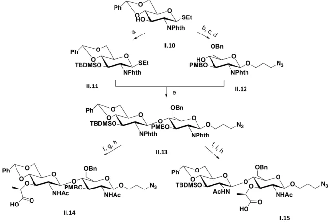
Figure 2.4 Synthesis summary to accomplish II.14 an II.15 reported by Boons. ... 22
Figure 2.5 Synthesis summary to accomplish II.21 reported by Mobashery. ... 24
Figure 2.6 Synthesis summary to accomplish II.27 reported by Walker. ... 27
Figure 2.7 Synthesis summary to accomplish II.30 reported by Fukase. ... 29
Figure 2.8 Synthesis summary to accomplish II.35 reported by Fukase. ... 31
Figure 2.9 Synthesis summary to accomplish II.38 reported by Marques. ... 33
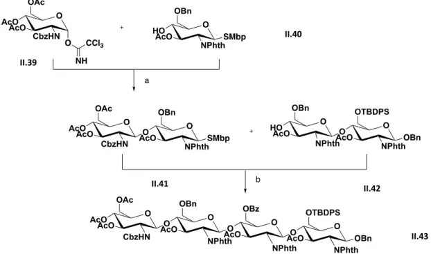
Figure 2.10 Synthesis summary to accomplish II.43 reported by Beau. ... 36
Figure 2.11 Synthesis summary to accomplish II.46 reported by Beau. ... 37
Figure 2.12 Synthesis summary to accomplish II.48 reported by Beau. ... 37
Figure 2.13 Synthesis summary to accomplish II.51 reported by Beau. ... 38
Figure 2.14 Schematic representation of NHTFA oligosaccharides synthesis starting with chitin. ... 38
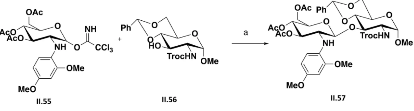
Figure 2.15 Synthesis summary to accomplish II.57 ... 39
Figure 2.16 Schematic summary of the previous reported synthesis towards NAG-NAM containing oligosaccharides. ... 40
Figure 2.17 Schematic synthetic plan towards NAG-NAM containing oligosaccharides... 41
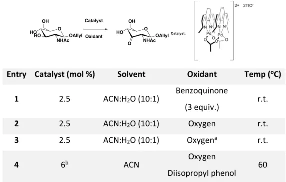
Figure 2.18 Regioselective hydroxy group oxidation catalyzed by palladium. ... 42
Figure 2.19 Proposed synthetic route to achieve a NAM-NAG precursor. ... 43
Figure 2.20 Synthesis of the catalyst for the oxidation reaction. ... 44
Figure 2.21 Monosaccharide O-3 oxidation reaction... 45
Figure 2.22 Disaccharide O-3 oxidation reaction. ... 46
Figure 2.23 Synthetic plan to form NPhthchitobiose (II.74) using Schwartz’s reagent. ... 48
Figure 2.24 Conversion of the NHAc group into other functional groups by NAc-NO modification. ... 49
Figure 2.25 Proposed conversion of N-peracetylated chitobiose into other functional groups using the methodology developed by Crich. ... 49
Figure 2.26 Conversion of NHAc into other functional groups using Crich methodology applied on II.44. ... 50
Figure 2.27 Proposed reaction mechanism to convert II.44 into II.83. ... 51
Figure 2.28 Reaction mechanism to afford glucose pentaacetate II.84 from II.81. ... 51
Figure 2.29 N(H)HSQC experiment of II.53 and II.78. ... 53
Figure 2.30 Versatile fully functionalized molecule that could be used as both acceptor and donor... 54
Figure 2.31 Synthetic route designed to synthesize II.85. ... 55
Figure 2.32 Synthetic route designed to convert NHAc into NPhth peracetylated chitobiose. ... 55
Figure 2.36 Second approach towards NAG-NAM precursor ... 60
Figure 2.37 Versatility of the NAG-NAM precursor II.103 and its wide applications. ... 61
Figure 3.1 Molecular skeleton of chitosan (III.1) (R=H > 50%) and chitin (III.2) (R=acetyl). 85 Figure 3.2 Antiparallel chain arrangement in a-chitin(A); parallel arrangement in b -chitin(B) ... 85
Figure 3.3 Regio- and chemoselective main modifications of chitosan (III.1). ... 88
Figure 3.4 Most commonly used N-protecting groups. ... 89
Figure 3.5 Selective pivaloylation. ... 90
Figure 3.6Examples of O- and N-carboxyalkylation. ... 91
Figure 3.7 Some recently described strategies for preparation of N,N,N-trimethyl chitosan. ... 93
Figure 3.8 Quaternized derivatives described by Sahariah et al... 95
Figure 3.9 Selective sulfation strategies. ... 97
Figure 3.10 Sulfation methods employing O- or N-protected derivatives. ... 98
Figure 3.11 Protocols developed to convert the amino group into azide. ... 99
Figure 3.12 Examples of phosphoryl and phosphonation methods. ... 100
Figure 3.13 General scheme for O-modification... 101
Figure 3.14 Protection of O-6 with trityl group. ... 103
Figure 3.15 Example a versatile O-6 modification. ... 104
Figure 3.16 Synthesis of 6-amino-6-deoxy-chitosan. ... 105
Figure 3.17 Comparison between Murein and Chitin. ... 106
Figure 3.18 Synthetic plan towards NAG-NAM containing oligosaccharides... 107
Figure 3.19 Molecular clamp generic representation, highlighting the different O-3 accessibilities. ... 108
Figure 3.20 Reaction of N-Phthaloylation of chitosan. ... 109
Figure 3.21 Synthesis of the dicarboxylic acids C1 and C2. ... 109
Figure 3.22 Coupling reaction of the dicarboxylic acids to the N-Phth chitosan. ... 110
Figure 3.23 FT-IR comparison between III.3-III.3’ and III.65-III.65’ (medium (left side) and high (right side) molecular weight chitosan); 13C CP-MAS comparison between III.3’ and III.65’ (medium molecular weight chitosan). ... 111
Figure 3.24 Reaction of O-6 Silylation of III.64-III.67. ... 112
Figure 3.25 FT-IR comparison between III.65-III.65’ and III.69-III.69’ (medium (left side) and high (right side) molecular weight chitosan); Comparison of the 13C CP-MAS of III.65’ and III.69’ (medium molecular weight chitosan). ... 112
Figure 3.26 Lactyl moiety insertion reaction scheme. ... 113
Figure 3.27 FT-IR comparison between III.69-III.69’ and III.73-III.73’ (medium (left side) and high (right side) molecular weight chitosan); Comparison of the 13C CP-MAS of III.69 and III.73 (medium molecular weight chitosan). ... 114
Figure 3.28 Synthetic sequence for removal of the protecting groups and N-acetylation. ... 115
Figure 3.29 Comparison of the 13C CP-MAS and FT-IR comparison of medium (left side) and high (right side) molecular weight chitosan along the synthetic sequence. ... 116
Figure 3.32 A – Lysozyme negative control (under the same conditions: III.79, buffer, temperature and reaction time without enzyme) – black; B – Lysozyme digestion – purple;
C – Mutanolysin negative control... 122
Figure 3.33 LC-MS spectra at m/z 521. ... 125
Figure 4.1 PGN synthetic strategies summary. ... 154
Figure 4.2 Summary of the two approaches investigated towards NAG-NAM precursors and mimetics ... 155
Figure S5.1: III.78 digestion ... 162
Figure S5.2: III.79 chromatogram after mutanolysin digestion. ... 164
Table 2.1 Glycosylation conditions regarding the N-protecting groups and their selectivity 18 Table 2.2 Monosaccharide optimization conditions for the O-3 oxidation reaction ... 45 Table 2.3 Disaccharide optimization conditions for the O-3 oxidation reaction with 3 equiv. of Benzoquinone as oxidant ... 47 Table 2.4 Monosaccharide optimization conditions for the NHAc transformation reaction . 52 Table 3.1 List of solvents and ionic liquids used to dissolve chitin and chitosan ... 87 Table 3.2 NAM:NAG ratio obtained by different synthetic strategies ... 118
Ac acetyl ACN acetonitrile Ala alanine Alloc allyloxycarbony aq. aqueous Ar aryl Arg arginine Bn benzyl Boc tert-butyloxycarbonyl Bu butyl C55-P undecaprenyl pyrophosphate C55-PP undecaprenyl pyrodiphosphate cat. catalyst CBz carboxybenzy CDI 1,1ʹ-carbonyldiimidazole COS chitooligosaccharide CP cross polarization
2-CPA (S)-(-)-2-chloropropionic acid
d doublet
DA degree of acetylation DAP 2,6-diaminopimelic acid
DBU 1,8-Diazabicyclo[5.4.0]undec-7-ene DCE 1,2-dichloroethane
DCM dichloromethane DD-TPases DD-transpeptidases
Ddiv 1-(4,4-dimethyl-2,6-dioxocyclohexylidene)isovaleryl DES deep eutectic solvent
DMAc N,N-dimethylacetamide
DMAP N,N-dimethyl-pyridin-4-amine
DMF N,N-dimethylformamide
DMM 2,3-dimethyl maleimide DMSO dimethyl sulfoxide
DMTST dimethyl(methylthio)sulfonium trifluoromethanesulfonate DP degree of polymerization
DS degree of substitution
DTBMP 2,6-di-tert-butyl-4-methylpyridine e.g. exempli gratia (for the sake of example)
EGF epidermal growth factor
eq equation
equiv. equivalent(s)
Fru-6-P fructose-6-phophate FtsW-RodA lipid II flippase
GFAT glutamine-fructose-6-phostate amindotransferase GlcN-1-P glucosamine-1-phosphate GlcN-6-P glucosamine-6-phosphate GlcNAc-1-P N-acetylglucosamine-1-phosphate GlcNAcases b-N-acetylglucosaminidase GlmM phosphoglucosamine mutase GlmS glucosamine-6-phosphate synthase
GlmU N-acetylglucosamine-1-phosphate transferase
Glu glutamic acid
GPC gel permeation chromatography GTases glycosyltransferases
h hour
HMDS hexamethyldisilane
HPLC high performance/pressure liquid chromatography HSQC heteronuclear single quantum coherence spectroscopy
Im imidazole
IR infrared
J coupling constant Lac D-Lactyl
LC liquid chromatography LDA lithium diisopropylamide Lig. ligand lit. literature Lys lysine m multiplet m meta M molar (concentration) M.p. melting point
MALDI matrix-assisted laser desorption/ionisation MAS magic angle spinning
mCPBA 3-chlorobenzoic acid MDP N-acetylmuramyl dipeptide
Me methyl
min minutes
Mpp N-acetylmuramyl pentapeptide
MraY phosphor-MurNAc-pentapetide translocase
Ms mesyl
MS molecular sieves
MW microwave Mw molecular weight NAG N-acetyl glucosamine
NAM N-acetyl muramic acid
n.d. nothing detected NIS N-Iodosuccionimide
NMR nuclear magnetic resonance
Nu nucleophile
o ortho
p para
PA pattern of acetylation PBPs penicillin-binding proteins PEG poly(ethylene glycol) Pg protecting group PGN peptidoglycan
PGRP peptidoglycan recognition proteins
Ph phenyl Phth phthalimide PMB p-methoxybenzyl Pyr pyridine quant. quantitative r.t. room temperature s singlet Ser serine SMbp methyl-5-tert-butylphenylthiol t triplet t terciary TA teichoic acids
TBAF tetrabutylammonium fluoride TBDMS tert-butyldimethylsilane
TBDPS tert-butyldiphenylsilane
TBS tributylsilane
TIC total ion chromatogram TCP tetrachlorophthalimide TEA triethylamine
TEMPO 2,2,6,6-tetramethylpiperidine-1-oxyl radical TES triethylsilane
Tf trifluoromethanesulfonyl (or triflyl) TFA trifluoroacetic acid
TMC N,N,N-trimethyl chitosan
TMG N,N,N-trimethyl-D–glucosamine
TMS trimethylsilane
TOF time of flight mass spectrometer
Tol tolyl
TPS triphenylsilane
Troc trichloroethoxycarbonyl
Ts tosyl
UDP uridine diphosphate UMP uridine-monophosphate
UppP undecaprenyl pyrophosphate phosphatase UppS undecaprenyl phosphate synthase
Val valine
The results presented in this thesis were developed during March 2015 – November 2018. This thesis is divided in three major chapters.
Chapter I – consists of a brief state of the art of the current knowledge on the biosynthesis of the bacterial peptidoglycan (PGN) and chitin.
Chapter II – covers the synthetic studies that were developed to modify glucosamine mono- and disaccharides in a chemo- and regioselective way towards NAG-NAM disaccharides and precursors of chitooligosaccharides (COSs).
This chapter describes a novel and simple synthetic scheme developed to obtain a versatile precursor of NAG-NAM oligosaccharides from a fully protected chitobiose. This process takes advantage of the chitobiose b-(1,4) glycosidic bond, which avoided the difficult enantioselective glycosylation reaction for the disaccharide assembly. Peracetylated chitobiose was used as starting material and the use of different protecting groups allowed the regioselective introduction of the critical lactyl unit of a versatile intermediate in 5 synthetic steps. This chapter also describes the synthetic studies performed to reach important intermediates by applying unconventional synthetic strategies to modify glucosamine derivatives.
Chapter III – reports the different approaches investigated to attain novel bacterial cell wall surrogates, in particular the oligosaccharides that were obtained from chitosan and that mimic the carbohydrate basic skeleton of most bacterial cell surfaces as they may be recognized by different molecular PGN recognition systems or bacterial enzymes involved in the maturation of PGN. In this chapter an innovative approach involving the chemical modification of chitosan, using a molecular clamp based strategy, was developed to produce
N-acetylglucosamine-N-acetylmuramic (NAG-NAM) containing oligomers. Intercalation of
NAM residues were confirmed through the analysis of oligosaccharide fragments released upon enzymatic digestion with a PGN hydrolase. This approach allowed us to determine that the developed synthetic route allowed the production of NAG-NAM containing oligosaccharides in 33% yield. This strategy combines steps of chemical modification and
enzymatic digestion and provides a novel and simple route for an easy access to bacterial cell wall fragments – biologically important targets.
General conclusions – this final chapter includes a summary of all results obtained and approaches investigated. The results obtained may serve as a conceptual framework to the design of novel synthetic strategies towards PGN oligosaccharides. In this chapter, I also propose procedures for the isolation and purification of these PGN oligosaccharides.
The results obtained in this thesis are published in two international and peer-reviewed journals:
• “A top-down chemoenzymatic approach towards N-cetylglucosamine-N-acetylmuramic oligosaccharides: chitosan as reliable template“, Fausto Queda, Gonçalo Covas, Tomé Silva, Cátia A. Santos, Maria R. Bronze, Francisco J. Canada, Marta C. Corvo, Sérgio R. Filipe, Maria M. B. Marques, Carbohydr. Polym., 2019, accepted. DOI: 10.1016/j.carbpol.2019.115133
• “From a natural polymer to relevant NAG-NAM precursors“, Luísa C. R. Carvalho, Fausto Queda, Cátia V. Almeida, Sérgio R. Filipe and M. Manuel B. Marques; Asian J.
Org. Chem., 2018, 7, 2544-2551.
• “Selective Modification of Chitin and Chitosan: on the route to tailored oligosaccharides”, Luísa C. R. Carvalho, Fausto Queda, Cátia V. Almeida Santos and M. Manuel B. Marques, Chem. Asian J., 2016, 11, 3468-3481.
And have been presented as oral/panel presentations in different national and international meetings:
Oral presentations:
• “Bacterial Cell Wall Surrogates from Chitosan: a new recognition system”, Fausto Queda, Gonçalo Covas, Cátia Almeida Santos, Tomé Silva, Maria R. Bronze, Francisco Javier Canada, Sérgio R. Filipe and M. Manuel B. Marques – 29th International Carbohydrate Symposium, ICS2018, Lisbon (Portugal), 14-19/07/2018.
• “Bacterial Cell Wall Surrogates from Chitosan: a new molecular recognition system”, Fausto Queda, Gonçalo Covas, Cátia Almeida Santos, Tomé Silva, Maria R. Bronze, Francisco Javier Canada, Sérgio R. Filipe and M. Manuel B. Marques – 6th Portuguese Young Chemists Meeting, 6PYCHEM, Setúbal (Portugal), 15 – 18/05/2018.
Panel presentations:
• “From chitin to bacterial PGN fragments”, Fausto Queda, Cátia Santos, Luísa C. R. Carvalho, M. Manuel B. Marques – 2nd International Caparica Christmas Conference on Translational Chemistry, IC3TC, 4-7/12/17.
• “An innovative approach towards bacterial cell Wall oligosaccharides”, Fausto Queda, Tomé Silva, Cátia Santos, Gonçalo Covas, Maria R. Bronze, Francisco Javier Canada, Sérgio R. Filipe and M. Manuel B. Marques – XXV Encontro Nacional da SPQ, Lisboa (Portugal), 16-19/07/2017.
• “An innovative approach towards bacterial cell wall oligosaccharides”, Fausto Queda, Tomé Silva, Cátia Santos, Gonçalo Covas, Sérgio Filipe, Francisco Canada, Maria Bonze, Maria Marques, 19th European Carbohydrate Symposium, EUROCARB, Barcelona (Spain), 2-6/07/2017.
• “Studies towards modified chitosan: A new approach to NAG-NAM“, Fausto Queda, Gonçalo Covas, Sérgio R. Filipe, Maria Manuel Marques – 11th International Meeting of the Portuguese Carbohydrate Group and 6th Iberian Carbohydrate Meeting, GLUPOR11, Viseu (Portugal), 6-10/09/2015.
• “Chitosan as renewable resource for the synthesis of NAG-NAM moiety”, Fausto Queda, Gonçalo Covas, Cátia Almeida Santos, Tomé Silva, Maria R. Bronze, Francisco Javier Canada, Sérgio R. Filipe and M. Manuel B. Marques – 2nd EuChemMS Congress on Green and Sustainable Chemistry, 2nd EuGSC, Lisbon (Portugal) 4-7/10/2015.
1.1
Introduction
1.1.1 Carbohydrate Polymers of b-(1,4) linked N-acetylglucosamine (NAG)
Carbohydrates, the most abundant class of organic compounds in nature, are essential components of the cell surface of both bacteria and mammalian cells, and are involved in different biological phenomena such as cell-cell communication and pathogen infection.1-10
In particular, 2-amino-sugars play an important role on biological cell surfaces and are, therefore, attractive targets for medicinal chemistry and biological research. Most of these carbohydrates exist as polysaccharides, glycoconjugates or glycosides linked to other carbohydrate units via O-glycosidic bonds. Among the biologically relevant carbohydrates are the glycoconjugates possessing residues of 2-amino-2-deoxy-b-D-glucopyranosyl (D-glucosamine) series. The most representative examples of natural oligosaccharides containing D-glucosamine and exhibiting a relevant biological role are depicted in Figure 1.1: chitin and chitosan, the natural and abundant biopolymers that are present in the crustaceans shells, fungi and other cephalopod, and that consists of b-(1,4) linked N-acetylglucosamine (NAG) repeating units;11, 12 the Nod13 (chitooligosaccharides – COS) and the Myc factors 14(lipochitooligosaccharides), that are involved in the signalization and root nodule formation
and that contain a sequence of three to four NAG units coupled to an N-substituted unit at the non-reducing end, linked by b-(1,4) glycosidic bonds between NAG units. Peptidoglycan (PGN), also known as murein, a major component of the bacterial cell wall, is made of repeating N-acetylglucosamine (NAG)-N-acetylmuramic (NAM) disaccharide units, linked via [NAG-(b-1,4)-NAM] linkage, with stem peptides attached to the D-lactyl (Lac) moiety of each NAM.15
Indeed, the presence of the NAG moiety is recurrent in biologically important oligosaccharides and glycoconjugates since they play a key role in a wide range of biological processes. However, the molecular details of carbohydrate-mediated recognition events are still not completely unraveled.
Figure 1.1 Structures of oligosaccharides containing N-acetyl-D-glucosamine (NAG)
1.1.2 Peptidoglycan biosynthesis
N-acetyl glucosamine (NAG or GlcNAc) is a major constituent of the PGN that is present in the
bacterial cell wall and of chitin that is present in the fungal cell wall and of invertebrates. The activated form of this amino sugar, the UDP-GlcNAc, is frequently used by these different organisms for biosynthesis purposes. In the production of PGN, three enzymes16 are used in
four sequential steps onto UDP-GlcNAc biosynthesis, Figure 1.2. The first step is the conversion of fructose-6-phophate (Fru-6-P) into glucosamine-6-phosphate (GlcN-6-P) by glucosamine-6-phosphate synthase (GlmS).17 Next step GlcN-6-P is converted by
phosphoglucosamine mutase (GlmM) in glucosamine-1-phosphate (GlcN-1-P).18 The two final
steps of this pathway involve the transfer of an acetyl and uridyl groups leading to the formation of N-acetylglucosamine-1-phosphate (GlcNAc-1-P) and finally UDP-GlcNAc, respectively. These final two steps are catalyzed by the same enzyme, N-acetylglucosamine-1-phosphate transferase (GlmU).19
Figure 1.2 Schematic representation of UDP-N-acetylglucosamine synthesis. Glk, glucokinase; Pgi, phosphoglucoseisomerase; GlmS, glucosamine-6-phosphate synthase; GlmM, phosphoglucosamine mutase; GlmU,
glucosamine-1-phosphate acetyltransferase/N-acetylglucosamine-1-phosphate uridyltransferase.20
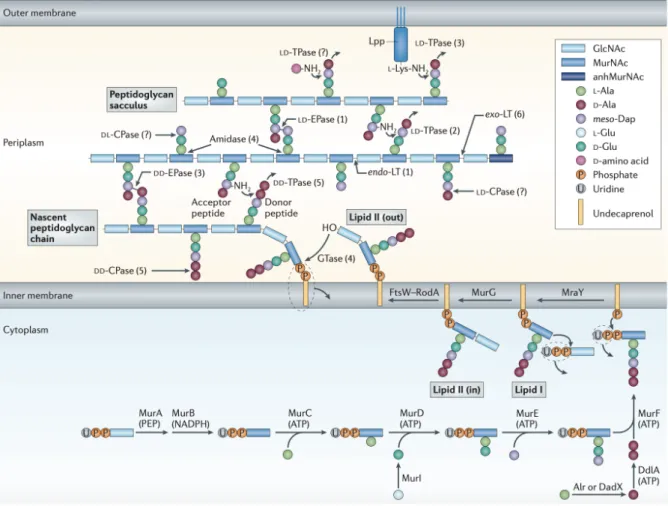
The synthesis of PGN takes place in three separated bacterial compartments: in the cytoplasm, at the membrane and in periplasmic/external space. It starts with the previously described UDP-GlcNAc synthesis in the cytoplasm site, Figure 1.3. Then the UDP-N-acetylmuramyl pentapeptide (UDP-Mpp) is synthesized, using as UDP-GlcNAc21, 22 as the
initial substrate, by the addition of different aminoacids that is carried out by the action of Mur enzymes (MurA, MurB, MurC, MurD, MurE and MurF). The final cytoplasmic PGN precursor is then embedded into the membrane and linked to undecaprenyl phosphate molecules whose synthesis is assisted by the undecaprenyl phosphate synthase (UppS). This enzyme catalyzes successive condensation reactions of farnesyl pyrophosphate (FPP) and eight isopentenyl pyrophosphates (IPP) to produce the molecule of undecaprenyl pyrodiphosphate (C55-PP). Then a dephosphorylation takes place by the undecaprenyl pyrophosphate phosphatase (UppP) to produce undecaprenyl pyrophosphate (C55-P).23, 24
The first membrane reaction step of PGN synthesis is catalyzed by the phospho-MurNAc-pentapetide translocase (MraY) that uses the two substrates of UDP-Mpp and C55-P.
Figure 1.3 The synthesis and attachment of a new peptidoglycan stand to the existing.25
Phospho-MurNAc-pentapetide moiety is transferred from UDP-Mpp to C55-P forming uridine-monophosphate (UMP) and undecaprenyl-pyrophophoryl-MurNAc-pentapetide, also known as Lipid I.26, 27
Then a GlcNAc moiety from a soluble UDP-GlcNAc precursor is transfered to Lipid I by the glycosyltransferase MurG to produce Lipid II. Consequently, Lipid II is transported from the cytoplasm to the outer membrane space by the action of the Lipid II flippase. This step was originally thought to be carried by the FtsW/RodA enzime28 but more recently growing
evidence have been produce to associate this activity to the MurJ like enzymes.29
In the final steps of PGN synthesis, glycosyltransferases (GTases) are required to polymerize the glycans chains and DD-transpeptidases (DD-TPases), or L,D-transpeptidases, to crosslink the peptides present in neighboring glycan chains.24 Bacteria produce different
bacterial PGN, an that can be divided in three types: the bifunctional GTase-TPases (A PBPs), the monofunctional TPases (B PBPs) and the monofunctional GTases.24 Three bifunctional
synthases (PBP1A, PBP1B, and PBP1C), a GTase (MgtA) and two TPases (needed for cell elongation (PBP2) or cell division (PBP3)) are found in E. coli.30 PBP1B is responsible for glycan
polymerization, 28 disaccharide units, and peptide crosslinking 40-50% of peptides. PBP1A generates shorter glycan strands (20 disaccharide units) and 22% of peptide crosslinking.31
Through labeling experiments, it was confirmed that PGN grows by insertion of fresh synthesized glycans, in the existing PGN chains.32, 33 The fact that no oligomers intermediates
were found in the cell suggests the polymerization and transpeptidation steps occur at the same time, which supports the existence of bifunctional enzymes.34
1.1.3 Bacterial cell wall
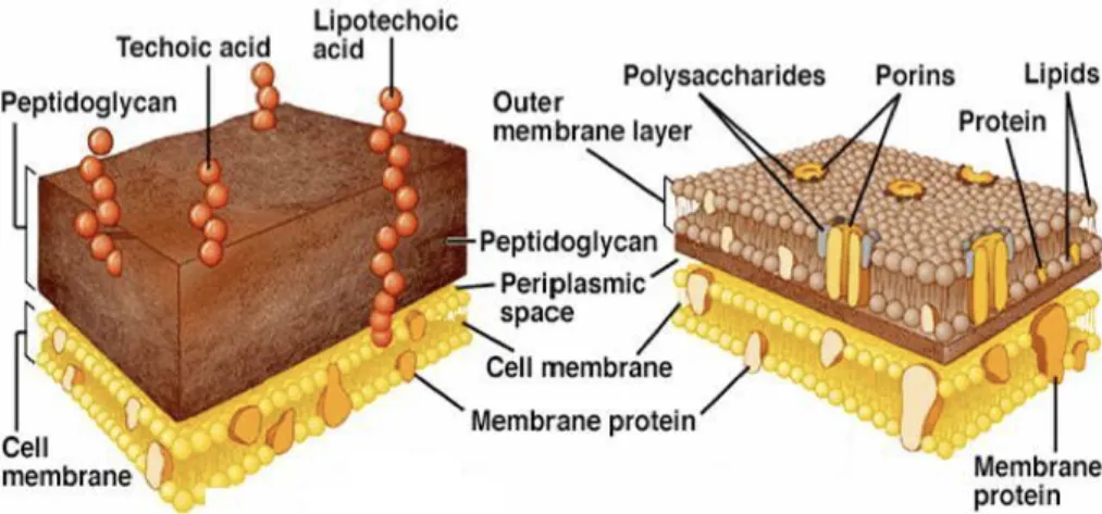
Bacteria can be divided in two major groups according to the composition and organization of their bacterial cell surface: the Gram-positive bacteria, frequently producing a lysine-type PGN, and the Gram-negative bacteria, usually with a DAP-type PGN, as it is shown in Figure 1.4. The bacteria cell wall present in Gram-positive bacteria has in its composition a cytoplasmatic lipid membrane, the macromolecule of PGN, also called murein, which may be covered by teichoic acids (TA) or other polysaccharides. The bacterial cell surface of Gram-negative bacteria has in its composition a cytoplasmatic membrane, a thin layer of PGN, which is concealed by an outer membrane that carries porins and lipoproteins. Different polysaccharides, such as the LPS (also termed as endotoxin), are also linked to the outer membrane.
Figure 1.4 Bacteria PGN atomic/skeleton structure representation highlighting the NAM unit. The repeating unit of the PGN produce by S. aureus (Gram-positive bacteria) (Left) and E. coli (Gram negative bacteria) (Right) are shown.
L-Ala D-GluNH2 L-Lys D-Ala (Gly)5 O OH O NHAc HO O O O NHAc O OH OH O NHAc HO O O D-Ala L-Lys D-GluNH2 L-Ala L-Ala D-GluNH2 m-Dap D-Ala O OH O NHAc HO O O O NHAc O OH OH O NHAc HO O O D-Ala m-Dap D-GluNH2 L-Ala D-Ala D-Ala
The organization of bacterial cell wall frequently explains the different characteristics and properties, such as thickness and permeability of the cell-wall, associated with these two groups of bacteria, Figure 1.5.
PGN is a major component of the bacteria cell wall, is normally composed by a disaccharide, monomer, of NAG linked via b-(1,4) glycosidic bond to a NAM, which is connected to a penta-peptide that may include aminoacids such as L-Ala, D-iso-Gln, L-Lys, D-Ala (frequently present in Gram-positive bacteria) or L-Ala, D- iso-Glu, m-DAP, D-Ala (observed in several Gram-negative bacteria). Despite this minimal chemical composition, several modifications in both the glycan structure, and the aminoacids included in the cross-linking bridge and the peptide stem, have been reported leading to numerous new structures of PGN. These modifications can be as varied as a glycine in the first position, a D-iso-glutamate in the second position or a D-serine in the fifth position.35
Figure 1.5 Bacteria cell-wall representation. Gram-negative and Gram-positive respectively.26
1.1.3.1 Modifications on PGN carbohydrate backbone
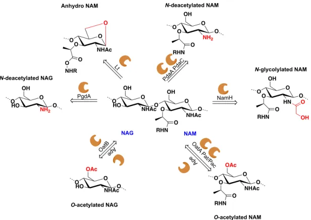
Some bacteria have developed strategies to protect their own PGN from the action of PGN hydrolases present in the growth medium, such as the host lysozyme-like enzymes. Chemical modifications of the NAG and NAM residues may help bacteria to evade the host immune system. Figure 1.6 summarizes the major changes in the PGN sugar backbone that may protect bacteria from the host immune system.36
The O-acetylation in NAM units in Gram-negative bacteria gives lysozyme resistance and resistance to autolysis (lysis induced by the activity of the PGN hydrolases produced by the bacteria).37 In the case of Gram-positive bacteria, the same modification gives lysozyme
resistance, resistance to macrophages killing and penicillin resistance.38, 39 Contrary to NAM,
O-acetylation in NAG units is not frequently observed in the PGN of bacteria. Although it seems to give no resistance to the activity of lysozyme, it provides resistance to the action of
N-acetyl-glucosaminidase Acm2, an enzyme produced by L. plantarum.40
The N-deacetylation of NAG units also provides resistance to the activity of lysozyme and to neutrophil killing.41 When present in the NAM unit, N-deacetylation helps bacteria to evade
the immune system since the NAc group from the NAM unit seems to be required in the recognition of this PGN fragment by NOD2.42
In certain bacteria, such as Mycobacterium tuberculosis, PGN may be modified by reaction of N-glycolylation in NAM units, which gives bacteria the ability to resist to lysozyme, b-lactam resistance and promotes the cytokine response.43, 44
Figure 1.6 Schematic representation of various modifications among PGN sugar backbone, highlighting the structural
modification.45
1.1.4 Chitin biosynthesis
Chitin, one of the most abundant polymers in nature and the most abundant amino polymer, is synthesized by fungi, nematodes and arthropods. Chitin is a poly–b(1,4)–NAG polymer that has the role of maintaining the strength of the cell wall of fungi (it can be found in 1-15% of the cell wall mass) and the exoskeletons of other organisms. Chitin biosynthesis carried out
O O HO NHAcO OH O O NHAc O OH RHN O O O O NHAc O O O NHR O O NH2 O OH RHN O O O O HN O OH RHN O O O OH O O NHAc O OAc RHN O O O O HO NHAcO OAc O O HO NH2 O OH NAG NAM
Anhydro NAM N-deacetylated NAM
N-deacetylated NAG N-glycolylated NAM O-acetylated NAM O-acetylated NAG Lt PgdA OatBe Ap PdaA Pd aC NamH O atA Pa t/Pa c Ap e
by membrane-bound chitin synthases. UDP-GlcNAc is used as substrate by these enzymes to form poly–b(1,4)–NAG. Chitin is first elongated by adding GlcNAc to the nonreducing end of the already present polymer and then extruded to the cell wall space by the reducing end. The polymeric chitin present within the cell wall matrix can be assembled into microfibrils, through interchain hydrogen bonding, and arranged in antiparallel pattern.46
Different enzymes are involved in the overall synthesis of chitin. In Figure 1.7, Glycogen phosphorylase (1) converts glycogen (a polymer of glucose residues that is used as a form of energy source) in Glc-1-P, which can be then used by the enzymes involved in glycolysis or trehalose synthesis after the action of a phosphoglucomutase (4). On the other hand, glucose residues are converted into fructose-6-P by the action of hexokinase (3), phosphoglucomutase (4) and glucose-6-P isomerase (5). The chitin pathway synthesis begins with glutamine-fructose-6-phostate amindotransferase (6, GFAT) and fructose-6-P. The transference of ammonia from 1-glutamine to fructose-6-P and then isomerization form fructosamine-6-P. After, an acetyl group from coenzyme A is transferred to fructosamine-6-P by gluctosamine-6-P acetyltransferase (7) to afford GlcNAc-6-P. By the action of phosphoacetylglucosamine mutase (8) the phosphate group is transferred from the C6 to C1. By the action of UDP-GlcNAc pyrophosphorylase (9) the uridiyl group transferred to obtain UDP-GlcNAc which is the substrate to the chitin synthase (10). Assembled chitin polymers can be hydrolyzed by chitinases (11) and N-acetylglucosamidases (12) to generate GlcNAc, which can be reused to a new chitin biosynthesis, Figure 1.7.47
Figure 1.7 Chitin synthesis pathway in fungi and insects.47
The selective construction of NAG glycosides has been a significant task in carbohydrate chemistry, and despite the synthetic advances remains a major challenge.48-52 Due to the
remarkable biological role of the NAG-containing oligosaccharides, special efforts have been dedicated to search for efficient synthetic approaches to such complex molecules, involving efficient, simple, stereo- and region- selective methods.53-55 One of the most efficient
strategies to prepare oligosaccharides consists in the preparation of key building blocks of di-, tridi-, and higher-oligosaccharidesdi-, that can be applied to assemble complex molecules.50, 53-58
Thus, a critical limitation for further PGN investigation, is the limited availability of PGN in pure form from natural sources, and the need of ligand purification in reliable amounts. Indeed, the extraction and purification process of PGN presents several difficulties, due to the presence of TA and proteins that can contaminate the PGN fragments.59-62
In this chapter both NOD and Myc factors were not covered since the main focus is PGN and chitin, synthesis and modifications.
1.2
References
1. Bertozzi, C. R.; Kiessling, L. L., Chemical glycobiology. Science 2001, 291 (5512), 2357-2364.
2. Helenius, A.; Aebi, M., Intracellular functions of N-linked glycans. Science 2001, 291 (5512), 2364-2369.
3. Alper, J., Searching for medicine's sweet spot. Science 2001, 291 (5512), 2338-2343. 4. Simon, P. M., Pharmaceutical oligosaccharides. Drug Discovery Today 1996, 1 (12), 522-528.
5. Dwek, R. A., Glycobiology: Toward understanding the function of sugars. Chemical
Reviews 1996, 96 (2), 683-720.
6. Varki, A., Biological Roles Of Oligosaccharides - All Of The Theories Are Correct.
Glycobiology 1993, 3 (2), 97-130.
7. Herzner, H.; Reipen, T.; Schultz, M.; Kunz, H., Synthesis of glycopeptides containing carbohydrate and peptide recognition motifs. Chemical Reviews 2000, 100 (12), 4495-4537. 8. Zhu, X. M.; Schmidt, R. R., New Principles for Glycoside-Bond Formation. Angewandte
Chemie-International Edition 2009, 48 (11), 1900-1934.
9. Cecioni, S.; Imberty, A.; Vidal, S., Glycomimetics versus Multivalent Glycoconjugates for the Design of High Affinity Lectin Ligands. Chemical Reviews 2015, 115 (1), 525-561. 10. Suresh, R.; Mosser, D. M., Pattern recognition receptors in innate immunity, host defense, and immunopathology. Advances in Physiology Education 2013, 37 (4), 284-291. 11. Pillai, C. K. S.; Paul, W.; Sharma, C. P., Chitin and chitosan polymers: Chemistry, solubility and fiber formation. Progress in Polymer Science 2009, 34 (7), 641-678.
12. Carvalho, L. C. R.; Queda, F.; Santos, C. V. A.; Marques, M. M. B., Selective Modification of Chitin and Chitosan: En Route to Tailored Oligosaccharides. Chemistry-an
Asian Journal 2016, 11 (24), 3468-3481.
13. Lerouge, P.; Roche, P.; Faucher, C.; Maillet, F.; Truchet, G.; Prome, J. C.; Denarie, J., symbiotic host-specificity of Rhizobium-meliloti is determined by a sulfated and acylated glucosamine oligosaccharide signal. Nature 1990, 344 (6268), 781-784.
14. Maillet, F.; Poinsot, V.; Andre, O.; Puech-Pages, V.; Haouy, A.; Gueunier, M.; Cromer, L.; Giraudet, D.; Formey, D.; Niebel, A.; Martinez, E. A.; Driguez, H.; Becard, G.; Denarie, J., Fungal lipochitooligosaccharide symbiotic signals in arbuscular mycorrhiza. Nature 2011, 469 (7328), 58-U1501.
15. Wang, L. H.; Weber, A. N.; Atilano, M. L.; Filipe, S. R.; Gay, N. J.; Ligoxygakis, P., Sensing of Gram-positive bacteria in Drosophila: GNBP1 is needed to process and present peptidoglycan to PGRP-SA. Embo Journal 2006, 25 (20), 5005-5014.
16. van Heijenoort, J., Recent advances in the formation of the bacterial peptidoglycan monomer unit. Natural Product Reports 2001, 18 (5), 503-519.
17. Durand, P.; Golinelli-Pimpaneau, B.; Mouilleron, S.; Badet, B.; Badet-Denisot, M. A., Highlights of glucosamine-6P synthase catalysis. Archives of Biochemistry and Biophysics 2008, 474 (2), 302-317.
18. Li, S.; Kang, J.; Yu, W. D.; Zhou, Y.; Zhang, W. L.; Xin, Y.; Ma, Y. F., Identification of M-tuberculosis Rv3441c and M-smegmatis MSMEG_1556 and Essentiality of M-smegmatis MSMEG_1556. Plos One 2012, 7 (8).
19. Barreteau, H.; Kovac, A.; Boniface, A.; Sova, M.; Gobec, S.; Blanot, D., Cytoplasmic steps of peptidoglycan biosynthesis. Fems Microbiology Reviews 2008, 32 (2), 168-207.
20. Rani, C.; Khan, I. A., UDP-GlcNAc pathway: Potential target for inhibitor discovery against M-tuberculosis. European Journal of Pharmaceutical Sciences 2016, 83, 62-70.
21. Lovering, A. L.; Safadi, S. S.; Strynadka, N. C. J., Structural Perspective of Peptidoglycan Biosynthesis and Assembly. Annual Review of Biochemistry, Vol 81 2012, 81, 451-478.
22. Bouhss, A.; Trunkfield, A. E.; Bugg, T. D. H.; Mengin-Lecreulx, D., The biosynthesis of peptidoglycan lipid-linked intermediates. Fems Microbiology Reviews 2008, 32 (2), 208-233. 23. Chang, H. Y.; Chou, C. C.; Hsu, M. F.; Wang, A. H. J., Proposed Carrier Lipid-binding Site of Undecaprenyl Pyrophosphate Phosphatase from Escherichia coli. Journal of Biological
Chemistry 2014, 289 (27), 18719-18735.
24. Vollmer, W.; Bertsche, U., Murein (peptidoglycan) structure, architecture and biosynthesis in Escherichia coli. Biochimica Et Biophysica Acta-Biomembranes 2008, 1778 (9), 1714-1734.
25. Typas, A.; Banzhaf, M.; Gross, C. A.; Vollmer, W., From the regulation of peptidoglycan synthesis to bacterial growth and morphology. Nature Reviews Microbiology 2012, 10 (2), 123-136.
26. Chung, B. C.; Zhao, J. S.; Gillespie, R. A.; Kwon, D. Y.; Guan, Z. Q.; Hong, J. Y.; Zhou, P.; Lee, S. Y., Crystal Structure of MraY, an Essential Membrane Enzyme for Bacterial Cell Wall Synthesis. Science 2013, 341 (6149), 1012-1016.
27. Suginaka, H.; Strominger, J. L.; Blumberg, P. M., Biosynthesis of peptidoglycan of bacterial cell-walls .28. Multiple penicillin-binding components in subtilis,
Bacillus-cereus, Staphylococcus-aureus, and Escherichia-coli. Journal of Biological Chemistry 1972, 247
(17), 5279-+.
28. Liu, Y.; Breukink, E., The Membrane Steps of Bacterial Cell Wall Synthesis as Antibiotic Targets. Antibiotics-Basel 2016, 5 (3).
29. Henrichfreise, B.; Brunke, M.; Viollier, P. H., Bacterial Surfaces: The Wall that SEDS Built. Current Biology 2016, 26 (21), R1158-R1160.
30. Yousif, S. Y.; Broomesmith, J. K.; Spratt, B. G., Lysis of escherichia-coli by beta-lactam antibiotics - deletion analysis of the role of penicillin-binding protein-1a and protein-1b.
Journal of General Microbiology 1985, 131 (OCT), 2839-2845.
31. Born, P.; Breukink, E.; Vollmer, W., In vitro synthesis of cross-linked murein and its attachment to sacculi by PBP1A from Escherichia coli. Journal of Biological Chemistry 2006,
281 (37), 26985-26993.
32. Burman, L. G.; Park, J. T., Molecular-model for elongation of the murein sacculus of
Escherichia-coli. Proceedings of the National Academy of Sciences of the United States of America-Biological Sciences 1984, 81 (6), 1844-1848.
33. Glauner, B.; Holtje, J. V., GROWTH-PATTERN OF THE MUREIN SACCULUS OF ESCHERICHIA-COLI. Journal of Biological Chemistry 1990, 265 (31), 18988-18996.
34. Goodell, E. W.; Markiewicz, Z.; Schwarz, U., Absence of oligomeric murein intermediates in Escherichia-coli. Journal of Bacteriology 1983, 156 (1), 130-135.
35. Vollmer, W.; Blanot, D.; de Pedro, M. A., Peptidoglycan structure and architecture.
Fems Microbiology Reviews 2008, 32 (2), 149-167.
36. Davis, K. M.; Weiser, J. N., Modifications to the Peptidoglycan Backbone Help Bacteria To Establish Infection. Infection and Immunity 2011, 79 (2), 562-570.
37. Dillard, J. P.; Hackett, K. T., Mutations affecting peptidoglycan acetylation in Neisseria gonorrhoeae and Neisseria meningitidis. Infection and Immunity 2005, 73 (9), 5697-5705.
38. Crisostomo, M. I.; Vollmer, W.; Kharat, A. S.; Inhulsen, S.; Gehre, F.; Buckenmaier, S.; Tomasz, A., Attenuation of penicillin resistance in a peptidoglycan O-acetyl transferase mutant of Streptococcus pneumoniae. Molecular Microbiology 2006, 61 (6), 1497-1509. 39. Shimada, T.; Park, B. G.; Wolf, A. J.; Brikos, C.; Goodridge, H. S.; Becker, C. A.; Reyes, C. N.; Miao, E. A.; Aderem, A.; Gotz, F.; Liu, G. Y.; Underhill, D. M., Staphylococcus aureus Evades Lysozyme-Based Peptidoglycan Digestion that Links Phagocytosis, Inflammasome Activation, and IL-1 beta Secretion. Cell Host & Microbe 2010, 7 (1), 38-49.
40. Bernard, E.; Rolain, T.; Courtin, P.; Guillot, A.; Langella, P.; Hols, P.; Chapot-Chartier, M. P., Characterization of O-Acetylation of N-Acetylglucosamine a novel structural variation of bacterial peptidoglycan. Journal of Biological Chemistry 2011, 286 (27), 23950-23958. 41. Fittipaldi, N.; Sekizaki, T.; Takamatsu, D.; Dominguez-Punaro, M. D.; Harel, J.; Bui, N. K.; Vollmer, W.; Gottschalk, M., Significant contribution of the pgdA gene to the virulence of Streptococcus suis. Molecular Microbiology 2008, 70 (5), 1120-1135.
42. Melnyk, J. E.; Mohanan, V.; Schaefer, A. K.; Hou, C. W.; Grimes, C. L., Peptidoglycan Modifications Tune the Stability and Function of the Innate Immune Receptor Nod2. Journal
of the American Chemical Society 2015, 137 (22), 6987-6990.
43. Raymond, J. B.; Mahapatra, S.; Crick, D. C.; Pavelka, M. S., Identification of the namH gene, encoding the hydroxylase responsible for the N-glycolylation of the mycobacterial peptidoglycan. Journal of Biological Chemistry 2005, 280 (1), 326-333.
44. Coulombe, F.; Divangahi, M.; Veyrier, F.; de Leseleuc, L.; Gleason, J. L.; Yang, Y. B.; Kelliher, M. A.; Pandey, A. K.; Sassetti, C. M.; Reed, M. B.; Behr, M. A., Increased NOD2-mediated recognition of N-glycolyl muramyl dipeptide. Journal of Experimental Medicine 2009, 206 (8), 1709-1716.
45. Yadav, A. K.; Espaillat, A.; Cava, F., Bacterial Strategies to Preserve Cell Wall Integrity Against Environmental Threats. Frontiers in Microbiology 2018, 9.
46. Ruiz-Herrera, J.; Elorza, M. V.; Valentin, E.; Sentandreu, R., Molecular organization of the cell wall of Candida albicans and its relation to pathogenicity. Fems Yeast Research 2006,
6 (1), 14-29.
47. Merzendorfer, H., The cellular basis of chitin synthesis in fungi and insects: Common principles and differences. European Journal of Cell Biology 2011, 90 (9), 759-769.
48. Enugala, R.; Carvalho, L. C. R.; Pires, M. J. D.; Marques, M. M. B., Stereoselective Glycosylation of Glucosamine: The Role of the N-Protecting Group. Chemistry-an Asian
Journal 2012, 7 (11), 2482-2501.
49. Paulsen, H., Advances in selective chemical syntheses of complex oligosaccharides.
Angewandte Chemie-International Edition in English 1982, 21 (3), 155-173.
50. Banoub, J.; Boullanger, P.; Lafont, D., Synthesis of oligosaccharides of 2-amino-2-deoxy sugars. Chemical Reviews 1992, 92 (6), 1167-1195.
51. Bongat, A. F. G.; Demchenko, A. V., Recent trends in the synthesis of O-glycosides of 2-amino-2-deoxysugars. Carbohydrate Research 2007, 342 (3-4), 374-406.
52. Yang, Y.; Yu, B., Recent advances in the synthesis of chitooligosaccharides and congeners. Tetrahedron 2014, 70 (5), 1023-1046.
53. Otsuka, Y.; Yamamoto, T.; Fukase, K., Syntheses of N-aryl-protected glucosamines and their stereoselectivity in chemical glycosylations. Tetrahedron Letters 2017, 58 (31), 3019-3023.
54. Xolin, A.; Losa, R.; Kaid, A.; Tresse, C.; Beau, J. M.; Boyer, F. D.; Norsikian, S., Stereocontrolled glycoside synthesis by activation of glycosyl sulfone donors with scandium(III) triflate. Organic & Biomolecular Chemistry 2018, 16 (2), 325-335.
55. Gouasmat, A.; Lemetais, A.; Solles, J.; Bourdreux, Y.; Beau, J. M., Catalytic Iron(III) Chloride Mediated Site-Selective Protection of Mono- and Disaccharides and One Trisaccharide. European Journal of Organic Chemistry 2017, (23), 3355-3361.
56. Despras, G.; Alix, A.; Urban, D.; Vauzeilles, B.; Beau, J. M., From Chitin to Bioactive Chitooligosaccharides and Conjugates: Access to Lipochitooligosaccharides and the TMG-chitotriomycin. Angewandte Chemie-International Edition 2014, 53 (44), 11912-11916. 57. Arya, P.; Ben, R. N., Combinatorial chemistry for the synthesis of carbohydrate libraries. Angewandte Chemie-International Edition in English 1997, 36 (12), 1280-1282. 58. Paulsen, H., Progress in oligosaccharide synthesis through a new orthogonal glycosylation strategy. Angewandte Chemie-International Edition in English 1995, 34 (13-14), 1432-1434.
59. Zhang, Y.; Fechter, E. J.; Wang, T.-S. A.; Barrett, D.; Walker, S.; Kahne, D. E., Synthesis of heptaprenyl-lipid IV to analyze peptidoglycan glycosyltransferases. Journal of the American
Chemical Society 2007, 129 (11), 3080-+.
60. Inamura, S.; Fukase, K.; Kusumoto, S., Synthetic study of peptidoglycan partial structures. Synthesis of tetrasaccharide and octasaccharide fragments. Tetrahedron Letters 2001, 42 (43), 7613-7616.
61. Meroueh, S. O.; Bencze, K. Z.; Hesek, D.; Lee, M.; Fisher, J. F.; Stemmler, T. L.; Mobashery, S., Three-dimensional structure of the bacterial cell wall peptidoglycan.
Proceedings of the National Academy of Sciences of the United States of America 2006, 103
(12), 4404-4409.
62. Filipe, S. R.; Tomasz, A.; Ligoxygakis, P., Requirements of peptidoglycan structure that allow detection by the Drosophila Toll pathway. Embo Reports 2005, 6 (4), 327-333.
2
Synthetic studies towards
a NAG-NAM disaccharide
2.1
Introduction
2.1.1 Glucosamine -a challenging scaffold
The most important classes of natural oligosaccharides and glycoconjugates contain residues of 2-amino-2-deoxy-b-D-glucopyranosyl (D-glucosamino) moieties. D-Glucosamines exist in several biological systems, such as the cell membrane of living organisms, acting as bacterial receptors or signaling molecules, as antibody recognition sites, as tumor markers, or as hormonal and enzymatic glycoproteins.1 Glucosamines are also part of important structural
oligosaccharides.
One of the most critical limitations in investigating the biological functions of these complex glycostructures is their limited availability and purity from natural sources. Consequently, there has been an increasing demand towards synthetic approaches that provide pure material of these oligosaccharides and glycoconjugates, or modified structures, for biological investigations.
The majority of naturally abundant glycoconjugates contain N-acetylated D-glucosamino moiety that are connected through a 1,2-trans linkage. Thus, it would be desirable to use the commercially available N-acetyl D-glucosamine for syntheses towards these glycoconjugates. However, the N-acetyl group poses several limitations to the glycosylation reaction with respect to both donor and acceptor units.
Indeed, the glycosylation of complex aglycones with glycosyl donors bearing a 2-acetamido-2-deoxy functionality is usually impractical due to the formation of a 1,2-O,N-oxazoline intermediate during glycosylation, which is a stable intermediate, Figure 2.1 A, and drastically decreases the rate and yield of the glycosylation step, especially when the chosen glycosyl acceptors have poor nucleophilicity or are sterically hindered. Also, concerning 2-acetamido-2-deoxy functionality as acceptor the amide group can establish a hydrogen bond with the hydroxy group O-4 decreasing the nucleophilicity at this position, Figure 2.1 C.
In order to avoid the formation of the stable 1,2-O,N oxazoline intermediate and improve the formation of 1,2-trans glycoside several disubstituted 2-amino2-deoxyglycosyl donors have been developed, Figure 2.1 B. The use of these protecting groups result on the blocking of the amino group either by using two different protecting groups or a bivalent protecting group.
Therefore, donor activation results in the formation of an unstable oxazolinium intermediate that blocks the a-face.2
Figure 2.1 Influence of the C-2 group on the glycosylation stereoselectivity.(adapted from2)
To overcome the problems associated with the use of NAG, Figure 2.1, many protecting groups have been employed. Figure 2.2 depicts some of the most used N-protecting groups applied in glucosamine derivatives.
Figure 2.2 Most common amino protecting groups. O RO RO NHAc LG OR O RO RO HN OR O RO RO N OR O O HO O RO RO NHAc OR O O Promotor O
Stable intermediate Major diastereomertrans glycoside
O RO RO N LG OR X O O Y O RO RO N OR O RO RO N OR Y O O HO O RO RO NHAc OR O O Promotor Y O
Unstable intermediate Only diastereomertrans glycoside X O X O A B slow C O O NH O H Hydrogen bound O N O O N-Phth O NH O O CCl3 N-Troc O N3 N3 O NH CCl3 O N-TCA O NH O O N-Alloc O NH O O N-Boc O N O O N-DMM O NH O CF3 N-TFA O N O O N-Ac2 O NH OBn O N-Cbz as donor as donor as acceptor Hydrogen bond
Bivalent protecting groups block the stability of the 1,2-O,N-oxazoline however, usually turn the glucosamine moiety more bulky. Monovalent protecting groups can provide well define selectivity towards the glycosylation reaction and also improve the solubility in organic solvents, comparing with N-acetyl D-glucosamine. The choice of the protecting group must be planned to take into consideration the synthetic steps involved, and reaction conditions used. Thus, most synthetic plans designed to afford glucosamine conjugated or oligosaccharides rely on the use of properly protected glucosamine moieties, in which the N-protecting group is modified during the synthetic sequence.3
Table 2.1 Different glycosylation conditions and their selectivity.
Entry Donor Acceptor Promotor Solvent T
(oC) Yield (%) Selectivity (a:b) 14 TMSOTf DCM -20 91 b 25 TMSOTf DCM 0 76 b 36 NIS, TfOH DCM - 20 86 b 47 DMTST, DTBMP DCM 0 -20 94 b 58, 9 PhCN) Ni(4-F-4(OTf)2 DCM 25 93 12:1 O R3O R4O NPG R 1 OR2 O HO Promotor Selective Disaccharide O AcO AcO NPhthO OAc CCl3 HN O HO MPMO NPhthOMPM OMPM O AcO AcO NHTroc O OAc CCl3 HN O HO HO NHTrocOTBDMS OTBDPS O AcO AcO NHTCA SEt OAc O OBz HO OBz OAllyl OBz O AcO O NAc SEt OAc O O BnO BnO OBn OMe HO O AcO AcO N SEt OAc MeO O O O OBn HO
The 2-amino protecting groups play a key role in glycosydic bond formation involving glucosamine derivatives acting both as donors and acceptors. Accordingly, several N-protecting groups have been developed not only with regard to their compatibility with diverse carbohydrate synthetic methods but also to their ability to direct the stereochemical outcome of the glycosylation reaction. Table 2.1, summarizes some examples on the importance of the N-protecting group regarding the desired selectivity of the new glycosylic bond.
2.1.2 Synthetic routes towards peptidoglycan fragments
The synthesis and composition of the PGN is associated with expression of bacterial resistance to different antibiotics and with a variety of host/bacteria interactions. The determination of the role of PGN in host disease has been hampered by the lack of pure and homogeneous polymerized PGN.10 The major limitation encountered in these studies is the limited
availability of pure PGN fragments obtained through purification procedures from natural sources. Thus, there is a need to have pure and well-defined PGN fragments in order to have viable biological studies. To circumvent this bottleneck, we and others have been developing strategies for the synthesis of muropeptides from glucosamine residues.
In 2001, Fukase and his co-workers reported the synthesis of PGN fragments.11 On the
follow-up of their previous report from the on the synthesis of partial structures of cell wall PGN,12
the authors developed an orthogonal synthesis in order to create building blocks to form the complex octasaccharide (NAG-NAM)4 (II.9) with two amino acid strands linked to every NAM
Figure 2.3 Synthesis of PGN fragment II.9 reported by Fukase11; (a) i) AllocCl, NaHCO3, H2O; ii) AllylOH, DOWEX 50 W X8
200–400 mesh, 80 °C; iii) PhCH(OMe)2, p-TsOH, 57%; (b) NaH then trifluoromethanesulfonyl-L-(S)-2-propionic acid benzyl
ester, 98%; (c) Pd(PPh3)4, AcOH, DCM, then TrocCl, 58%; (d) Me3N·BH3, BF3·Et2O, ACN, 87%; (e) BnBr, AgO, DCM, 87% (f) Ir
complex, H2, THF, then I2, H2O, 99%; (g) CCl3CN, Cs2CO3, DCM, quantitative; (h) TMSOTf (0.1 equiv.), MS 4 Å, DCM, −15°C,
88%; (i) Me3N·BH3, BF3·Et2O, ACN, 52%; (j) Ir complex, H2, THF, then I2, H2O, 91%; (k) CCl3CN, Cs2CO3, DCM, 85%; (l) TMSOTf (0.1 equiv.), MS 4 Å , DCM, −15 °C, 79%; (m) Me3N·BH3, BF3·Et2O, ACN, 60%; (n) Ir complex, H2, THF, then I2, H2O, 81%; (o) CCl3CN, Cs2CO3, DCM, 83%; (p) TMSOTf (0.1 equiv.), MS 4 Å , DCM, −15 °C, 70%; (q) Zn–Cu, AcOH, then Ac2O, Py, 82%; (r) 1 M NaOMe, THF, H2O: LiOH, dioxane, THF, H2O, 97%; (s) HCl·H-L-Ala-D-Glu(OBn)-NH2, WSCI·HCl, HOBt, TEA, DCM, 31%; (t) Pd(OH)2, AcOH, H2 (10 atm), 48%.
The first step of this synthesis consisted in the protection of the amine group of D-glucosamine with the allyloxycarbonyl (Alloc) group, by treatment of the glucosamine with AllocCl in the presence of NaHCO3 as base.
O HO HO NH3Cl OH OH O HO O TrocHN OAllyl OBn O EtO O O HO TrocHN OAllyl O Ph O O BnO TrocHN O Ph O O O TrocHN OAllyl OBn O EtO O O BnO TrocHN O Ph O O O TrocHN OBn O EtO O HO BnO TrocHN OBn O O O TrocHN OAllyl OBn O EtO O NH Cl Cl Cl O O BnO TrocHN OBn O O O TrocHN OAllyl OBn O EtO O O BnO TrocHN O Ph O O O NHTroc OBn O EtO O O HO NHAc OH O O O AcHN O OH O O O HO HO NHAc OH O O O NHAc OH O O L-Ala D-isoGln L-Ala D-isoGln 2 a-d e, f, g h i j, k l m-u O O BnO TrocHN O O Ph NH Cl Cl Cl II.1 II.2 II.3 II.4 II.5 II.6 II.7 II.8 II.9


![Figure 2.8 Synthesis summary to accomplish II.35 reported by Fukase. 20 a) [Ir(cod)H-(MePh2P)2]PF6, THF, 1.5 h; b) I2/H2O, 30 min, 81%; c) CCl3CN/Cs2CO3, DCM, 30 min, quantitative; d) Me3N BH3/BF3·Et2O, ACN, 1 h, 83%; e) TMSOTf, DCM, 158](https://thumb-eu.123doks.com/thumbv2/123dok_br/19196607.952072/57.892.114.779.108.765/figure-synthesis-summary-accomplish-reported-fukase-quantitative-tmsotf.webp)