THE
3-D
IMENSIONAL
S
HOULDER
K
INEMATICS OF
2
S
HOULDER
R
OTATION
T
ESTS
Augusto Gil Pascoal, PhD,
aand Nuno Morais, MSc
bA
BSTRACTObjectives: The purpose of this study was to compare shoulder external rotation range of motion (ROM) during the hand-behind-neck (HBN) test and a standard shoulder external rotation test and to describe the 3-dimensional scapular motion during the HBN test.
Methods: An electromagnetic tracking device was used to assess the dominant shoulder of 14 healthy participants while performing active full ROM in a standard shoulder external rotation test in an elevated position (EREP) and in the HBN test. The humeral and scapular 3-dimensional positions at the end of EREP and HBN were compared using a paired-sample t test. A correlation analysis was performed between humeral and scapular angles to assess the contribution of scapular motion to the full shoulder ROM during the HBN test.
Results: No significant differences were found between the HBN test and the EREP at the end-range of the glenohumeral external rotation (HBN: 15.6° ± 6.3° vs EREP: 23.4° ± 4.7°; P = .08) and on scapular internal-external rotation (HBN test: 21.2° ± 6.3° vs EREP: 15.6° ± 1.8°; P = .23). Significant differences were found in scapular upward rotation (HBN: 21.2° ± 6.3° vs EREP: 15.6° ± 1.8°; Pb .01) and scapular spinal tilt (HBN: −0.4° ± 2.3° vs EREP: 8.1° ± 2.1°; P b .01). There was a positive correlation between the humeral angles and scapular internal and posterior spinal tilt angles with the HBN test. Conclusions: The results of the present study showed that, in young asymptomatic participants with no known shoulder pathology, the end-range of shoulder rotation was similar in the HBN test and in a standard shoulder rotation test. During the HBN test, the scapula assumed a more internal and anterior spinal tilted position at the end-range of active shoulder external rotation. These results suggest that the HBN test may be used to assess the end-range of glenohumeral external rotation. (J Manipulative Physiol Ther 2015;38:288-294)
Key Indexing Terms: Shoulder; Range of Motion; Articular; Biomechanical Phenomena
C
linical examination of the shoulder complex commonly includes the assessment of axial rota-tions. The patient is placed in a supine or sitting position with the shoulder at 90° of abduction while the examiner passively moves the arm to an extreme position (end-range) of internal and/or external rotation.1,2To restrict motion to the glenohumeral joint, the scapular contribution is limited through a posterior force applied by the examiner to the coracoid process and the clavicle. If a seated position isused, the examiner holds or supports the patients elbow at their side while the arm is passively rotated around the long axis of the humerus.3,4In both ROM testing positions, the joint end-range is determined by the examiner according to multiple criteria, including the capsular end-feel,5–7 the scapular lift-off,8or the presence of pain.9
Shoulder external rotation range of motion (ER-ROM) is clinically assessed using different methodologies and instruments such as goniometry,10,11inclinometry,12visual estimation,10,13and photography.10Several studies showed that these measurement systems might provide, in general, satisfactory reliability and agreement results for clinical purposes, particularly if assessed over time by the same rater and instrument.10–13 The assessment of shoulder ER-ROM is clinically important for (1) the diagnosis of glenohumeral disorders, for example, frozen shoulder,14 and instability15,16; (2) the assessment of the severity of disability17; (3) the identification of risk factors for developing shoulder pain, particularly in overhead throw-ing athletes18 (eg, tightness or excessive ROM); (4) the assessment of treatment progression and effectiveness17,19; and (5) quantifying and monitoring the amount of change in movement quality occurring over time.19,20
a
Professor, Sports & Health Department of the Human Kinetics Faculty, University of Lisbon, Lisbon, Portugal.
b
Adjunct-Professor, School of Health Sciences, Polytechnic Institute of Leiria, Leiria, Portugal.
Submit requests for reprints to: Augusto Gil Pascoal, PhD, Universidade Lisboa, Faculdade Motricidade Humana, Estrada da Costa, Dafundo, Cruz Quebrada, 1499-002 Lisboa, Portugal. (e-mail:[email protected]).
Paper submitted February 11, 2014; in revised form October 21, 2014; accepted October 31, 2014.
0161-4754
Copyright © 2015 by National University of Health Sciences.
http://dx.doi.org/10.1016/j.jmpt.2014.10.017 Open access under CC BY-NC-ND license.
The use of an active self-end-range determination has been suggested as advantageous for shoulder assessment to collect information close to shoulder functional motion, namely, arm throwing cycle or daily-living activities.10,21
To address this issue, shoulder function-related tests were proposed as indirect methods to assess shoulder rotation ROM.22,23 These tests include the so-called hand-behind-back for internal rotation, the hand-over-other-scapula for horizontal adduction, and the behind-head or hand-behind-neck (HBN) for external rotation and abduction. These indirect methods of measuring shoulder rotations are part of the standardized shoulder assessment form adopted by the American Shoulder and Elbow Surgeons.22,24The HBN is described as a shoulder function-related test that combines glenohumeral elevation (abduction) and external rotation combined with scapular motion at the scapulothoracic, acromioclavicular, and sternoclavicular joints.22,23It there-fore reflects an essential action for daily-living activities such as combing hair or throwing an object. Yang et al23showed that the HBN is a highly reliable test when using a 5-point functional scale to identify the individual inability to perform actions above the head. They have also suggested that it could be incorporated into the standard clinical assessment of patients with various shoulder pathologies.
For the HBN test, it is assumed that the end-range of glenohumeral external rotation is achieved when the hand placed behind the head reaches down the back with the thumb as far as possible. As scapular motion is not constrained during the HBN test, it seems questionable whether the ER-ROM of the glenohumeral joint is close to its maximum and could be comparable with other examination procedures, namely, those used in goniometry. In addition, a clarification about the relative contribution of the glenohumeral and scapulothoracic joints to full shoulder ROM during the HBN test is needed. Knowledge about the pattern of shoulder rotation during the HBN is useful for the examination of the shoulder complex and when planning rehabilitation protocols for patients with functional shoulder ER-ROM deficits as well.
Therefore, the purpose of this study was 2-fold: (1) to compare shoulder ER-ROM during the HBN test with a standard shoulder external rotation test and (2) to describe the 3-dimensional (3D) scapular motion during the HBN test. We hypothesized that the ER-ROM would be similar during the HBN test and the standard shoulder external rotation test.
M
ETHODS ANDM
ATERIALSParticipants
The dominant shoulder of 14 healthy young adults (male = 7, age = 21.1 ± 2.2 years, height = 166.3 ± 10.8 cm, body mass = 65.8 ± 12.6 kg) with no history of shoulder pain or pathology was studied.
All participants were informed about the purposes and procedures of the study, having signed the informed consent. The study had the approval of the Scientific Council of the Interdisciplinary Centre for the Study of Human Performance at the Faculty of Human Kinetics, Technical University of Lisbon, Portugal (number: CC-CE-162009), regarding the protection of the rights of the participants and the confidentiality of the data.
Experimental Protocol.
Participants were asked to remain in the sitting position and perform full active ROM of their dominant shoulder by 2 different methods: (a) the HBN test and (b) the external rotation in an elevated position (EREP). Participants performed the tests in a random order.To perform the HBN, participants were asked to place their hand above their head and then reach down the back with the thumb as far as possible (Fig 1A). With the EREP, the end-range of humeral external rotation ROM was actively reached while the arm was supported by one investigator at 90° of abduction with the elbow kept at 90° of flexion (Fig 1B). Participants were encouraged to maintain both their scapula and clavicle stable during the test and to hold the arm on the scapular plane.
Instrumentation.
Humeral and scapular 3D positions were recorded by means of a 6DOF electromagnetic tracking device (hardware: Flock of Birds System Ascension Technology; software: Motion Monitor v 7.0) that allowed simultaneous tracking and registration of the position and orientation of several sensors when they are inserted in an extended electromagnetic field. The static accuracy of the sensors with an extended range transmitter is up to 0.76 cm RMS/0.5° RMS at a 1.52-meter distance from the transmitter. Results of a previous calibration of the electromagnetic field revealed a translational residual measurement error of about 3 mm for each coordinate and a rotational root mean square area of less than 2° for each axis of rotation. Recordings were made at 100 Hz with a 3-sensors setup (Fig 1): the thorax sensor, firmly attached to skin over the spinous process of the first thoracic vertebrae (T1); the humerus sensor attached to the external side of the arm by means of a cuff just below the deltoid attachment; and the scapular sensor placed on the superior flat surface of the acromion process. A fourth sensor, mounted on a handheld stylus (± 6.5 cm), was used for bony landmarks digitalization to link sensors to the local anatomical coordinate systems and to subsequently calcu-late segments and joint rotations by combining the local anatomical coordinate systems with the sensor motions. Using a palpation method, selected shoulder bony land-marks were manually identified in the thorax and scapula and then digitized using the stylus. The error associated to palpation has been estimated to be approximately 2°.25 Orientation errors of shoulder bones resulting from the measurement inaccuracy have been estimated to be less than 2°.26 For bony landmarks digitalization, participants assumed an elevated shoulder position with elbow flexion289
Pascoal and Morais
Journal of Manipulative and Physiological Therapeutics
Hand-Behind-Neck Test Kinematics Volume38, Number 4
(± 90°) and the arm artificially supported in a standard position in a way that the humerus was aligned with the plane of the scapula. Given our digitalization protocol, the zero (0°) or neutral rotation was defined as the position when the participant’s forearm was perpendicular to the floor.
Kinematics.
Joint angles were calculated and expressed as Euler angle decompositions of the relative orientation of the distal segment with respect to a proximal segment. To obtain joint angles, anatomical (local) coordinates systems were defined on thorax, scapula, and humerus based on standard shoulder bony landmarks (Table 1). To describe the 3D motions of a bone, at least 3 angles (rotations) are needed; and it has to be taken into account that the order of rotation is essential.30The rotation order used in this study was based on the International Society of Biomechanics standardization proposal for the upper extremity.27Scapular rotations were defined with respect to the thorax using a Y, X’, Z” Euler sequence. The first rotation was defined as a rotation around the y-axis (sY), scapular internal/external rotation, where a positive rotation refers to scapular internal rotation. This scapular rotation is also known as scapular protraction (internal rotation) and retraction (external rotation). The second rotation was defined as a rotation around the rotated scapular x-axis (sX), upward rotation (positive), and downward rotation (nega-tive). The third rotation was defined as a rotation around the scapular z-axis (sZ), anterior spinal tilt (negative), and posterior spinal tilt (positive). In other words, anterior tilting occurs when the angulus inferior of the scapula moves away from the thorax. The humerus angles were defined both as thoracohumeral angles with respect to the
thorax, and as scapulohumeral or glenohumeral angles with respect to the scapula. For the glenohumeral angles, the first rotation was defined as a rotation around the y-axis also defined as the plane of arm elevation; the second rotation was defined around the x-axis that corresponded to the angle of arm elevation; and the third rotation around the moved y-axis (axial arm rotation) defined as external rotation (negative) and internal rotation (positive).
Variables and Statistics.
The glenohumeral angles as well as the scapular 3D position recorded at the end-range of external arm motion were compared between the HBN test and the EREP using a paired-sample t test. To describe the scapular contribution to arm motion, correlations were performed between glenohumeral angles and scapular 3D position recorded at the ER-ROM. All statistical analyzes were performed with SPSS (version 16; SPSS, Chicago, IL), and theα level was set at .05.R
ESULTSNo significant differences were found between the HBN test and the EREP at the end-range of the glenohumeral external rotation (HBN: 15.6° ± 6.3° vs EREP: 23.4° ± 4.7°; P = .08) and on scapular internal-external rotation (HBN test: 21.2° ± 6.3° vs EREP: 15.6° ± 1.8°; P = .23). Significant differences were found in scapular upward rotation (HBN: 21.2° ± 6.3° vs EREP: 15.6° ± 1.8°; Pb .01) and scapular spinal tilt (HBN:−0.4° ± 2.3° vs EREP: 8.1° ± 2.1°; P b .01). During the HBN test, the scapula assumed a more downward and anterior tilted position (Fig 2) than in the EREP. The results showed that, during the HBN Fig 1.Experimental setup.
test, the scapula was in a downward and anterior tilt position in comparison to EREP. This suggests that there is a significant contribution of the scapulothoracic, acromiocla-vicular, and sternoclavicular joints during the HBN test in comparison to EREP.
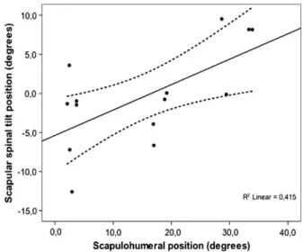
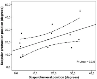
In the HBN test, a significant positive correlation was found between the end-range position of shoulder external rotation and scapular internal-external (r = 0.582; P = .02) and scapular spinal tilt (r = 0.644; P = .01) rotations. No significant correlations were found between humerus and scapula positions at the end-range of external arm rotation. According to these results, the high values of external arm rotation are significant but moderately related with scapular internal rotation (glenoid faces forward) and posterior spinal tilt (scapular inferior angle approaches to the rib cage) (Figs 3and4).
D
ISCUSSIONThe clinical examination of the shoulder complex commonly includes the assessment of shoulder axial rotations ROM with patients placed in a supine or sitting position with the arm at 90° of abduction.1,2 In a supine position, the shoulder end-range rotation (internal and/or external) is reached with the arm fully supported by the table. Shoulder motion is mostly the result of the glenohumeral joint motion, as the force applied by the examiner on the coracoid process and clavicle constrains scapulothoracic motion.
In a sitting position, the patient holds or supports the arm with elbow at 90° of flexion. The examiner restricts scapulothoracic motion by a force applied over the inferior angle of the scapula while the arm is rotating around the long axis of the humerus.3,4 With both ROM testing positions, the end-range of shoulder rotation could be actively determined by the patients, with31,32or without33 the effects of gravity, or by the examiner following a standard goniometry procedure by which the patient’s arm is passively positioned and limited by capsular end-feel,5–7,32 scapular lift-off,8,34or pain.35
Recently, function-related tests, such as the HBN test, were proposed to assess shoulder external rotation ROM given that these tests are more related with daily-living activities such as toileting and dressing.22,24,36 Function-related tests are easy to administer and interpret, and have a straightforward application in patients with various shoul-der pathologies. According to Yang et al,23those tests have demonstrated adequate reliability for clinical purposes and can be easily incorporated either individually or as a battery of functional-related tests in the clinical assessment of the shoulder. Restricted or painful HBN is commonly reported as an indicator of limiting function in patients with painful shoulder dysfunction, whereas an improvement in the ROM is associated with an increase in shoulder functional capacity.22–24
Previous research has shown that these shoulder function-related tests present satisfactory reliability results.23,37In a study involving participants with stiff and painful shoulder dysfunction, Green et al37 demonstrated that the hand-Table 1.Bony Landmarks Used and the Anatomical (Local) Coordinates Systems Definition, According to Wu et al27
Segments Bony Landmarks Anatomical (Local) Coordinates System Definition
Thorax C7: processus spinosus (spinous process) of the 7th cervical vertebra
tY: the line connecting the midpoint between
PX and T8 and the midpoint between IJ and C7 pointing upward T8: processus spinosus (spinal process)
of the 8th thoracic vertebra
tZ: the line perpendicular to the plane formed by IJ,
C7 and the midpoint between PX and T8 pointing to the right IJ: deepest point of incisura jugularis
(suprasternal notch)
tX: the common line perpendicular to Zt and Yt-axis pointing forward PX: processus xiphoideus (xiphoid process),
most caudal point on the sternum
tO: the origin coincident with IJ Scapula TS: trigonum spinae scapulae (root of the spine),
the midpoint of the triangular surface on the medial border of the scapula in line with the scapular spine
sY: the common line perpendicular to Xs and Zs-axis pointing upward
AI: angulus inferior (inferior angle), most caudal point of the scapula
sZ: the line connecting TS and AA pointing to AA AA: angulus acromialis (acromial angle),
most laterodorsal point of the scapula
sX: the line perpendicular to the plane formed by AI, AA, and TS, pointing forward.
sO: The origin coincident with AA Humerus GH: glenohumeral rotation center,
estimated calculating the pivot point of instantaneous helical axes of GH motion28,29
hY: the line connecting thigh and the midpoint of EL and EM, pointing to GH
EL: most caudal point on lateral epicondyle hZ: the common line perpendicular to the Yh and Zh-axis pointing to the right
EM: most caudal point on medial epicondyle hX: the line perpendicular to the plane formed by EL, EM, and GH pointing forward
hO: the origin coincident with GH
291
Pascoal and Morais
Journal of Manipulative and Physiological Therapeutics
Hand-Behind-Neck Test Kinematics Volume38, Number 4
behind-back test has a good interrater (intraclass correla-tion coefficient, 0.75) and an excellent intrarater (intra-class correlation coefficient, 0.90) reliability. More recently, Yang et al23 found a high intratester (k = 0.80) and intertester (k = 0.90) reliability associated with the HBN test in patients with shoulder pathologies.
No information is currently available to demonstrate if the end-range of active glenohumeral external rotation is actually reached during the HBN. Because scapular motion is not restricted during the HBN test, scapulothoracic joint contribution could be increased, thereby reducing the relative contribution of the glenohumeral joint to the full ROM.
The results of the present study suggest that the HBN test accurately reproduces the end-range of the active shoulder external rotation. In fact, no differences were found between glenohumeral position recorded during HBN test and during EREP. Of course, analyses were restricted to the active shoulder motion, which means that these results might not be directly extrapolated to goniometry or passive assessment of the end-range of shoulder external rotation. This is important for clinical reasoning and decision-making processes, as passive assessment of the end-range of shoulder external rotation is used to assess the function of the passive anterior retainers of the glenohumeral joint (capsuloligamentous complex), whereas active end-range of shoulder external rotation is used to assess the dynamic restraining capacity of the rotator cuff.16 The present findings suggest that, besides its usefulness for assessing function from the patient’s and clinician’s perspectives,23 the HBN test might also provide information about the dynamic restraining capacity of the rotator cuff and thus be of value in the assessment of rotator cuff dysfunction. Less
ability to reach actively behind the neck should alert the clinician to assess specific tissue dysfunction, for example, rotator cuff. Moreover, a scapular pattern different from the one found in the HBN test may also suggest pathology.
Regarding scapular contribution during the HBN test, we have hypothesized that the glenohumeral external rotation ROM would be amplified by scapular anterior spinal tilt rotation. These expectations were not confirmed after analyzing the differences of scapular spinal tilt position between HBN test and the EREP (Fig 2). Our results suggest that, during HBN test, the scapula assumes a more anterior spinal tilt position; that is, the acromion moves forward around an axis oriented with the scapular spine. This scapular motion is in the same direction of the external humeral movement at the glenohumeral joint, meaning that the amplitude of external rotation ROM at the glenohumeral joint is partially due to both humerus external rotation and scapular anterior spinal tilt. According to these results, the contribution of the scapulothoracic, acromio-clavicular, and sternoclavicular joints to arm motion is an important component of the HBN test.
Another difference found in scapular behavior between both ROM testing positions refers to the scapular upward-downward rotation angles. Scapular upward rota-tion is particularly related to the angle of humeral elevarota-tion, which reflects the so-called scapulohumeral rhythm. During the HBN test, the scapula was in a less upward position when compared with the EREP. These results suggest that, in the HBN test, the end-range of glenohumeral external rotation was associated with lower angles of arm elevation. It seems that the motion patterns of the HBN test start by an external rotation of the arm followed by a progressive Fig 3.Scatter plots of scapulohumeral position vs scapular spinal tilt position on HBN test both recorded at the end-range of shoulder external rotation. Linear fit (solid line) and the 95% confidence interval (dashed lines) are also represented.
Fig 2.Scapular position (degrees) at the end-range of shoulder rotation. sX : upward rotation (+) | downward rotation (−); sY: internal rotation (+) | external rotation (−);sZ : anterior spinal tilt (+) | posterior spinal tilt (−).
increase in the angle of elevation, which may have implications concerning the quality of shoulder movement. The correlation matrix demonstrated a level of associ-ation between scapulohumeral angles and 3D scapular position (internal rotation and posterior spinal tilt) recorded at the end-range position in the HBN test. According to these results (Figs 3and4), the high values of external arm rotation are significantly but moderately related with scapular internal rotation (glenoid faces forward) and posterior spinal tilt (inferior angle of the scapula is compressed against the rib cage).
Limitations
Although it is believed that the HBN test consists of movement components that are fundamental to daily-living activities, a direct relationship should not be assumed because there are no studies to support such assumption. Another limitation refers to methodological constraints in data acquisition. Scapular motion is the result of move-ments occurring at the sternoclavicular, acromioclavicular, and scapulothoracic joints. Because no sensor was coupled to the clavicle, the real contribute of each joint to the overall scapular motion could not be measured. However, to fully characterize shoulder girdle movements, this should be addressed in the future. In addition, the component of shoulder horizontal abduction was not considered in the present study. Further investigations regarding the HBN test should consider and control this component. Finally, this study was conducted in a small sample of young adults with normal ROM and upper quadrant posture. Caution is needed when generalizing these findings to other popula-tions such as older adults or children.
C
ONCLUSIONSThe results of the present study showed that, in young asymptomatic participants with no known shoulder pathology, the end-range of shoulder rotation was similar in the HBN test and in a standard shoulder rotation test. During the HBN test, the scapula assumed a more internal and anterior spinal tilted position at the end-range of active shoulder external rotation. These results suggest that the HBN test may be used to assess the end-range of glenohumeral external rotation.
F
UNDINGS
OURCES ANDC
ONFLICTS OFI
NTERESTNo funding sources or conflicts of interest were reported for this study
C
ONTRIBUTORSHIPI
NFORMATIONConcept development (provided idea for the research): A.G.P.
Design (planned the methods to generate the results): A.G.P. and N.M.
Supervision (provided oversight, responsible for organiza-tion and implementaorganiza-tion, writing of the manuscript): A.G.P. Data collection/processing (responsible for experiments, patient management, organization, or reporting data): A.G.P. and N.M.
Analysis/interpretation (responsible for statistical analysis, evaluation, and presentation of the results): A.G.P. and N.M. Literature search (performed the literature search): N.M. Writing (responsible for writing a substantive part of the manuscript): A.G.P. and N.M.
Critical review (revised manuscript for intellectual content; this does not relate to spelling and grammar checking): A.G.P.
Fig 4.Scatter plots of scapulohumeral position vs scapular internal rotation on HBN test both recorded at the end-range of shoulder external rotation. Linear fit line (solid line) and the 95% confidence interval (dashed lines) are also represented.
Practical Applications
• This study described the 3D shoulder kine-matics of the HBN test and compared it with active shoulder external rotation at 90° of abduction (EREP).
• There were no significant differences in shoulder external ROM between the HBN test and standard active EREP, but scapular kinematics was different.
• This study contributed to understand the usefulness of the HBN test to shoulder assessment and rehabilitation process.
293
Pascoal and Morais
Journal of Manipulative and Physiological Therapeutics
Hand-Behind-Neck Test Kinematics Volume38, Number 4
R
EFERENCES1. Myers JB, Laudner KG, Pasquale MR, Bradley JP, Lephart SM. Glenohumeral range of motion deficits and posterior shoulder tightness in throwers with pathologic internal impingement. Am J Sports Med 2006;34:385-91.
2. Yamamoto N, Itoi E, Minagawa H, et al. Why is the humeral retroversion of throwing athletes greater in dominant shoulders than in nondominant shoulders? J Shoulder Elbow Surg 2006;15:571-5.
3. Boon AJ, Smith J. Manual scapular stabilization: its effect on shoulder rotational range of motion. Arch Phys Med Rehabil 2000;81:978-83.
4. Ellenbecker TS, Roetert EP, Piorkowski PA, Schulz DA. Glenohumeral joint internal and external rotation range of motion in elite junior tennis players. J Orthop Sports Phys Ther 1996;24:336-41.
5. Awan R, Smith J, Boon AJ. Measuring shoulder internal rotation range of motion: a comparison of 3 techniques. Arch Phys Med Rehabil 2002;83:1229-34.
6. Barlow JC, Benjamin BW, Birt P, Hughes CJ. Shoulder strength and range-of-motion characteristics in bodybuilders. J Strength Cond Res 2002;16:367-72.
7. Reagan KM, Meister K, Horodyski MB, Werner DW, Carruthers C, Wilk K. Humeral retroversion and its relationship to glenohumeral rotation in the shoulder of college baseball players. Am J Sports Med 2002;30:354-60.
8. Warner JJ, Micheli LJ, Arslanian LE, Kennedy J, Kennedy R. Patterns of flexibility, laxity, and strength in normal shoulders and shoulders with instability and impingement. Am J Sports Med 1990;18:366-75.
9. Andrews AW, Bohannon RW. Decreased shoulder range of motion on paretic side after stroke. Phys Ther 1989;69: 768-72.
10.Hayes K, Walton JR, Szomor ZR, Murrell GA. Reliability of five methods for assessing shoulder range of motion. Aust J Physiother 2001;47:289-94.
11.Yasin MN, Naqui SZ, Muir LTSW. The reliability of the Constant-Murley shoulder scoring system. Shoulder Elbow 2010;2:259-62.
12.Mullaney MJ, McHugh MP, Johnson CP, Tyler TF. Reliability of shoulder range of motion comparing a goniometer to a digital level. Physiother Theory Pract 2010;26:327-33.
13.Terwee CB, de Winter AF, Scholten RJ, Jans MP, Devillé W, van Schaardenburg D. Interobserver reproducibility of the visual estimation of range of motion of the shoulder. Arc Phys Med Rehabil 2005;86:1356-61.
14.Rundquist PJ, Anderson DD, Guanche CA, Ludewig PM. Shoulder kinematics in subjects with frozen shoulder. Arch Phys Med Rehabil 2003;84:1473-9.
15.Kuhn JE, Huston LJ, Soslowsky LJ, Shyr Y, Blasier RB. External rotation of the glenohumeral joint: ligament restraints and muscle effects in the neutral and abducted positions. J Shoulder Elbow Surg 2005;14:39S-48S.
16.Magarey ME, Jones MA. Dynamic evaluation and early management of altered motor control around the shoulder complex. Man Ther 2003;8:195-206.
17.Yang J-l, Chang C-w, Chen S-y, Lin J-j. Shoulder kinematic features using arm elevation and rotation tests for classifying patients with frozen shoulder syndrome who respond to physical therapy. Man Ther 2008;13:544-51.
18.Braun SKDMPJ. Shoulder injuries in the throwing athlete. J Bone Joint Surg 2009;91:966-78.
19.Yang J-l, M.-H. Jan, Chang C-w, Lin J-j. Effectiveness of the end-range mobilization and scapular mobilization approach in
a subgroup of subjects with frozen shoulder syndrome: a randomized control trial. Man Ther 2012;17:47-52.
20. Vermeulen HM, Stokdijk M, Eilers PH, Meskers CG, Rozing PM, Vliet Vlieland TP. Measurement of three dimensional shoulder movement patterns with an electromagnetic tracking device in patients with a frozen shoulder. Ann Rheum Dis 2002;61:115-20.
21. Ellenbecker TS, Roetert EP. Effects of a 4-month season on glenohumeral joint rotational strength and range of motion in female collegiate tennis players. J Strength Cond Res 2002; 16:92-6.
22. Fusco A. The shoulder in sport: management, rehabilitation and prevention. Edinburgh: Churchill Livingstone; 2008.
23. Yang JL, Lin JJ. Reliability of function-related tests in patients with shoulder pathologies. J Orthop Sports Phys Ther 2006; 36:572-6.
24. Ginn KA, Cohen ML, Herbert RD. Does hand-behind-back range of motion accurately reflect shoulder internal rotation? J Shoulder Elbow Surg 2006;15:311-4.
25. Groot JH. The variance of the shoulder motions recorded by means of palpation. In: Groot JH, editor. The shoulder kinematics and dynamic analysis of motion and loading. Delft: Delft University of Technology; 1998. p. 23-37.
26.Meskers CG, Fraterman H, van der Helm FC, Vermeulen HM, Rozing PM. Calibration of the "Flock of Birds": electromag-netic tracking device and its application in shoulder motion studies. J Biomech 1999;32:629-33.
27. Wu G, van der Helm FC, Veeger HE, et al. ISB recommendation on definitions of joint coordinate systems of various joints for the reporting of human joint motion— part II: shoulder, elbow, wrist and hand. J Biomech 2005;38: 981-92.
28. Veeger HE. The position of the rotation center of the glenohumeral joint. J Biomech 2000;33:1711-5.
29.Stokdijk M, Nagels J, Rozing PM. The glenohumeral joint rotation centre in vivo. J Biomech 2000;33:1629-36.
30.Karduna AR, McClure PW, Michener LA. Scapular kinemat-ics: effects of altering the Euler angle sequence of rotations. J Biomech 2000;33:1063-8.
31.Braun S, Kokmeyer D, Millett PJ. Shoulder injuries in the throwing athlete. J Bone Joint Surg Am 2009;91:966-78.
32.Macedo LG, Magee DJ. Differences in range of motion between dominant and nondominant sides of upper and lower extremities. J Manipulative Physiol Ther 2008;31:577-82.
33.Dwelly PM, Tripp BL, Tripp PA, Eberman LE, Gorin S. Glenohumeral rotational range of motion in collegiate overhead-throwing athletes during an athletic season. J Athl Train 2009;44:611-6.
34.Conte AL, Marques AP, Casarotto RA, Amado-Joao SM. Handedness influences passive shoulder range of motion in nonathlete adult women. J Manipulative Physiol Ther 2009; 32:149-53.
35.McCully SP, Kumar N, Lazarus MD, Karduna AR. Internal and external rotation of the shoulder: effects of plane, end-range determination, and scapular motion. J Shoulder Elbow Surg 2005;14:602-10.
36.Lluch E, Benitez J, Duenas L, et al. The shoulder medial rotation test: an intertester and intratester reliability study in overhead athletes with chronic shoulder pain. J Manipulative Physiol Ther 2014;37:198-205.
37.Green S, Buchbinder R, Forbes A, Bellamy N. A standardized protocol for the measurement of the range of movement of the shoulder using the Plurimeter-V inclinometer and assessment of its intrarater and interrater reliability. Arthritis Care Res 1998;11:43-52.