UNIVERSIDADE DE LISBOA
FACULDADE DE CIÊNCIAS
DEPARTAMENTO DE BIOLOGIA VEGETAL
Crosstalk between the miRNA and the SnRK1 signalling pathways
Diana Reis Barata
Mestrado em Biologia Molecular e Genética
Dissertação orientada por:
Doutora Elena Baena-González
Professor Doutor Jorge Silva
I
ACKNOWLEDGEMENTS
First of all, I want to thank Elena Baena for giving me the opportunity of working in the Plant Stress Signalling (PSS) group. It was an incredible journey and it was such an honour to meet and work with you. You’re so enthusiastic about science and your work is remarkable. You’re the kind of person whose thoughts are always running in high speed through your head, trying to connect dots between loose ends and you always have a question in mind that helps us finding new and better ways to achieve results. Thank you so much for all the support, motivation and knowledge transmitted during this period.
I would also like to thank my Professor, Jorge Silva, for accepting to be my co-supervisor and for all shown availability and help during this thesis.
My dear Ana Confraria, I was so blessed to have you as my mentor. You are amazing!! Thank you for your patience (I acknowledge that it’s necessary lots of it to teach me), for your kindness and dedication and for guiding me and boosting my confidence in the lab. You are such a smart girl! You are optimistic, perfectionist, humble, extremely focused and you even manage to be really funny. One day I aspire to be a researcher just like you! “Obrigada” é uma palavra demasiado pequena para expressar o quão grata me sinto por tudo o que me tens ensinado.
To all the other PSS members, Leonor, Mattia, Alex, Américo and Borja, thank you for all the knowledge shared, for helping me whenever I had questions and doubts and for all the amusing moments in the lab. I wish to also thank our lab neighbours, the Plant Molecular Biology group, for sharing knowledge, experience and lab material. A special thank to Rui and Pipa, for all the support and for your priceless friendship.
To Vera, our plant technician, and my 10-years-apart twin sister, for taking such a great care of our plants and plant growth facilities. Thank you also for the laugher moments and for your accurate sense of humour.
Last but not least, I would like to acknowledge my friends, my parents, my sister and my boyfriend. They were a fundamental piece in this chapter of my life. Their support was invaluable!
Obrigada à minha mãe, ao meu pai, à minha irmã, ao Jota e aos meus amigos do coração pelas oportunidades que me deram e por porem sempre o meu bem-estar na primeira linha das suas prioridades. Obrigada pelo carinho, por tomarem conta de mim nos momentos mais difíceis, por me encorajarem a perseguir uma carreira e por acreditarem em mim.
II
ABSTRACT
As sessile organisms, plants are constantly exposed to environmental stresses that limit photosynthesis and/or respiration, thereby compromising ATP production, growth, and ultimately survival. To cope with these conditions, plants have evolved mechanisms that promote stress and defence responses at the expense of growth until the environmental conditions become favourable again.
A core component of stress signalling pathways is the evolutionarily conserved sucrose non-fermenting 1 (SNF1)-related protein kinase 1 (SnRK1), the plant ortholog of yeast SNF1 and mammalian AMP-activated protein kinase (AMPK). SnRK1 is activated in response to declining energy levels during stress, implementing a vast metabolic and transcriptional reprogramming that restores homeostasis and thereby promotes plant survival.
How SnRK1 regulates gene expression, and in particular how it exerts gene repression is poorly understood, but several lines of evidence suggest the involvement of microRNAs (miRNAs) in this process. miRNAs are 20–24nt non-coding RNAs that regulate gene expression posttranscriptionally mainly through cleavage and/or translation repression of complementary mRNA targets. MiRNAs have been extensively implicated in plant growth and development, but also in responses to environmental stress. SnRK1 has been shown to repress particular targets in a miRNA-dependent manner, suggesting that SnRK1 and miRNAs signalling pathways may be interconnected, even though the mechanisms underlying this connection are currently unclear.
The aim of this thesis was to explore the mechanisms underlying this crosstalk by testing two non-mutually exclusive hypotheses: a) SnRK1 affects miRNA biogenesis and b) SnRK1 affects miRNA activity.
To test the first hypothesis, I compared the levels of two specific miRNAs, miR156 and miR319, between wild-type Arabidopsis plants and SnRK1 partial loss-of-function mutants. Results showed that the partial inactivation of SnRK1 is accompanied by reduced levels of both miRNAs, suggesting that SnRK1 may be required for miRNA accumulation.
To address whether SnRK1 affects miRNA activity and based on preliminary results from the Baena-González Lab, I asked whether SnRK1 could interact with ARGONAUTE 1 (AGO1), the major effector of the RNA-induced silencing complex (RISC). MiRNA activity is dependent on the correct loading of mature miRNAs into appropriate AGO proteins. In Arabidopsis, there are ten members of the AGO family, but most miRNAs are loaded into AGO1. I started by testing for a physical interaction between SnRK1 and AGO1 using yeast-two-hybrid (Y2H) pairwise assays, followed by co-immunoprecipitation from plant extracts. Results showed that SnRK1α1 physically interacts with AGO1 in yeast; on the other hand, by co-immunoprecipitation I was unable to detect in planta interactions between SnRK1α1 and AGO1 in mature leaves. However, to confidently discard or confirm a physical interaction in planta, it would be important to repeat the co-immunoprecipitation in tissues or conditions with higher AGO1 expression and, eventually, to optimise further the co-immunoprecipitation conditions.
In parallel, I asked whether there were genetic interactions between AGO1 and SnRK1. For that,
ago1-27 mutants were crossed to SnRK1α1 loss- and gain-of-function mutants and their genetic
interaction was evaluated using both a phenotypic characterisation of early seedling development under increasingly high concentrations of glucose or ABA and a flowering time assessment in long day conditions. Regarding seedling development, ago1-27 enhances the hypersensitivity of the SnRK1α1 overexpressor (OE) to stress derived from high concentrations of glucose, whilst under high
III concentrations of ABA, SnRK1α1OE enhances the hypersensitive phenotype of ago1-27, suggesting that AGO1 and SnRK1 may negatively regulate each other. Moreover, the SnRK1 pathway seems to predominate under high sugar stress whilst AGO1 seems to be more important for ABA responses. Interestingly, the snrk1α1-3 mutation partially reverted the delayed flowering phenotype of ago1-27, further reinforcing that AGO1 is a negative regulator of SnRK1.
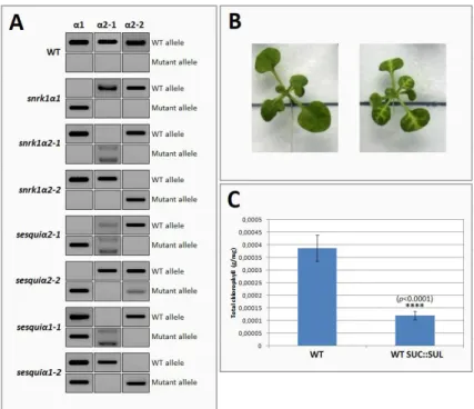
Finally, to explore the functional interaction between SnRK1 and AGO1 in the future, I developed important genetic tools based on a SUC::SUL reporter line that will allow to test whether changes in SnRK1 levels and activity affect AGO1-mediated silencing activity. The SUC::SUL reporter is based on the vasculature-specific silencing of a gene involved in chlorophyll biosynthesis (SULPHUR), through small interfering RNAs derived from its sense and antisense expression under the SUC2 promoter. New plant lines for different snrk1α mutants harbouring this reporter were generated and are ready to be used in future assays, for which the experimental setup was also optimised here.
Taken together, the results presented in this thesis suggest that SnRK1 may indeed regulate the miRNA pathway at different levels, affecting miRNA accumulation and potentially AGO1 function to control growth, development and plant stress responses. Further work will be required to confirm the extent and mechanism by which SnRK1 impacts on miRNA biogenesis and/or stability and to dissect the details of its interaction with AGO1.
IV
RESUMO ALARGADO
As plantas, devido à sua incapacidade de locomoção, estão confinadas ao local onde germinaram sendo, por isso, vulneráveis a condições ambientais que restringem o seu crescimento e desenvolvimento. Temperaturas extremas, seca, inundações, elevada salinidade, exposição a metais pesados, lesões mecânicas, condições de luz inadequadas e infeção por patogéneos estão entre as maiores causas de perda de produtividade agrícola a nível mundial.
Para fazer face a estas flutuações ambientais, as plantas desenvolveram, por um lado, estratégias adaptativas de sobrevivência de carácter específico, que lhes permitem responder a um tipo particular de stress e, por outro, mecanismos gerais responsáveis pelo ajuste metabólico e pela reprogramação da expressão de genes, permitindo, assim, rapidamente reparar os componentes celulares danificados e alocar nutrientes para os processos adequados, de forma a restaurar a homeostasia.
Em condições normais, as plantas convertem a luz em energia química sob a forma de açúcares que são depois distribuídos pelos vários órgãos da planta permitindo o seu correto crescimento e desenvolvimento. Contudo, condições desfavoráveis com impacto deletério nos processos de fotossíntese e respiração resultam, frequentemente, na diminuição dos níveis de energia celular da planta e afetam a alocação de açúcares para os órgãos em crescimento, levando à ativação da proteína cinase SnRK1. Esta, por sua vez, para restabelecer a homeostasia, ativa processos catabólicos e inibe processos anabólicos através da fosforilação de diversas enzimas metabólicas e de uma extensa reprogramação do transcriptoma, permitindo, assim, a aclimatação e sobrevivência das plantas. A SnRK1 pertence a uma família altamente conservada de cinases e partilha semelhanças estruturais e funcionais com as proteínas ortólogas “AMP-activated protein kinase” (AMPK), nos mamíferos, e “Sucrose non-fermenting 1” (SNF1), nas leveduras, funcionando como um complexo heterotrimérico composto por uma subunidade catalítica α e duas subunidades regulatórias, β e γ. Em Arabidopsis, existem três diferentes isoformas de SnRK1α, apesar de apenas duas (codificadas pelos genes
SnRK1α1/KIN10 e SnRK1α2/KIN11) serem expressas constitutivamente em todos os tecidos da planta,
três diferentes isoformas de SnRK1β (codificadas pelos genes SnRK1β1, SnRK1β2 e SnRK1β3) e apenas uma isoforma da subunidade γ, SnRK1βγ.
Não obstante a sua enorme importância a nível da resposta a diferentes tipos de stress, a via de sinalização da SnRK1 vai além do mero ajuste metabólico nessas condições, estando igualmente implicada na sinalização de açúcares, associada a vias de sinalização mediadas por várias hormonas vegetais e envolvida na modulação do crescimento e desenvolvimento das plantas.
Contudo, apesar do seu papel central, são poucos os mecanismos descritos, até à data, capazes de explicar a reprogramação transcripcional desencadeada pela SnRK1. O aumento da expressão de determinados genes, por seu lado, é atribuído em grande parte a fatores de transcrição “basic leucine Zipper” (bZIP) bem estabelecidos enquanto efetores a jusante da via de sinalização da SnRK1. Por outro lado, quanto à repressão de genes, os mecanismos subjacentes continuam maioritariamente desconhecidos, embora os microRNAs (miRNAs) tenham sido implicados na repressão de alguns dos alvos de SnRK1, ainda que através de mecanismos desconhecidos.
Os miRNAs correspondem a uma classe de pequenos RNAs endógenos não codificantes com 20-24 nucleótidos que regulam a expressão de genes pós-transcricionalmente, através da clivagem e/ou do bloqueio da tradução de transcritos complementares. Desta forma, os miRNAs têm sido extensivamente implicados tanto no crescimento e desenvolvimento das plantas como na resposta a fatores de stress biótico e abiótico.
V O trabalho desenvolvido nesta tese de mestrado teve como principal objetivo aumentar o conhecimento sobre os mecanismos envolvidos na comunicação entre a via de sinalização de SnRK1 e os miRNAs. Para isso, foram testadas duas hipóteses não mutuamente exclusivas: a) SnRK1 afeta a biogénese de miRNAs e b) SnRK1 afeta a atividade de miRNAs.
A biogénese de miRNAs ocorre no núcleo, englobando várias processos interdependentes desempenhados por componentes organizados num complexo. Em linhas gerais, a RNA Polimerase II (Pol II) é recrutada para o gene MIR, promovendo a sua transcrição e dando origem a um miRNA primário (pri-miRNA) que é processado pela proteína “DICER-like 1” (DCL1) com o auxílio das proteínas “Hyponastic-leaves 1” (HYL1) e “Serrate” (SE), originando um percursor de miRNA (pré-miRNA). Este pré-miRNA é novamente processado pela DCL1, originando uma pequena cadeia dupla formada por miRNA/miRNA*, que constituem respectivamente a cadeia guia e a cadeia passageira. A extremidade 3’ do duplex miRNA/miRNA* é metilada pela metil-transferase “Hua-Enhancer 1” (HEN1). Ainda no núcleo, a cadeia passageira é normalmente alvo de degradação e a cadeia guia, que constitui o miRNA maduro, é reconhecida e incorporada no complexo “RNA-induced Silencing Complex” (RISC). Este complexo tem como principal efetor uma proteína da família “ARGONAUTE” (AGO), responsável pelo reconhecimento, já no citoplasma, de transcritos-alvo com sequência complementar à do miRNA, e pela sua subsequente clivagem ou bloqueio de tradução. Problemas na biogénese de miRNAs refletem-se, normalmente, num aumento de pri-miRNAs e numa acumulação deficiente de miRNAs maduros. Deste modo, para testar a primeira hipótese, comecei por analisar a acumulação de dois miRNAs específicos – miR156 e miR319 – em plantas Arabidopsis tipo silvestre e em mutantes com perda parcial de função de SnRK1. Os resultados mostraram que a inativação parcial de SnRK1 levou à redução dos níveis de expressão de ambos os miRNAs testados. Ainda que não se consiga apurar por agora se este é um efeito específico para os miRNAs testados ou se se trata de um mecanismo geral, a confirmar-se futuramente estes mesmos resultados para outros miRNAs, pode potencialmente significar que a atividade de SnRK1 é uma condição necessária para a correta acumulação de miRNAs.
Para perceber se SnRK1 afeta a atividade de miRNAs, e com base em resultados preliminares do laboratório da Baena-González, procurei avaliar se SnRK1 poderia eventualmente estar a interagir com a proteína AGO1, o maior efector do complexo de silenciamento RISC. O correto reconhecimento dos miRNAs pela proteína AGO adequada e a sua subsequente incorporação no complexo RISC representa a etapa final da biogénese de miRNAs e é crítico para a sua interação com os respetivos transcritos-alvo. Em Arabidopsis, a família AGO é constituída por dez membros, mas os miRNAs são, na sua grande maioria, incorporados na AGO1. Assim, comecei por testar a interação física entre SnRK1 e AGO1 através de ensaios par-a-par de dois híbridos em levedura (“Yeast Two Hybrid” - Y2H) seguido de uma co-imunoprecipitação. Apesar de ter sido detetada uma interação positiva entre SnRK1α1 e AGO1 em levedura, através da co-imunoprecipitação não foi possível detetar interações entre as duas proteínas in planta em folhas maduras de roseta. Este resultado poderá ser talvez devido ao carácter transiente ou fraco da interação ou à sua ocorrência específica em determinados tecidos ou fases de desenvolvimento. No futuro, seria importante repetir esta experiência em tecidos ou condições em que a expressão de AGO1 seja mais elevada, eventualmente, otimizando ainda as condições da co-imunoprecipitação, para que se possa confiantemente descartar ou confirmar a ocorrência de interação física entre SnRK1 e AGO1 in planta.
Paralelamente a esta abordagem bioquímica, procurei ainda perceber se existia alguma interação genética entre AGO1 e SnRK1. Para tal, mutantes ago1-27, deficientes no silenciamento pós-translacional de genes, foram cruzados com mutantes com ganho e perda de função de SnRK1α1. A
VI interação genética foi avaliada com base na caracterização fenotípica dos processos de germinação e enverdecimento dos cotilédones na presença de elevados níveis de glucose ou ácido abscísico (ABA) e com base no tempo de floração em condições de dias longos. Relativamente à germinação e enverdecimento dos cotilédones, enquanto que, sob elevados níveis de glucose, a mutação ago1-27 parece potenciar a hipersensibilidade do sobreexpressor de SnRK1α1, sob elevadas concentrações de ABA é o sobreexpressor de SnRK1α1 que parece aumentar o fenótipo hipersensível do mutante
ago1-27. Estes resultados sugerem uma possível regulação negativa de AGO1 sobre SnRK1 e vice-versa
cuja intensidade e sentido podem variar consoante o tipo de stress. Curiosamente, a mutação
snrk1α1-3 reverteu parcialmente o fenótipo de floração atrasado do mutante ago1-27, com o duplo mutante a
florir em média dois dias antes do que as plantas ago1-27 e com menos folhas de roseta do que as plantas do tipo silvestre, reforçando ainda mais o potencial papel de AGO1 enquanto regulador negativo de SnRK1.
Finalmente, para explorar a interação funcional entre SnRK1 e AGO1, desenvolvi importantes ferramentas genéticas baseadas numa linha repórter SUC::SUL que irão permitir, no futuro, testar até que ponto diferentes níveis de SnRK1 influenciam a atividade silenciadora de AGO1. No sistema repórter SUC::SUL, um RNA de cadeia dupla do gene SULPHUR, é expresso sob o controlo do promotor SUC2, específico das células de companhia do floema, dando origem a pequenos RNAs de interferência que são incorporados na AGO1, reprimindo, assim, o transcrito SUL (envolvido na biossíntese de clorofila) e causando a clorose das células silenciadas na vasculatura. Novas linhas de plantas homozigóticas para este repórter contendo diferentes mutações das subunidades catalíticas de SnRK1 foram geradas e estão prontas para ser usadas em ensaios futuros para os quais a configuração experimental foi também aqui otimizada.
Em suma, os resultados apresentados nesta tese fornecem novas evidências que apontam para uma possível ação de SnRK1 em diferentes níveis de regulação sobre a via de sinalização dos miRNAs, nomeadamente na acumulação de miRNAs e na atividade de AGO1, para controlar o crescimento, desenvolvimento e respostas das plantas a stress. Estudos futuros serão necessários para confirmar a extensão e mecanismo de impacto de SnRK1 na biogénese de miRNAs e para dissecar em detalhe a interação com AGO1.
VII
TABLE OF CONTENTS
ACKNOWLEDGEMENTS ... I ABSTRACT ... II RESUMO ALARGADO ... IV INDEX OF FIGURES ... VIII INDEX OF TABLES ... VIII ABBREVIATION LIST... IX
I. INTRODUCTION ... 1
1. Environmental stress and SnRK1 activation ... 1
2. SnRK1 structure and function ... 1
3. miRNAs and ARGONAUTE1 ... 2
II. MATERIALS AND METHODS ... 5
1. Plant material and growth conditions ... 5
2. RNA extraction and cDNA synthesis ... 5
3. Quantitative real-time RT-PCR (RT-qPCR) ... 5
4. Plasmid construction and Yeast-Two-Hybrid assay ... 5
5. SnRK1α1 immunoprecipitation and western-blotting ... 6
6. Phenotypical analyses... 7
7. Introgression of the SUC::SUL transgene into the SnRK1 sesquiα mutants ... 7
8. Selection of sesquiαSUC::SUL mutants for phenotypic analyses ... 8
9. Chlorophyll extraction and quantification ... 8
III. RESULTS ... 9
1. Does SnRK1 affect miRNA accumulation? ... 9
2. Does SnRK1 affect miRNA activity?... 10
2.1. Physical interaction between SnRK1 and AGO1 ... 10
2.2. Genetic interaction between SnRK1 and AGO1 ... 12
2.3. Functional interaction between SnRK1 and AGO1 ... 17
IV. DISCUSSION ... 20
V. CONCLUSIONS ... 27
VI. REFERENCES ... 28
VIII
INDEX OF FIGURES
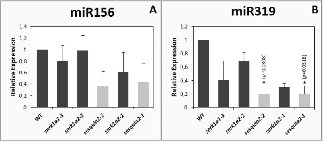
Figure 1. Relative abundance of miR156 (A) and miR319 (B) in leaves of wild type and
SnRK1 loss-of-function mutants. 9
Figure 2.1. AtAGO1 amplification and plasmid construction. 10
Figure 2.2. SnRK1α1 interacts with AGO1 in a pairwise yeast two hybrid assay. 11
Figure 2.3. AGO1 does not co-immunoprecipitate with SnRK1α1 in mature
Arabidopsis rosettes. 12
Figure 2.4. SnRK1α1 overexpressor (OE) is hypersensitive to glucose and epistatic to
ago1-27 during germination. 13
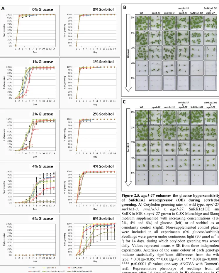
Figure 2.5. ago1-27 enhances the glucose hypersensitivity of SnRK1α1 overexpressor
(OE) during cotyledon greening. 14
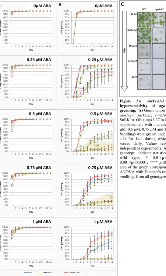
Figure 2.6. snrk1α1-3 partially reverts the ABA hypersensitivity of ago1-27 during
germination and greening. 16
Figure 2.7. snrk1α1-3 partially reverts the delayed flowering of ago1-27 mutants. 17
Figure 2.8. SUC::SUL silencing reporter system. 17
Figure 2.9. Schematic representation of the selection process of SnRK1 loss-of-function
mutants homozygous for SUC::SUL. 18
Figure 2.10. Strategy steps to assess the functional interaction between SnRK1 and
AGO1. 19
Figure 3.1. Schematic representation of the possible mode of action of SnRK1 and AGO1 during germination/early seedling development under high glucose stress or
ABA. 24
Figure 3.2. AGO1 is a negative regulator of SnRK1 during flowering. 25
Supplementary Figure 1. SnRK1α2 and β3 do not interact with AGO1 in a pairwise
yeast two hybrid assay. 38
INDEX OF TABLES
Table 1.1. Summarised phenotypic characterization of germination and cotyledon
greening under high concentrations of glucose or ABA. 23
Table 1.2. Summarised phenotypic characterisation of flowering under normal long day
conditions. 25
IX
ABBREVIATION LIST
ABA Abscisic acid
AGO1 ARGONAUTE1
AMPK AMP-activated protein kinase
bZIP Basic leucine Zipper
DCL1 DICER-like 1
HEN1 HUA-ENHANCER 1
HYL1 HYPONASTIC LEAVES 1
KO Knockout
mRNA Messenger RNA
miRNA MicroRNA
Pol II RNA Polymerase II
pre-miRNA Precursor miRNA
pri-miRNA Primary miRNA
SE SERRATE
siRNA Small interfering RNA
SNF1 Sucrose non-fermenting 1
SnRK1 Sucrose non-fermenting 1-related protein kinase 1
sRNA Small RNA
RISC RNA-induced silencing complex
RNA Ribonucleic Acid
WT Wild-type
1
I. INTRODUCTION
1. Environmental stress and SnRK1 activation
As sessile organisms, plants are constantly exposed to changing environmental conditions that threaten homeostasis, fitness and ultimately plant survival. Extreme temperatures, drought, flooding, high salinity, exposure to heavy metals, mechanical wounding, inadequate light conditions and infection by pathogens are amongst the major causes of crop losses worldwide1.
To cope with these conditions, plants rely on stress-specific mechanisms that promote tolerance to particular types of stress, such as the induction of a specific Na+ transporter to extrude salt from tissues2 or the induction of localised cell death in the root cortex to promote airflow and respiration during flooding3. However, general mechanisms that reprogram metabolism and gene expression are also essential to mount a successful stress response by allowing the repair of damaged cellular components, the redistribution of resources to appropriate processes, and ultimately by restoring homeostasis4.
One important general stress signal is the depletion of cellular energy. Under normal conditions, plants convert light into chemical energy in the form of sugars, which then need to be distributed throughout the different organs to allow proper growth and development. However, hostile conditions often lead to reduced rates of photosynthesis and/or respiration, affecting the overall energy status of the plant, the allocation of sugars to the growing organs, and ultimately limiting growth and development5,6. When ATP production is compromised and carbon levels decline, the energy sensing Snf1-related protein kinase 1 (SnRK1) is activated. To re-establish homeostasis, SnRK1 turns on energy-producing processes and inhibits energy-consuming pathways through a vast transcriptional reprogramming and through phosphorylation of a number of metabolic enzymes, thus promoting stress tolerance and survival7–9.
2. SnRK1 structure and function
SnRK1 belongs to a family of evolutionarily conserved kinases and shares structural and functional features with its ortholog proteins, the mammalian AMP-activated protein kinase (AMPK) and the yeast Sucrose non-fermenting 1 (Snf1)10. It functions as a heterotrimeric complex composed by a catalytic α subunit and two regulatory subunits, β and γ.
The α subunit comprises a catalytic domain harbouring a conserved T-loop whose phosphorylation is essential for activity, and a regulatory domain that is important for the interaction with the β and γ subunits. The β subunit regulates subcellular localization and kinase activity and acts as a scaffold between α and γ subunits9,11,12
. In mammals AMP and ADP bind to the AMPK γ subunit, thereby stimulating T–loop phosphorylation and activation of AMPK13,14. In plants, however, the only γ-like subunit present in SnRK1 heterotrimeric complexes is the highly atypical βγ, which does not appear to bind AMP or ADP15,16.
In Arabidopsis, there are three different SnRK1α isoforms [encoded by the SnRK1α1/KIN10,
SnRK1α2/KIN11 and SnRK1α3/KIN12 genes – although SnRK1α3 is poorly expressed in most plant
tissues)17 and three different SnRK1β isoforms (encoded by the SnRK1β1, SnRK1β2, SnRK1β3 genes), whereas SnRK1βγ is encoded by a single gene. Given this, it is likely that different SnRK1 αβγ heterotrimers assemble through various combinations of α1/α2/β1/β2/β3/βγ subunits and that
2 consequently SnRK1 complex composition may differ between particular subcellular compartments, tissues or developmental stages9. The number of possible combinations is further increased when considering that most SnRK1 transcripts can be alternatively spliced.
A complete SnRK1α1/α2 knockout is lethal in higher plants7 and plants with compromised SnRK1 function display several metabolic and developmental defects, such as defective seed storage, maturation and dormancy in pea18, male sterility in barley19, and impaired starch degradation, severe growth reduction, and early senescence in Arabidopsis7. Moreover, antisense mediated silencing of the β-subunit StubGAL83 in potato, affects tuber and root development20
. Conversely, SnRK1α1 overexpression in Arabidopsis delays flowering and senescence, alters significantly seedling growth, and promotes tolerance to stresses such as nutrient-deprivation or hypoxia21,22.
In brief, SnRK1 functions as a sugar signalling hub, being activated under carbon starvation and inhibited by sugars like trehalose-6-phosphate (T6P)23,24 by mechanisms not yet fully understood9. Supporting the regulation of SnRK1 by the carbon status, SnRK1α1 overexpression generates a starvation transcriptomic signature opposite to that occurring in response to supply of exogenous sugar7.
The SnRK1 pathway has also been shown to interact with several hormonal pathways. Leaf senescence driven by ethylene signalling is in part regulated by the SnRK1-mediated repression of the transcription factor ETHYLENE INSENSITIVE3 (EIN3)25. A physical interaction between SnRK1α1 and MYC2, a master regulator of jasmonate (JA) signalling, has also been established26. Importantly, SnRK1 connection to abscisic acid (ABA) signalling has been demonstrated through phenotypical and biochemical analyses of SnRK1 mutants27 and further supported by the identification of two established repressors of the ABA pathway (ABI1 and PP2A) as negative regulators of SnRK1 signalling28.
SnRK1 implements vast metabolic changes during stress, partly through the direct phosphorylation of biosynthetic enzymes such as sucrose phosphate synthase (SPS)29,30, HMG-CoA reductase (HMGR)29, nitrate reductase (NR)29,30, 6-phosphofructo-2-kinase/2,6-fructose-bis-phosphatase (F2KP)31 and trehalose-6-phosphate synthase (TPS)30,32.
On the other hand, SnRK1 also induces an extensive transcriptional reprogramming through mechanisms mostly still unknown. bZIP transcription factors are well described in the literature as downstream effectors of the SnRK1 signalling cascade in response to low-energy stress, salt stress, or during dark-induced senescence7,33,34, but other factors involved in SnRK1-dependent gene regulation, and in particular in gene repression, remain largely unidentified.
In the last decade, miRNAs have been increasingly implicated in the response to environmental stress35 and previous work from the Plant Stress Signalling Group has shown that miRNAs can mediate the repression of specific mRNA targets by SnRK1 in response to energy deprivation36. Nevertheless, the underlying mechanisms remain elusive.
3. miRNAs and ARGONAUTE1
MicroRNAs (miRNAs) are a class of endogenous non-coding 20- to 24-nucleotide RNAs involved in post-transcriptional gene repression. In plants, miRNAs act either through cleavage or translation attenuation of complementary mRNA targets37.
In Arabidopsis, miRNAs biogenesis takes place in the nucleus through a series of interdependent steps involving cooperation of several proteins. More specifically, RNA Polymerase II (Pol II) is recruited
3 to the promoters of MIR genes, transcribing them into primary-miRNAs (pri-miRNAs)38. Then, pri-miRNAs fold into stem-loop structures that are processed through cleavage by DICER-LIKE 1 (DCL1) into miRNA precursors (pre-miRNAs). In this first step, central components like HYPONASTIC LEAVES1 (HYL1) and SERRATE (SE) interact and collaborate with DCL1 to promote the accurate processing of pri-miRNAs39. Pre-miRNAs are further processed by DCL1 into miRNA/miRNA* duplexes that are stabilised through 3´-terminal 2´-O-methylation by HUA ENHANCER 1 (HEN1)40. Recent evidence suggests that, while in the nucleus, mature miRNAs (miRNA single guide strands) are recognised and loaded into the RNA-induced Silencing Complex (RISC) containing ARGONAUTE1 (AGO1) proteins41. The passenger strands (miRNAs*) are often targeted for degradation and the miRNA-loaded RISC complexes are exported to the cytoplasm where AGO1 recognises target mRNAs complementary to the loaded miRNA, either slicing the transcript or blocking its translation42–49.
Mutations that compromise the function of the miRNA biogenesis machinery or the RISC complex often lead to aberrant plant phenotypes with pleiotropic developmental defects, such as abnormal embryogenesis, short stature, altered leaf shape, defects in floral meristems and reduced fertility50–52, reinforcing the importance of these small RNAs as crucial developmental regulators.
Well-known examples of this are miR156 and miR172, two independent but interconnected miRNAs involved in flowering time control, whose expression levels are oppositely correlated53. Other examples include miR319, which targets TEOSINTE BRANCHED1, CYCLOIDEA,
and PROLIFERATING CELL NUCLEAR ANTIGEN BINDING FACTOR (TCP) transcription factors that regulate leaf shape and senescence54, miR159, which targets MYB transcription factors with a role in seed germination55 and miR396, which targets GROWTH-REGULATING FACTORs (GRF) involved in the regulation of cell division56.
Besides their established role in plant growth and development, miRNAs are also involved in the response to multiple stresses57. For example, in Arabidopsis, miR160 promotes tolerance to heat stress58 and miR394 acts as a negative regulator of resistance to Botrytis cinerea59. Additionally, the expression levels of several miRNAs are differentially influenced by distinct types of stress, sometimes, in a tissue-dependent manner. In Arabidopsis, the expression of miR169 increases upon exposure to salt60 but decreases during drought treatment61 and, in wheat, at least six miRNAs, including miR159, miR172, miR319, miR399, miR528, and miR4393 showed an induced expression in leaves but were inhibited in roots, in response to drought62. Moreover, miRNA biogenesis mutants are often hypersensitive to ABA, a key regulator of abiotic stress tolerance, and display aberrant responses to different types of stresses, including salt and osmotic stress63–66.
Over the past years many factors involved in miRNA signalling have been discovered, suggesting that miRNAs are tightly regulated at several levels, such as transcriptional induction, splicing, processing, loading, export, activity and degradation37,67.
The precise recognition of miRNAs by the appropriate ARGONAUTE protein and their subsequent loading to the RISC complex represents the final step of miRNA biogenesis and is critical for their interaction with targets. In Arabidopsis, the ARGONAUTE family comprises 10 member proteins, all harbouring N-terminal, PAZ, MID and PIWI domains68, and the sorting of each miRNA into a specific AGO relies mainly, but not exclusively69, on the identity of the 5'-sequence position of mature miRNAs70–72. From the 10 AGO family members, ARGONAUTE 1 is particularly important since the majority of the mature miRNAs are sorted into it, given their 5’-terminal uridine. By contrast, miRNAs bearing a 5’ terminal adenosine are preferentially loaded into AGO2 whereas those carrying
4 a cytosine at the 5’ terminus are sorted to AGO570,72,73
.
AGO1 is a nucleo-cytosolic shuttling protein41 with slicer activity that selectively recruits miRNAs for post-transcriptional gene silencing (PTGS)74, being able to regulate its own expression via a negative feedback loop that involves miR168 activity75.
In addition to its well-described role in PTGS, AGO1 was recently implicated in the co-transcriptional down-regulation of MIR genes67 and in the transcriptional induction of jasmonate signaling genes through direct chromatin binding at the target loci76.
Given the importance of SnRK1 signalling for stress tolerance and the increasing number of reports linking miRNAs to stress responses, the Baena-González group proposed a possible cross-talk between these two pathways. The finding that the repression of specific transcripts by SnRK1 requires miRNAs confirmed this hypothesis, establishing miRNAs as downstream effectors of SnRK1 signalling36. However, the mechanisms by which SnRK1 affects miRNA signalling pathways remained unknown.
Therefore, the aim of this thesis was to gain mechanistic insight into how SnRK1 activity results in miRNA-mediated gene repression. To this end, two non-mutually exclusive hypotheses were tested: a) SnRK1 affects miRNA biogenesis and b) SnRK1 affects miRNA activity, possibly through an interaction with AGO1.
5
II. MATERIALS AND METHODS
A list of all primers used in this study is provided in Table S1.
1. Plant material and growth conditions
All Arabidopsis thaliana plants used are in the Columbia (Col-0) background except
35S::SnRK1α1-HA, which is in Landsberg erecta7. The snrk1α1-333, 35S::SnRK1α127, snrk1α2-177, ago1-2778 and
SUC::SUL plant lines79,80 have been previously described. snrk1α2-2 is a T-DNA insertion mutant (WiscDsLox384F5), which had been previously isolated and preliminarily characterised in the Baena-González lab (A. Confraria, unpublished). These single mutants had previously been crossed to generate partial SnRK1α loss-of-function mutants (snrk1α1-/- snrk1α2+/- and snrk1α1+/- snrk1α2-/- ; A.
Confraria, unpublished), hereafter designated as sesquiα2 and sesquiα1, respectively, depending on the
snrk1α mutation that is segregating. For plant growth seeds were surface-sterilised for 1 min in 70%
(V/V) ethanol followed by 10 min in 20% (V/V) bleach under gentle rocking, and then washed at least 3 times in sterile water. Sterilised seeds were stratified in the dark at 4°C for 2-3 days and sowed on plates containing half strength Murashige and Skoog medium (Duchefa Biochemie) with 0.1% MES and 0.8% phytoagar. Unless otherwise specified, plants transferred to soil were grown under a 16h-light (100μmol m-2 s-1), 22ºC / 8h-dark, 18ºC regime.
2. RNA extraction and cDNA synthesis
Total RNA was extracted from rosette leaves of 5 week-old plants grown in soil under a 12h-light (110 μmol m-2 s-1, 22ºC)/12h-dark (18ºC) regime with TRIzolTM (ThermoFisher Scientific) according to the manufacturer’s instructions. RNA was then treated with RQ1 RNase-Free DNase (Promega; 2 DNase units per μg of RNA) at 37°C for 30 min and recovered by a phenol/chloroform extraction and subsequent precipitation with isopropanol. DNase-treated RNA (1 μg) was reverse transcribed in a total reaction volume of 40μL using Super Script III Reverse Transcriptase (InvitrogenTM), following the manufacturers’ instructions. cDNA was synthesised using a mix of OligoDT (for the housekeeping genes) and miRNA-specific primers (Supplementary Table 1).
3. Quantitative real-time RT-PCR (RT-qPCR)81
qRT-PCR analyses were performed in 384-well reaction plates using the QuantStudio™ 7 Flex Real-Time PCR System (Thermo Fisher). The reactions were prepared in a total volume of 10μL containing 1μL of cDNA (diluted 1:10) corresponding to 2.5ng of RNA, 5μL of iTaq Universal SYBR Green Supermix (BioRad) and 0.8μL of each gene-specific 5μM primer. No-template and -RT controls were included for each gene in comparative gene expression analyses.
The 2−∆∆Ct method was used for relative quantification82. Expression values were normalised to the geometric mean of Ct values obtained for the following reference genes: EIF4 (At3g13920), UBQ10 (At4g05320) and UBC21 (At5g25760)83.
4. Plasmid construction and Yeast-two-hybrid assay
The full AGO1 coding sequence was amplified from the plasmid pHBT95-AGO1 already existing in the Baena-González lab, using primers containing attB1 and attB2 sites. The PCR product was extracted and purified from a 0.8% agarose-TAE gel, using a Macherey-Nagel NucleoSpin® Gel and PCR Clean-up, following the manufacturer’s instructions. The purified PCR product was thereafter recombined into the pDONR221 vector through a BP reaction. Recombination and incorporation of
6 the AGO1 insert into the pDONR221 vector was further confirmed using two restriction enzymes – XbaI and BamHI – to digest the resultant pENTR clones and the full-length AGO1 insert was sequenced to confirm the absence of PCR-derived mutations. This entry clone was then recombined by an LR reaction into a GatewayTM-modified pGADT7 destination vector (pDEST-GADT7) kindly provided by Nasser Rusan (NIH, Maryland, USA) and the recombination sites flanking AGO1 were sequence-verified before proceeding to the next step. All SnRK1 subunits (α1, α2, β1, β2, β3 and βγ), cloned into pGBKT7 vectors, were already available in the Baena-González lab.
Competent yeast cells (Y2HGold) were co-transformed with pDEST-GADT7-AGO1 and pGBKT7 vectors containing the SnRK1 subunits. Each construct was also co-transformed with the complementary empty vector, to control for growth due to auto-activation. Three independent colonies of each co-transformation were inoculated into liquid synthetic dropout (SD) medium and grown to saturation, at 28ºC, shaking at 180rpm. Ten-fold serial dilutions of saturated yeast cultures were spotted onto plates containing SD medium with increasing stringency, lacking 3 H) or 4 (-L-W-H-A) amino acids. When indicated, 25 mM amino-triazole (AT) was added to SD medium to further increase stringency.
Positive interactions were assayed three times in independent replicates, while negative interactions were assayed twice.
5. SnRK1α1 immunoprecipitation and western-blotting
SnRK1α1-HA was immunoprecipitated from 5-week old rosettes of 35S::SnRK1α1-HA plants treated for 6h in darkness, or kept in the light as controls. Wild type 5-week old rosettes of Landsberg erecta (Ler) plants were used as controls. Samples were harvested, flash-frozen and reduced to a fine powder in liquid nitrogen. Proteins were extracted with immunoprecipitation (IP) buffer [2mL buffer/g fresh weight; 50mM Tris-HCl pH 7.5, 100mM NaCl, 10mM EDTA, 0.002% (v/v) phosphatase inhibitor cocktail #2 (Sigma P572G), 0.002% (v/v) phosphatase inhibitor cocktail #3 (Sigma P0044), 0.3% (V/V) IGEPAL, 2mM DTT and 2X cOmpleteTM protease inhibitor cocktail (Roche; 1 tablet/25mL)] and incubated at 4°C for 10 min. After discarding insoluble material by centrifugation, total protein concentration of the supernatant was quantified using the Bradford protein assay. For the IP, 1.2 ml of extract containing 3.5 mg protein were incubated with 40μL of anti-HA affinity matrix (Roche) with gentle rocking for 3h30min at 4ºC. Agarose beads containing immunoprecipitated proteins were recovered by a brief centrifugation at a maximum speed of 2000 rpm and washed five times with IP buffer. To elute immuno- and co-immunoprecipitated proteins, 20μL of 4X Laemmli buffer84 were added to the beads and samples were boiled for 5 min at 95°C. For all samples, aliquots of cleared tissue lysates before the immunoprecipitation (general input) and of the supernatant after immunoprecipitation (pass-through) were kept as controls. These samples were denatured with Laemmli buffer and boiled for 5 min at 95°C, similarly to the immunoprecipitated proteins.
Proteins were resolved by SDS-PAGE, wet-transferred to a 0.45μm PVDF membrane (100 V, 70min, 4°C), and analysed by immunoblotting with anti-AGO1 (1/10000, Agrisera, AS09527) antibody, followed by an incubation with anti-HA (1/1000, Roche, #11867423001) to ensure the effectiveness of the IP.
Primary antibodies were diluted according to the manufacturers’ instructions and incubated with the membrane over night at 4°C under gentle rocking. Secondary antibodies (anti-rabbit for AGO1 and anti-rat for HA) conjugated to horseradish peroxidase were diluted 1:10000 in 1% non-fat milk in TBS and incubated with the membrane for 1 h at room temperature. For detection, we used SuperSignal™
7 West Femto Maximum Sensitivity Substrate (Thermo Fisher). Images were acquired with a Bio-Rad ChemiDoc, using ImageLab software.
6. Phenotypical analyses
For phenotypic assays, seeds of wild type, ago1-27, snrk1α1-3, snrk1α1-3 x ago1-27, SnRK1α1OE and SnRK1α1OE x ago1-27 were sterilised and imbibed as previously described. A total of 22 seeds of each genotype were sown per plate, containing half strength Murashige and Skoog medium with 0.1% MES, 0.8% phytoagar and supplemented or not with increasing concentrations of glucose (1%, 2%, 4% or 6%), sorbitol (1%, 2%, 4% or 6%) or ABA (0.25μM, 0.5 μM, 0.75 μM or 1 μM). Plates were sealed with hypoallergenic adhesive (3M) and kept in dark at 4ºC for 2 days. After stratification the plates were incubated at 22ºC under continuous low light (70μmol m-2 s-1) during 14 days.
Germination and cotyledon greening were scored every day except on the 6th and 13th days and all the assays were carried out in triplicate. The criterion for scoring germination was the rupture of the outer seed coat (“testa rupture”), whereas for scoring cotyledon greening the day when cotyledons greened was considered, independently of being or not expanded. Seeds that had not germinated by the end of the assay (14 days) were not scored. Germination and cotyledon greening were expressed as percentage of the total germinated seeds. Average percentages were calculated with standard error of the triplicates.
For the flowering assays, 9 seeds of each genotype were treated as previously described and after stratification were sown in soil and grown under a 16h-light (100μmol m-2 s-1), 22ºC / 8h-dark, 18ºC regime. To quantify the flowering phenotypes, the number of rosette leaves from each individual plant was counted every two days until bolting. The number of days until bolting was also recorded. This assay was carried out in triplicate. Average numbers of days and rosette leaves to flowering were calculated for each genotype with standard error of the triplicates.
7. Introgression of the SUC::SUL transgene into the SnRK1 sesquiα mutants
SUC::SUL transgenic Arabidopsis plants79 were previously crossed to SnRK1 sesquiα2 and sesquiα1 mutants (A. Confraria, unpublished). From the F1 progeny of these crosses, double heterozygous mutants for snrk1α1 and snrk1α2, displaying the “chlorotic vein” phenotype associated with the
SUC::SUL transgene were selected by genotyping the snrk1α1 and snrk1α2 mutations. Progeny of
single F1 plants were sown on 0.5X Murashige and Skoog medium supplemented with 1% Sucrose and 10 ug/mL BASTA in order to select SUC::SUL homozygous plants. Resistant plants displaying the chlorotic vein phenotype were transferred to soil. Later, gDNA was extracted and genotyping analyses were performed for the snrk1α1 and snrk1α2 mutations (see below). In this way, we could ensure to recover plants of all the required snrk1α genotypes (sesquiα1, sesquiα2, and the corresponding single mutants). For the next round of selection, progeny of single F2 plants were sown on 0.5X Murashige and Skoog medium supplemented with 1% Sucrose and 10 ug/mL BASTA. After 10d of growth, plates with susceptible seedlings were discarded and plates with 100% resistant seedlings exhibiting the chlorotic vein phenotype characteristic of the SUC::SUL plants were selected. 12-18 seedlings from each plate were transferred to soil and grown in the same light conditions as before. Genotyping was performed by PCR using allele-specific primers (Supplementary Table 1) to select the plants with the genotypes of interest. This process was repeated until all plants bearing different SnRK1 loss-of-function mutations were homozygous for SUC::SUL.
8
8. Selection of sesquiαSUC::SUL mutants for phenotypic analyses
Given the fact that either the snrk1α1 or the snrk1α2 mutations are in heterozygosity in the sesquiα mutants and that the double snrk1α1 snrk1α2 mutant is lethal, the progeny of sesquiα mutants is always a mix of seeds corresponding to the sesquiα itself and to a single homozygous snrk1α mutant. When grown under normal conditions, it is impossible to distinguish these genotypes without using an invasive genotyping-based approach. However, for being able to quantitatively and reproducibly compare SUC::SUL silencing in a WT vs. sesquiα background it would be desirable to grow plants in controlled tissue culture conditions and to avoid their wounding. I therefore set to devise a non-invasive strategy that allowed me to grow plants of all genotypes in the same conditions and at the same time be able to distinguish the sesquiα mutants. Work related to another project in the Baena-González lab had shown that sesquiα mutants display reduced hypocotyl elongation when germinated and grown for 4-5 days in darkness (M. Adamo, unpublished). Therefore, I made use of this phenotype to distinguish in a non-invasive manner sesquiα mutants from the single snrk1α mutants in the segregating sesquiα progeny. Sterilised and stratified seeds from previously genotyped sesquiαSUC::SUL mutants were sown on 0.5X Murashige and Skoog and grown vertically. The plate was kept under normal light (100μmol m-2 s-1) for three hours and then covered with aluminium foil and left in the dark at 22ºC during four days. Seedlings with considerably shorter hypocotyls (corresponding to
sesquiα mutants) were transferred to a fresh plate containing 0.5X Murashige and Skoog medium, 1%
sucrose and grown horizontally under low light conditions (50 μmol m-2 s-1) for recovery.
9. Chlorophyll extraction and quantification
Wild type and SUC::SUL plants (12 of each genotype) grown for 14d under low light (50 μmol m-2 s-1) were harvested, quickly weighed, frozen with liquid nitrogen, and ground to a fine powder. Acetone [1.2 mL of 80% (V/V)] was added to each individual sample and tubes were briefly vortexed. Samples were covered with aluminium foil to prevent chlorophyll degradation and chlorophyll was extracted over night at room temperature, under rotation (40 rpm). After a centrifugation step (15 min, 4ºC, 3000 rpm), 200 μL of each supernatant were used to measure absorbance at 645 nm and 663 nm. Total chlorophyll levels were calculated as described 85 using the following equation (Equation 1):
Equation 1: Ca+b(mg/g) = [8.02×A663+20.20xA645]×V/1000×W (Chlorophyll a+b)
9
III. RESULTS
1. Does SnRK1 affect miRNA accumulation?
To evaluate whether SnRK1 affects miRNA accumulation, SnRK1 partial loss-of-function sesquiα2 mutants recently obtained in the Baena-González lab were used. This kind of analyses was not previously possible due to the lethality associated with complete loss of SnRK1 activity. These partial loss of function mutants have the α1-catalytic subunit knocked out and the α2-subunit knocked down [snrk1α1-/-/snrk1α2+/-], and despite showing significantly compromised SnRK1 signalling they are fully viable (A. Confraria, unpublished). These mutants are thereafter referred as sesquiα2-1 or
sesquiα2-2, depending on the snrk1α2 allele they bear (see Materials and Methods for details).
These initial analyses were focussed on two specific miRNAs: miR156 and miR319. miR156 targets SPL transcription factors and is hence involved in the regulation of flowering time53,86 whereas miR319 targets TCP transcription factors that are important for leaf development54. In addition, it was recently found that the inactivation of SnRK2 kinases, closely related to SnRK1 and core components of ABA and osmotic stress signalling pathways, leads to reduced accumulation of these specific miRNAs87.
To see whether our SnRK1 loss of function mutants had altered accumulation of miR156 and miR319, I extracted RNA from 5-week-old rosette leaves of WT, sesquiα2-1, sesquiα2-2 mutants and the corresponding parental lines and measured the levels of mature miR156 and miR319 using stem-loop quantitative RT-PCR (qPCR)81 and specific primers (Supplementary Table 1).
Although not statistically significant in the case of miR156 (only two biological replicates were processed), both sesquiα2 mutants appeared to accumulate less miR156 and miR319 (Figure 1) than the wild type, supporting that the partial inactivation of SnRK1 may have a negative impact in miRNA biogenesis. The single mutants, however, vary in their behaviour in what concerns the two miRNAs.
Figure 1. Relative abundance of miR156 (A) and miR319 (B) in leaves of wild type and SnRK1 loss-of-function mutants. miRNA levels were analysed by stem-loop qRT-PCR in 5-week-old rosette
leaves of wild type, snrk1α1-3, snrk1α2-2, sesquiα2-2, snrk1α2-1 and sesquiα2-1, using the geometric mean of reference genes EIF4, UBQ10 and UBC21 to normalise expression levels. Wild type was used as control sample. Bars represent SEM of two biological replicates. Asterisks indicate statistically significant differences from the wild type: * p<0.05 (p-value; one-way ANOVA with Dunnett’s test).
10
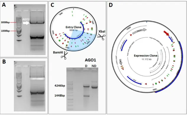
Figure 2.1. AtAGO1 amplification and plasmid construction. A) AGO1 was amplified from the plasmid
pHBT95-AGO1 with specific primers and B) the PCR product was extracted from the agarose gel to be recombined into the pDONR221 vector. C) Enzymatic digestion of the entry clone with two enzymes – BamHI and XbaI – revealed the expected band pattern with two bands of 4246bp and 1448bp. The sample identified as D was digested and the ND sample corresponds to the non-digested entry clone used as a control. D) Schematic representation of the expression clone pGADT7-AGO1.
2. Does SnRK1 affect miRNA activity?
Previous preliminary work in the Baena-González lab showed that, in response to stress treatments, increased SnRK1 activity and downstream miRNA-dependent gene repression was not accompanied by increased miRNA levels or increased miRNA incorporation into the silencing complex (Cláudia Martinho, PhD Thesis, UNL 2013), suggesting a potential regulation of the silencing complex itself. Given that most miRNAs are sorted into AGO1, which ultimately conducts the cleavage or translation attenuation of complementary mRNAs, regulation of this core component of the silencing complex by SnRK1 could influence miRNA activity by driving a differential gene repression. Therefore, I decided to test the interaction between SnRK1 and AGO1 at different levels.
2.1. Physical interaction between SnRK1 and AGO1 2.1.1. Pair-wise yeast-two hybrid assays
To test the physical interaction between SnRK1 and AGO1, I started by using a yeast-two-hybrid (Y2H) pairwise assay, which is a sensitive and cost-effective means to test protein-protein interactions.
The coding sequence of AtAGO1 was amplified from a plasmid available in the lab and the PCR product was loaded in an agarose-TAE gel. After electrophoresis, the gel showed a clear band with approximately 3150 bp, the expected size for AGO1 coding sequence (Figure 2.1.A), and an unspecific PCR product. The band corresponding to AGO1 was excised from the gel (Figure 2.1.B), purified and recombined into the pDONR221 vector using Gateway technology.
11 To evaluate if the recombination was successful, the entry clone pDONR221-AGO1 was then digested with the XbaI and BamHI restriction enzymes, having produced the expected restriction pattern. To ensure that the entry clone did not contain unexpected mutations, the full length AGO1 insert was sequenced. Following confirmation that the sequence was correct, the entry clone was recombined by an LR reaction into the destination vector pGADT7-Dest (Figure 2.1.D) and the recombination sites flanking AGO1 were sequenced to ensure that this last step yielded a functional non-mutated expression clone.
For the pairwise Y2H assay, competent yeast cells were co-transformed with pGADT7-AGO1 and pGBKT7 constructs for expressing the various SnRK1 subunits. As controls, pGBKT7-SnRK1 subunits and pGADT7-AGO1 were also co-transformed with the respective complementary empty vectors.
As shown in Figure 2.2., cells co-transformed with SnRK1α1 and AGO1 grew in synthetic dropout medium lacking three amino acids (-L-H-W), whereas there was no growth in either of the controls under the same conditions, supporting that these two proteins are able to physically interact in yeast. When stringency was increased by adding aminotriazole (AT) or by removing one further amino acid (-L-W-H-A), the interaction was disrupted, indicating that the SnRK1α1-AGO1 interaction is weak.
In fact, in pairwise Y2H assays, SnRK1α1 was the only subunit for which I could detect a positive interaction with AGO1. Cells co-expressing SnRK1α2 or SnRK1β3 and AGO1 failed to grow in medium with increased stringency (Supplementary Figure 1). For the remaining subunits, SnRK1β1, β2 and βγ, their individual expression was sufficient to support yeast growth in -L-W-H medium, yielding a very high background growth that precluded the assessment of a possible interaction between these subunits and AGO1 (Supplementary Figure 1).
2.1.2. Co-immunoprecipitation
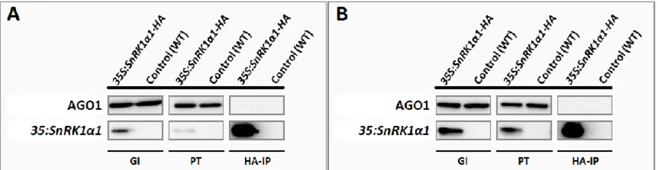
To further analyse the physical interaction between SnRK1α1 and AGO1, a co-immunoprecipitation assay was carried out using proteins extracted from rosettes of 5-week-old plants exposed to a SnRK1-activating dark treatment or kept in the light as a control. Under these conditions, we could not detect AGO1 in the fraction co-immunoprecipitating with SnRK1α1-HA either in the light or in the dark (Figure 2.3. A and B). This does not exclude that SnRK1α1 and AGO1 may still interact in planta in different conditions, developmental stages, or tissues.
Figure 2.2. SnRK1α1 interacts with AGO1 in a pairwise yeast two hybrid assay. Ten-fold serial
dilutions of saturated yeast cultures were spotted onto plates containing synthetic dropout medium with increased stringency. Growth in -L-W-H medium was observed only when cells were co-transformed with SnRK1α1 and AGO1. 25 mM amino-triazole (AT) was used to increase stringency of -L-W-H medium. This assay was repeated three times with similar results. The picture shows a representative experiment.
12
2.2. Genetic interaction between SnRK1 and AGO1
To further assess a potential connection between SnRK1 and AGO1, I evaluated their genetic interaction using a phenotypic characterisation of early seedling development under specific stress conditions. To this end I used ago1-27 mutants crossed to SnRK1 loss- and gain-of-function lines. The
ago1-27 mutant bears a single nucleotide mutation in the PIWI domain that partially impairs AGO1
function48,78. This mutant was previously crossed to the snrk1α1-3 knockout mutant33 and to a SnRK1α1 overexpressor (SnRK1α1OE)27 (N. Fernandes, unpublished). The two resulting double homozygous lines had been previously isolated.
SnRK1α1OE lines have been extensively described as hypersensitive to glucose in early seedling development7,27,28, whereas ago1-25 mutants were previously described as hyposensitive88. Glucose in high concentrations is known to induce stress responses and developmental arrest by a combination of specific glucose effects and by the consequent increase in osmotic pressure. I grew seedlings of all relevant genotypes (wild type, ago1-27, snrk1α1-3, snrk1α1-3 x ago1-27, SnRK1α1OE and SnRK1α1OE x ago1-27) under continuous low light in 0.5X Murashige and Skoog medium supplemented with increasing concentrations of glucose, scoring germination and greening rates for 14 days. In order to distinguish between the signalling and osmotic effects of glucose, the same experiment was repeated with sorbitol as an osmolarity control.
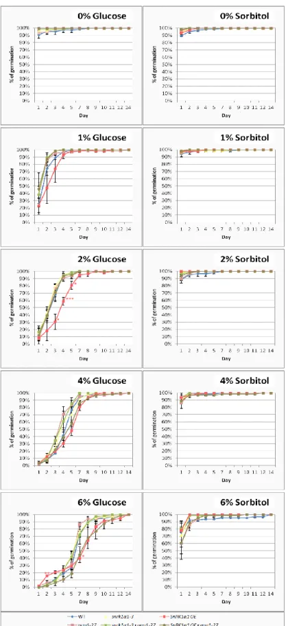
As shown in Figure 2.4., the presence of glucose in the medium delayed germination in all genotypes. As expected, SnRK1α1OE was hypersensitive, showing delayed germination in comparison to the wild type in most glucose concentrations. Interestingly, in the higher glucose concentrations (4% and 6%), SnRK1α1OE was epistatic to ago1-27. Under increasing sorbitol concentrations all the genotypes behaved similarly with regard to germination. Even though not statistically significant, in medium supplemented with 6% sorbitol, ago1-27 had slightly delayed germination when compared to the wild type.
All the seeds from all the tested genotypes were able to green in plates non-supplemented with glucose/sorbitol (Figure 2.5.A). On the contrary, for plates supplemented with 6% glucose, all genotypes were impaired in cotyledon greening. This was not the case for medium supplemented with 6% sorbitol, showing that the glucose effects are to a large extent independent of the osmotic pressure.
Figure 2.3. AGO1 does not co-immunoprecipitate with SnRK1α1 in mature Arabidopsis rosettes. SnRK1α1 was
immunoprecipitated with high-affinity HA-agarose beads from 5-week old rosettes of 35S::SnRK1α1-HA plants exposed to A) normal light conditions or exposed to B) a 6-hour dark treatment, using wild type as a control. Proteins co-immunoprecipitated with SnRK1α1-HA were separated by SDS-PAGE and transferred to a PVDF membrane. General input (GI), cleared tissue lysate before immunoprecipitation; Pass-through (PT), supernatant after immunoprecipitation; HA immunoprecipitate (HA-IP). AGO1 was not detected in the immunoprecitated fraction. There was no obvious difference between rosettes exposed to a SnRK1-activating dark treatment (B) and those kept in the light (A).
13
Figure 2.4.SnRK1α1 overexpressor (OE) is hypersensitive to glucose and epistatic to ago1-27 during germination. Germination
rates of wild type, ago1-27, snrk1α1-3, snrk1α1-3 x ago1-27, SnRK1α1OE and SnRK1α1OE x ago1-27 grown in 0.5X Murashige and Skoog medium supplemented with increasing concentrations (1%, 2%, 4% and 6%) of glucose (left) or of sorbitol as an osmolarity control (right). Non-supplemented control plates were included in all experiments (0% glucose/sorbitol). Seedlings were grown under continuous light (70 μmol m-2 s-1) for 14 days, during which germination was scored daily. Values represent means ± SE from three independent experiments. Asterisks indicate statistically significant differences from the wild type: * 0.01≤p<0.05; ** 0.001≤p<0.01; *** 0.001≤p<0.0001; **** p<0.0001 (p-value; one-way ANOVAwith Dunnett’s test).
14 Similarly to germination, and as predicted, the SnRK1α1OE seedlings were hypersensitive to glucose, displaying delayed cotyledon greening in the presence of 1%-4% glucose. On 2% glucose, SnRK1α1OE was epistatic to ago1-27, as in germination. Strikingly, on 4% glucose, the delay in cotyledon greening of the double mutant SnRK1α1OE x ago1-27 was more accentuated, suggesting that ago1-27 enhances the SnRK1α1OE delayed greening phenotype. Indeed, by day 14, SnRK1α1OE
Figure 2.5. ago1-27 enhances the glucose hypersensitivity of SnRK1α1 overexpressor (OE) during cotyledon greening.A) Cotyledon greening rates of wild type, ago1-27,
snrk1α1-3, snrk1α1-3 x ago1-27, SnRK1α1OE and SnRK1α1OE x ago1-27 grown in 0.5X Murashige and Skoog medium supplemented with increasing concentrations (1%, 2%, 4% and 6%) of glucose (left) or of sorbitol as an osmolarity control (right). Non-supplemented control plates were included in all experiments (0% glucose/sorbitol). Seedlings were grown under continuous light (70 μmol m-2 s
-1) for 14 days, during which cotyledon greening was scored
daily. Values represent means ± SE from three independent experiments. Asterisks of the same colour of each genotype indicate statistically significant differences from the wild type: * 0.01≤p<0.05; ** 0.001≤p<0.01; *** 0.001≤p<0.0001; **** p<0.0001 (P value; one-way ANOVA with Dunnett’s test). Representative phenotype of seedlings from all genotypes after 14 days of growth in B) glucose and in C) sorbitol.
15 and WT had greening rates of approximately 50% and 80%, respectively, which contrasted markedly with the 10% of the SnRK1α1OE x ago1-27 double mutant. This effect was glucose-specific, as in the 4% sorbitol plates all seedlings from all the tested genotypes developed green cotyledons well before the 14th day.
Even though the differences were not statistically significant, the snrk1α1-3 and the snrk1α1-3 x
ago1-27 mutants also seemed to be slightly affected under 4% glucose when compared to the wild type. By
day 14 these genotypes displayed a greening percentage of approximately 70% compared to 80% in the wild type. ago1-27 mutants behaved similarly to the wild type in all the glucose conditions tested. It is noteworthy that on 6% sorbitol, the ago1-27 mutants, as well as all other genotypes harbouring the ago1-27 mutation, displayed delayed cotyledon greening when compared to the wild type. This was particularly significant during days 3-6, but from then on there were no statistically significant differences. The most sensitive genotype appeared to be SnRK1α1OE x 27, followed by
ago1-27. On sorbitol, the greening rates of SnRK1α1OE were comparable to those of the wild type.
Figures 2.5.B and 2.5.C show that genotypes bearing the ago1-27 mutation displayed similar features
to the ago1-27 parental line in terms of leaf shape and size (leaves are smaller, greener and pointy), whereas the other genotypes were comparable to the wild type. Under progressively higher concentrations of glucose/sorbitol concentrations, all plants gradually decreased in size. Whilst on sorbitol all the genotypes were similarly affected, on high glucose concentrations both SnRK1α1OE and especially SnRK1α1OE x ago1-27 were severely impaired in cotyledon greening when compared to the remaining genotypes, in agreement with the quantification in the graphs.
In addition to glucose sensitivity, SnRK1 lines and ago1 mutants have also been described as having altered sensitivity to ABA27,66,89,90. Therefore, I decided to test the early seedling development of the same genotypes on increasing ABA concentrations, in a similar experimental setup to the one used to probe glucose sensitivity.
Results show that increasing concentrations of ABA equally affected the germination rates of all the tested genotypes, except ago1-27, which was hypersensitive (Figure 2.6.A). On low concentrations of ABA, ago1-27 was delayed in germination with major differences observed in the first counting day after stratification. After the first day these differences became progressively smaller and by the 14th day all seeds from all genotypes germinated on the ABA concentrations used. On 1μM the differences on the first day were not significant, as early germination of other genotypes was also affected.
Moreover, when analyzing the cotyledon greening rates (Figures 2.6.B and 2.6.C), I observed that all genotypes harbouring the ago1-27 mutation were hypersensitive even in the medium supplemented with the lowest ABA concentration, whereas the SnRK1α1OE and snrk1α1-3 parental lines behaved similarly to wild type.
For media supplemented with 0.25μM or 0.5μM ABA, SnRK1α1OE enhanced the ABA sensitivity of
27, whereas snrk1α1-3 partially reverted it. For ABA concentrations higher than 0.5μM all ago1-27 genotypes were severely impaired in the greening process and indistinguishable amongst them.
Late flowering phenotypes have also been described for SnRK1α1OE lines7,89 and ago1 mutants78,91. Therefore, to further assess the genetic interaction between SnRK1 and AGO1, sterilised and stratified seeds from the same genotypes used to probe glucose and ABA sensitivity were sown in soil and grown under long day conditions. Their flowering times were subsequently quantified, counting both
16 the number of days (“chronological age”, Figure 2.7.A) and of rosette leaves to flower (“developmental age”, Figure 2.7.B).
Figure 2.6. snrk1α1-3 partially reverts the ABA hypersensitivity of ago1-27 during germination and greening. A)Germination and B) greening rates of wild type, ago1-27, snrk1α1, snrk1α1 x ago1-27, SnRK1α1OE and SnRK1α1OE x ago1-27 in 0.5X Murashige and Skoog medium supplemented with increasing concentrations of ABA (0.25 μM, 0.5 μM, 0.75 μM and 1 μM) or without ABA, as a control. Seedlings were grown under continuous low light (70μmol m-2 s-1) for 14d, during which germination and greening were scored daily. Values represent means ± SE from three independent experiments. Asterisks of the same colour of each genotype indicate statistically significant differences from the wild type: * 0.01≤p<0.05; ** 0.001≤p<0.01; *** 0.001≤p<0.0001; **** p<0.0001; dots confined to the darker area of the graph correspond to a p<0.0001 (P value; one-way ANOVA with Dunnett’s test). C) Representative phenotype of seedlings from all genotypes after 14 days of growth in ABA.