UNIVERSIDADE DA BEIRA INTERIOR
Ciências da Saúde
Role of astrocytes in an in vitro model of ischemic
stroke
Cláudio André Martins Roque
Tese para obtenção do Grau de Doutor em
Biomedicina
(3º ciclo de estudos)
Orientador: Profª. Doutora Graça Maria Baltazar
Dedicatory
To all those whom might use this knowledge to take a step forward towards
something, that really helps people to recover from their illness
Acknowledgements
Esta tese é o culminar de 6 anos de muito trabalho, de muita resiliência, teimosia, insistência, paciência e perseverança. Muitas vezes tive que dar um passo atrás para poder dar dois em frente. Pelo caminho fui encontrando inúmeros obstáculos que me foram colocando á prova, mas no fim a grandiosidade desta conquista deve-se a isso mesmo, às dificuldades. Este trabalho contribuiu e muito para o meu crescimento profissional e académico, mas também para me conhecer um pouco melhor, para saber que os meus limites iam muito além daquilo que pensava. No entanto esta caminhada não teria sido possível se a tivesse feito sozinho, e como tal quero prestar o meu agradecimento público a essas pessoas.
Em primeiro lugar agradecer à grande companheira desta vida, à minha esposa Tânia. Pela amizade, companheirismo, cumplicidade e estabilidade emocional que me proporcionou para que este trabalho se tornasse uma realidade. Sei que houve momentos difíceis em que dediquei mais tempo a este trabalho que a ti, mas como sempre te disse era apenas uma fase. Uma parte dos méritos das minhas conquistas é também tua, e por isso te agradeço.
À minha Mãe, por ter lançado as fundações da minha personalidade. Tenho plena consciência que nem sempre os tempos foram fáceis, mas todas as dificuldades que enfrentámos se tornaram forças e motivação para todas estas conquistas. Provavelmente foram esses momentos que me deram a garra com que enfrento a vida e me dão esta vontade que tenho de vencer e de ir mais além. Agradeço também ao meu Pai, do jeito dele, demonstrou o máximo apoio e provavelmente a grande parte da “teimosia” que me fez terminar este trabalho devo tê-la herdado dele, sei também que tem o máximo orgulho em mim. Estou também grato à minha família de sangue e aqueles que adotei como família, os meus amigos mais próximos que de perto acompanharam esta luta, este trajeto. Nem sempre estive tão presente como queria e deveria, mas o trabalho assim o exigiu e termino sabendo que para vocês este trabalho é um motivo de orgulho.
Tenho também que deixar um agradecimento a todos os colegas de laboratório que se cruzaram comigo ao longo deste percurso e que de alguma forma contribuíram para este trabalho, nomeadamente: Rita Videira, Diogo Neto, Filipa Campos, Daniela Talhada, Catarina Chendo, Diogo Tomé, Joana Tomás e Emika Calado, etc.. Mas em especial à grande companheira desta viagem, a Julieta Oliveira, pela integração no laboratório, pelo companheirismo, pela transmissão de conhecimentos e pelo apoio que foi dando ao longo de todo o trabalho. Foste um dos pilares fundamentais deste trabalho e sem ti provavelmente não teria conseguido chegar até aqui, sinceramente espero que a vida te sorria e que consigas alcançar todos os teus objetivos.
Uma palavra também para uma pessoa que eu não conheço pessoalmente mas que esteve presente em todas as publicações que fizemos, a Doutora Ana Saavedra. Pela sua análise
critica aos artigos, pela revisão do inglês e pelas sugestões que fez em todos os artigos. Com a sua experiência contribuiu decisivamente para o aumento da qualidade de todos os trabalhos publicados.
Por fim, uma palavra muito especial também para a grande mentora deste trabalho e deste percurso de 6 anos, a minha orientadora, a Professora Doutora Graça Baltazar. Ambos sabemos que esta caminhada não foi fácil, com muitos percalços pelo caminho e muitas dificuldades enfrentadas. Sem a sua ajuda isto não teria sido possível, e como tal tenho a necessidade de lhe agradecer pela ajuda, pela compreensão, pela paciência (sobretudo a ler os meus extensos textos), mas acima de tudo pela sua humildade e pela forma que teve de me orientar, não se colocou no cimo do pedestal a orientar, mas sim a meu lado para ajudar. Tenho também que lhe dizer que provavelmente o que melhor aprendi consigo é o rigor e a exigência que coloca em cada um dos trabalhos que faz, muito obrigado.
Funding
We want to acknowledge the funding support by FEDER funds through the POCI - COMPETE 2020 - Operational Program Competitiveness and Internationalization in Axis I - Strengthening research, technological development and innovation (Project POCI-01-0145-FEDER-007491), by FCT-Foundation for Science and Technology (Project UID/Multi /00709/2013 and Project UID/Multi/00709/2019) and by ‘‘Programa Operacional do Centro, Centro 2020” through the funding of the ICON project (Interdisciplinary Challenges On Neurodegeneration; CENTRO-01-0145-FEDER-000013).
List of Publications
:
Roque, C. and Baltazar, G. (2017). Impact of astrocytes on the injury induced by in vitro
ischemia. Cellular and Molecular Neurobiology, 37, 1521-1528.
Roque, C., Mendes-Oliveira, J. and Baltazar, G. (2018). G protein-coupled estrogen receptor
activates cell type-specific signaling pathways in cortical cultures: relevance to the selective loss of astrocytes. Journal of Neurochemistry, 149, 27-40.
Roque, C. and Baltazar, G. (2019). G protein-coupled estrogen receptor 1 (GPER) activation
triggers different signaling pathways on neurons and astrocytes. Neural
Regeneration Research, 14, 2069-2070.
Roque, C., Mendes-Oliveira, J., Duarte-Chendo, C., and Baltazar, G. (2019). The role of G
protein-coupled estrogen receptor 1 on neurological disorders. Frontiers in
Resumo
O Acidente vascular cerebral isquémico representa uma das principais causas de incapacidade em países desenvolvidos, e mesmo após décadas de investigação ainda não existem abordagens terapêuticas eficazes, especialmente nas fases subaguda e crónica da doença. Atualmente, nestes estadios da patologia, não existe uma alternativa que promova a recuperação dos tecidos cerebrais que foram afetados pela isquemia. A maior parte dos tratamentos (fisioterapia, terapia da fala, terapia ocupacional, etc.) são aplicados com o objetivo de reduzir as sequelas ou de controlar os fatores de risco modificáveis (hipertensão, diabetes, coagulopatias, etc.). O que leva a que exista uma necessidade de desenvolver novas abordagens que possibilitem a recuperação desses tecidos, diminuam os défices neuronais e, se possível, promovam a melhoria das funções que são reguladas pelas regiões cerebrais afetadas.
Tendo isto em consideração, este trabalho tem como principal objetivo explorar a ação de duas abordagens distintas na recuperação de lesões isquémicas. A primeira está relacionada com os potentes efeitos fisiológicos do estrogénio no sistema nervoso central e a sua participação em diversos processos como a neurogénese, promoção da expressão de fatores neuroprotetores e ativação de mecanismos antioxidantes, mais precisamente através da avaliação dos potenciais efeitos benéficos induzidos pela ativação seletiva do recetor de estrogénio acoplado à proteína G (GPER). A segunda será através da avaliação dos efeitos induzidos pela estimulação magnética repetitiva de alta frequência (HF-rMS), uma abordagem que já foi descrita como tendo a capacidade de corrigir distúrbios ao nível da neurotransmissão e de melhorar a comunicação neuronal durante o processo de recuperação. Ambas as abordagens já foram descritas como tendo a capacidade de induzir neuroprotecção em patologias neurodegenerativos, como é o caso das doenças de Alzheimer e Parkinson e de perturbações de humor.
De forma a padronizar a lesão isquémica e avaliar os efeitos induzidos por estas duas abordagens, vários modelos in vitro foram desenvolvidos e caracterizados. Foram utilizados três tipos de culturas primárias do córtex (cultura de astrócitos, cultura de neurónios e cultura de neurónios e células gliais), as quais foram submetidas à privação de oxigénio e glucose, seguindo-se um período de reperfusão. A avaliação dos efeitos induzidos por estas duas abordagens foi feita através de vários parâmetros relacionados com a sobrevivência e proliferação celular, avaliação do cálcio intracelular, assim como da análise morfométrica das neurites e de modificações sinápticas.
Em relação ao papel do GPER na lesão isquémica, observamos que a privação de oxigénio e glucose não alterou os níveis de expressão deste recetor, nem em neurónios nem em astrócitos. A ativação seletiva do GPER não teve impacto na sobrevivência neuronal mas
promoveu a morte astrocitária através de um mecanismo que envolve a ativação da via da fosfolipase C e o subsequente aumento dos níveis de cálcio intracelular. Estes dados mostram um impacto direto do GPER na viabilidade dos astrócitos e que a ativação do GPER está associada a diferentes vias de sinalização em astrócitos e neurónios.
Os nossos resultados indicam também a HF-rMS reduz alguns dos efeitos negativos desencadeados pela lesão isquémica, tais como a morte neuronal, a degeneração inicial das neurites e a diminuição de marcadores sinápticos. Curiosamente, o efeito protetor da HF-rMS apenas é observável na presença de astrócitos. Estes dados sugerem que a HF-rTMS tem potencial para poder ser utilizada como uma abordagem terapêutica para reduzir a morte neuronal e os danos neuronais, limitando a degeneração das neurites e melhorando a conectividade funcional e a plasticidade sináptica nas áreas afetadas pela isquemia.
Os nossos resultados sugerem também que os astrócitos desempenham um papel crucial na lesão isquémica. Para além de serem mais resistentes a períodos de isquemia do que os neurónios, todos os dados experimentais obtidos mostraram que quando os astrócitos estavam presentes a lesão foi menor, o que indica um papel ativo na proteção neuronal contra a lesão induzida pela isquemia. Tendo em consideração o seu papel preponderante na fisiologia neuronal e o fato de a sua presença ser obrigatória para os efeitos benéficos induzidos pela HF-rMS, parece evidente que os astrócitos podem ter um impacto substancial na proteção e recuperação da lesão induzida por isquemia. Como tal os astrócitos devem ser encarados como potenciais alvos terapêuticos para o tratamento da isquemia cerebral e qualquer metodologia/abordagem que potencialize os seus efeitos protetores pode ser uma abordagem terapêutica bastante promissora.
Palavras-chave:
astrócitos; cálcio intracelular; culturas primárias do córtex; degeneração neurítica; estimulação magnética repetitiva de alta frequência; isquemia; neurónios; plasticidade sináptica; privação de oxigénio e glucose; receptor de estrogénio acoplado à proteína G (GPER ou GPR30);
Abstract
Ischemic stroke (IS) is the leading cause of complex and serious long-term disability in developed countries, and after decades of effort there are no effective clinical treatments for IS, especially in the subacute and chronic phases. Currently, in these stages of the IS there is no alternative to promote the recovery of brain tissues affected by the ischemic injury. Most of the treatments (e.g., physical therapy, speech therapy, occupational therapy) are applied with the aim of reducing the sequelae left, or to controlling modifiable risk factors (e.g., hypertension, diabetes, coagulopathies). This leads to a need to develop new approaches to recover those areas, reduce the neurological deficits and, if possible, enhance the functions regulated by the affected brain regions.
In this context, this work intends to explore two approaches that hypothetically could induce the recovery of the areas affected by ischemia. The first is related to the potent physiological effects of estrogens on central nervous system (CNS) and its participation in several processes such as, neurogenesis, the expression of neuroprotective factors and antioxidant mechanisms, through the evaluation of the potential beneficial effects induced by the selective activation of G protein–coupled estrogen receptor 1 (GPER or GPR30). The second, by evaluating the potential protective effects induced by high frequency repetitive magnetic stimulation (HF-rMS), an approach that has been described as having the ability to correct maladaptive brain plasticity and to enhance neuronal communication during rehabilitation. In both cases the ability to induce neuroprotection in neurodegenerative disorders, such as, Alzheimer´s disease, Parkinson’s disease, and mood disorders, was already demonstrated.
To standardize the ischemic damage and evaluate the potential beneficial effects induced by these two approaches several in vitro models of ischemia were developed and characterized. Neuron-enriched, neuron-glia, and astrocyte-enriched primary cortical cultures subjected to oxygen and glucose deprivation (OGD) followed by a reperfusion period, were used as models. The evaluation of the effects induced by GPER activation and by HF-rMS was performed through the assessment of several parameters related cell survival and proliferation, GPER expression, calcium imaging, as well as neurite morphometric and synaptic modifications. Concerning the role of GPER on the ischemic injury, we observe that ischemia did not change the levels of GPER in neurons and astrocytes. Moreover, GPER selective activation had no impact in neuronal survival, whereas it induced the apoptosis of astrocytes, being this effect meditated by the activation of phospholipase C pathway, and the subsequent intracellular calcium rise. These data indicate a direct impact of GPER on the viability of astrocytes, and the coupling of GPER to different signaling pathways in astrocytes and neurons.
Our data also shows that HF-rMS reduces the neuronal loss, the initial neurite degeneration and the loss of synaptic markers triggered by ischemia. Interestingly the
protective effect triggered by HF-rMS required the presence of astrocytes. Taken together the data obtained suggests that HF-rTMS has the potential to be used as a therapeutic approach to reduce neuronal death and neuronal damage, by limiting neurite degeneration and enhance functional connectivity and synaptic plasticity in the areas affected by the ischemia.
Furthermore, our results also suggest that astrocytes play a crucial role on ischemic injury. Astrocytes were more resistant to ischemic periods than neurons in all experiments performed and when they were present the injury was smaller, which indicate an active role in the neuronal protection against ischemia-induced injury. Taking into account their preponderant role in neuronal physiology and the fact that their presence is crucial for the observed beneficial effects induced by HF-rMS it seems evident that astrocytes could have a substantial impact on the protection and recovery of ischemia-induced lesion. Thereby, we hypothesize that astrocytes could be a potential therapeutic target for the treatment of cerebral ischemia and any methodology/approach that potentiate their beneficial effects may be a promising therapeutic approach.
Keywords:
astrocytes; G protein–coupled estrogen receptor 1 (GPER or GPR30); high frequency repetitive magnetic stimulation; intracellular calcium; ischemia; neurite degeneration; neurons; oxygen and glucose deprivation; primary cortical cultures; synaptic plasticity.
Index
Dedicatory
IIIAcknowledgements
VFunding
VIIList of Publications
IXResumo
XIAbstract
XIIIIndex
XVIndex of figures
XIXIndex of Tables
XXIIndex of Acronyms
XXIIIChapter I - General Introduction
11. - Ischemic Stroke
31.1. - Pathophysiology of IS 3
1.1.1. - Risk factors 4
1.1.2. - Prevention 7
1.2. - Signs and symptoms 7
1.3. - Post-Stroke impairments 7
1.4. - Epidemiology 8
1.5. - Effects of Ischemia on brain cells 9
1.5.1. - Cellular mechanisms triggered by ischemia 10
1.5.2. - Effects of ischemia on neurons 11
1.5.3. - Effects of ischemia on Glial cells 12
1.5.4. - Effects of ischemia on brain vasculature 12
1.6. - Models for the study of IS 13
1.6.1. - In vivo IS models 13
1.6.1.1. - Embolic models 14
1.6.1.2. - Intraluminal suture MCAO model 14
1.6.1.3. - Craniectomy models 14
1.6.1.4. - Photothrombosis models 15 1.6.1.5. - Endothelin-1 model 15
1.6.1.6. - Global models of ischemia 15
1.6.2. - In vitro IS models 15
1.6.2.1. - Cellular systems 16
1.6.2.2. - Oxygen and glucose deprivation 16
2. - G Protein-coupled estrogen receptor (GPER)
19 2.1. - Expression of GPER in the CNS 212.1.1. - Sex differences in GPER expression 22
2.1.2. - Regulation of cell proliferation and differentiation by GPER 22
2.2. - GPER and aging 23
2.3. - Signaling pathways triggered by GPER activation 24
2.4. - GPER and neurological disorders 26
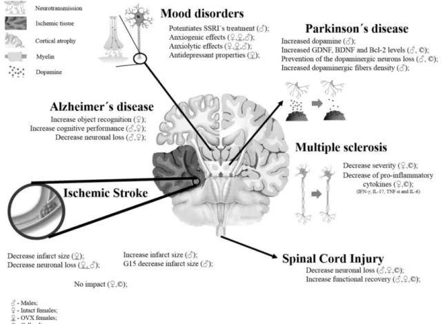
2.4.1. - Cerebral Ischemia 27 2.4.2. - Neurodegenerative disorders 31 2.4.2.1. - Alzheimer’s disease 31 2.4.2.2. - Parkinson’s disease 31 2.4.2.3. - Multiple sclerosis 33 2.4.3. - Mood disorders 35
2.4.4. - Autism spectrum disorder 37
2.4.5. - Spinal cord injury 38
3. - Repetitive transcranial magnetic stimulation (rTMS)
393.1. – Principles of rTMS 39
3.2. – Cellular and molecular effects of rTMS on brain cells 40
3.2.1. - Synaptic plasticity 40
3.2.2. - Neurotransmission 41
3.2.3. - Gene expression 42
3.2.4. - Neuroprotection and neurogenesis 43
3.2.5. - Prevention neuronal of cell death 43
3.2.6. - Glial cells 43
3.3. – Application of rTMS on neurological disorders 44
3.3.1. – Depression 44
3.3.2. – Obsessive–compulsive disorders 45
3.3.3. – Pain syndromes 45
3.3.4. - Movement disorders 46
3.3.5. – Epilepsy 46
3.3.6. – Spinal cord injury 47
3.3.7. – Ischemic Stroke 47
3.4. - Contraindications 48
3.5. - Side effects 48
Bibliography 51
Chapter III – Impact of astrocytes on the injury induced by in vitro
ischemia
71Abstract 73
1. – State of the art 75
2. - Material and methods
2.1. - Cell Cultures 77
2.2. - OGD and reperfusion 78
2.3. - Immunocytochemistry assays 78
2.4. - Cell counting 79
2.5. - Cell viability assessment 79
2.6. - Statistical analysis 80
3. - Results
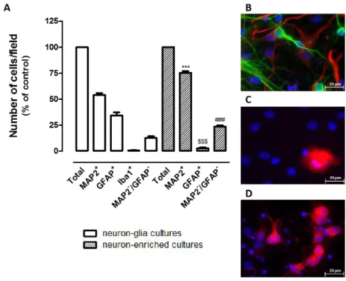
3.1. - Characterization of neuron-glia and neuron-enriched cortical cultures 81
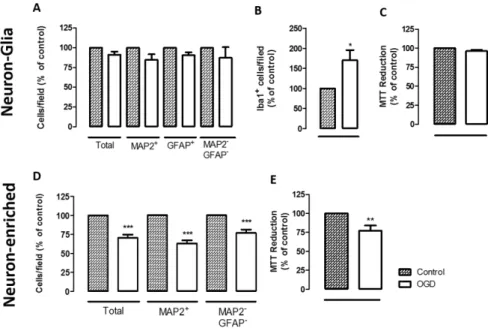
3.2. - The presence of astrocytes influences the extent of OGD-induced injury in cortical cultures
82
4. – Discussion 83
Bibliography 87
Chapter IV - G protein-coupled estrogen receptor activates cell type
specific signaling pathways in cortical cultures: relevance to the
selective loss of astrocytes
89
Abstract 91
1. – State of the art 93
2. - Material and methods
2.1. - Cell Cultures 95
2.2. - Cell culture treatments 95
2.3. - OGD and reperfusion 95
2.4. - Cell viability assessment 96
2.5. - Immunocytochemistry assays 96
2.6. - Cell counting 96
2.7. - Quantification of GPER expression 96
2.8. - Calcium imaging 97
2.9. - Statistical analysis 97
3. - Results
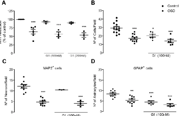
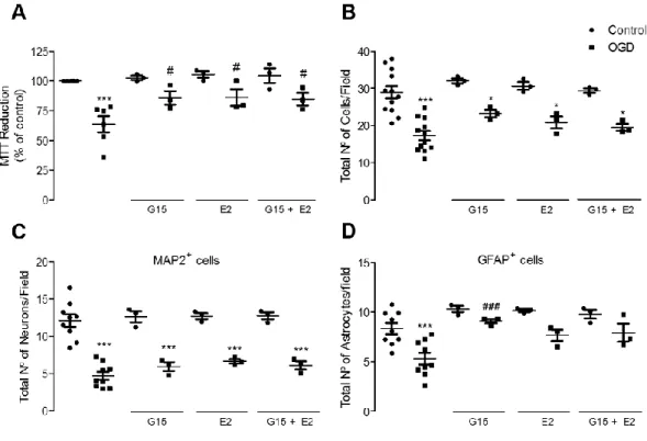
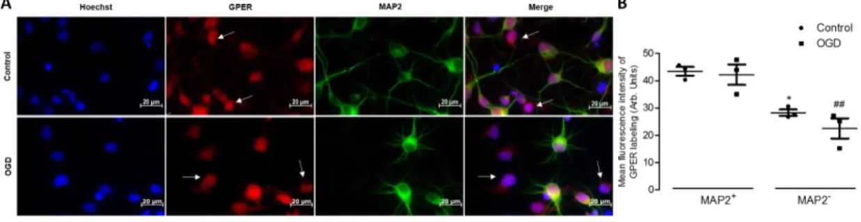
3.1. - GPER activation does not protect from an injury induced by OGD 99 3.2. - GPER blockade protects astrocytes from OGD-induced injury 100 3.3. - OGD does not induce modifications in GPER expression 101 3.4. - GPER activation promotes apoptosis in astrocytes 101
3.5. - Blockade of the PLC pathway prevents G1-induced apoptosis in astrocytes 104 3.6. - Exposure to G1 induces an increase in intracellular calcium levels in astrocytes
but not in neurons
104
4. – Discussion 107
Bibliography 111
Chapter V - High-frequency repetitive magnetic stimulation prevents
ischemia-induced neurite degeneration
117Abstract 119
1. – State of the art 121
2. - Material and methods
2.1. - Cell Cultures 123
2.2. -
OGD and reperfusion
1232.3. - HF-rMS protocol 123
2.4. - Cell viability assessment 124
2.5. - Immunocytochemistry assays 124
2.6. - Cell counting 124
2.7. - Morphometric analysis of neurons 125
2.8. - Evaluation of synapses 125
2.9. - Statistical analysis 125
3. - Results
3.1. - HF-rMS induces beneficial effects on neuron-glia cortical cultures after
ischemia
127 3.2. - Glial cells are required for HF-rMS modulation of ERK 1/2 and c-Fos 128 3.3. - HF-rMS applied after ischemia prevents neurite degeneration and increases
synaptic markers
131
4. – Discussion 135
Bibliography 139
Chapter VI - General conclusions and future perspectives
143Appendix
151Article: Impact of astrocytes on the injury induced by in vitro ischemia 153 Article: G protein-coupled estrogen receptor activates cell type specific signaling
pathways in cortical cultures: relevance to the selective loss of astrocytes
161 Article: G protein-coupled estrogen receptor 1 (GPER) activation triggers different
signaling pathways on neurons and astrocytes
175 Article: The role of G protein-coupled estrogen receptor 1 on neurological disorders 177
Index of Figures
:
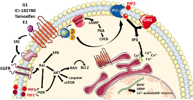
Figure 1: Schematic representation of the diversity of signaling pathways regulated by GPER
25 Figure 2: Effects induced by GPER selective activation on brain disorders 38 Figure 3: Characterization of neuron-glia cortical and neuron-enriched cortical
cultures
81 Figure 4: Effect of OGD on neuron-glia and neuron-enriched cortical cultures 82 Figure 5: Effect of G1 on rat primary neuron-glia cortical cultures exposed to 4
hours of OGD
99 Figure 6: Effect of GPER inhibition on the viability of primary neuron-glia
cortical cultures exposed to 4 hours of OGD
100 Figure 7: GPER staining in neurons and non-neuronal cells under control and
ischemic conditions
101 Figure 8: Evaluation of nuclei with apoptotic morphology in primary neuron-glia
cortical cultures exposed to OGD
102 Figure 9: Effect of GPER activation on annexin V labeling in primary neuron-glia
cortical cultures exposed to OGD
102 Figure 10A: Effect of GPER stimulation on caspase-3/7 activation in primary
neuron-glia cortical cultures exposed to OGD
103 Figure 10B: Representative images of immunocytochemistry for MAP2, caspase-3
and Hoechst 33342 staining in cultures exposed to OGD in the absence or in the presence of G1
103 Figure 11: Contribution of PLC and JNK pathways to the deleterious effect of
GPER activation in astrocytes
104 Figure 12: Cell type specific changes in intracellular calcium levels triggered by
G1
105 Figure 13: G protein-coupled estrogen receptor activates cell type specific
signaling pathways in cortical cultures: relevance to the selective loss of astrocytes
110 Figure 14: Effect of HF-rMS on rat primary neuron-glia cortical cultures exposed
to 6 hours of OGD
127 Figure 15: Effect of HF-rMS on rat primary neuron-enriched cortical cultures
exposed to 6 hours of OGD.
128 Figure 16: Effect of HF-rMS on the expression of ERK 1/2 in rat primary
neuron-glia cortical cultures exposed to 6 hours of OGD
129 Figure 17: Effect of HF-rMS on the expression of c-Fos in rat primary neuron-glia
cortical cultures exposed to 6 hours of OGD
129 Figure 18: Effect of HF-rMS on the number of cells expressing ERK 1/2 in rat
primary neuron-enriched cortical cultures exposed to 6 hours of OGD
Figure 19: Effect of HF-rMS on the levels of c-Fos in rat primary neuron-enriched cortical cultures exposed to 6 hours of OGD
130 Figure 20: Evaluation of neuronal morphometric changes triggered by HF-rMS in
rat primary neuron-glia cortical cultures exposed to 6 hours of OGD
132 Figure 21: Evaluation of synaptic modifications triggered by HF-rMS in rat
primary neuron-glia cortical cultures exposed to 6 hours of OGD
132 Figure 22: Evaluation of neuronal morphometric changes triggered by HF-rMS in
rat primary neuron-enriched cortical cultures exposed to 6 hours of OGD
133 Figure 23: Evaluation of synaptic modifications triggered by HF-rMS on rat
primary neuron-enriched cortical cultures exposed to 6 hours of OGD
133 Figure 24: Cell type-specific signaling pathways activated by GPER on neurons
and astrocytes
Index of Tables:
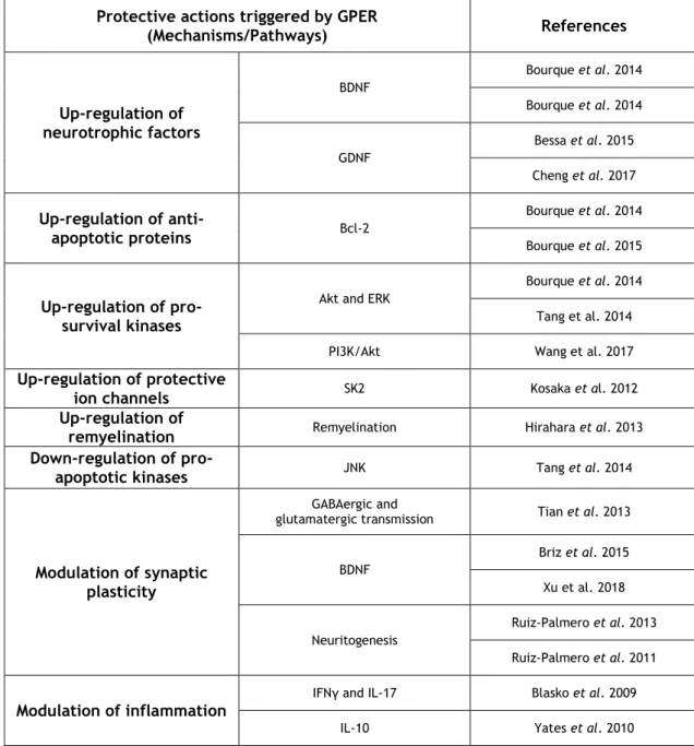
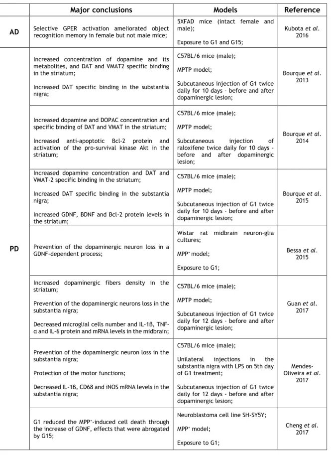
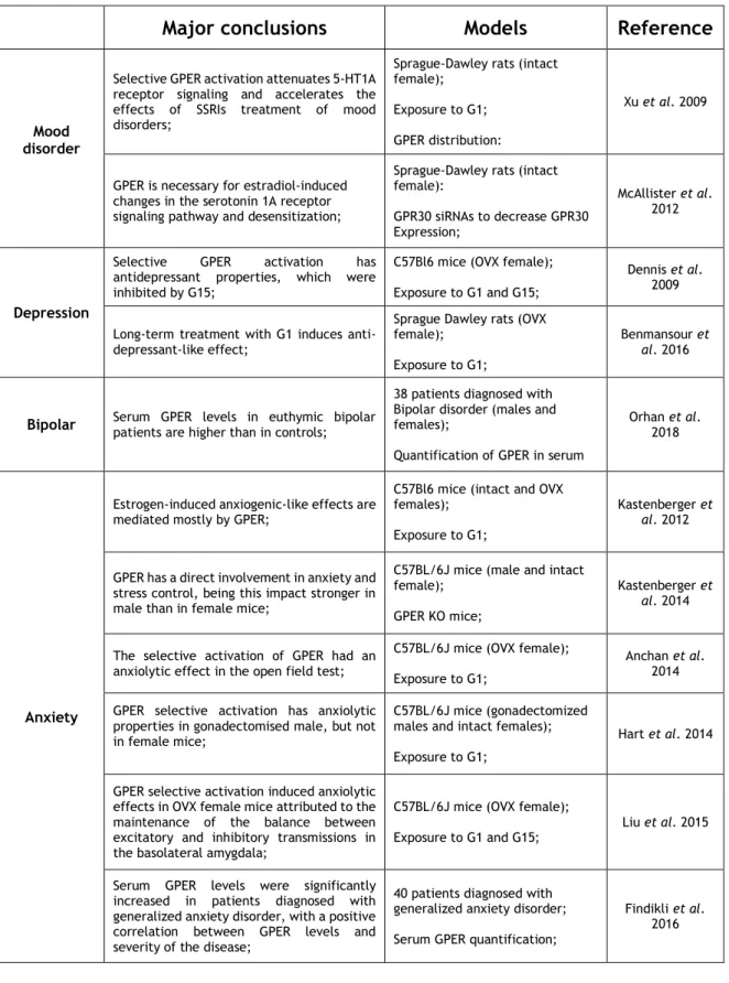
Table 1: Protective actions triggered by GPER activation in the brain 26 Table 2: Effects induced by GPER selective activation in brain ischemia 30 Table 3: Effects induced by GPER selective activation in neurodegenerative
disorders
34 Table 4: Effects induced by GPER selective activation in mood disorders 37 Table 5: Primary antibodies used on immunocytochemistry assays of impact of
astrocytes on the injury induced by in vitro ischemia
78 Table 6: Secondary antibodies used on Immunocytochemistry assays of impact of
astrocytes on the injury induced by in vitro ischemia
79 Table 7: Primary antibodies used on Immunocytochemistry assays to evaluate the
effects induced by GPER selective activation
96 Table 8: Secondary antibodies used on Immunocytochemistry assays to evaluate
the effects induced by GPER selective activation
96 Table 9: Primary antibodies used on Immunocytochemistry assays to evaluate the
effects induced by HF-rMS
124 Table 10: Secondary antibodies used on Immunocytochemistry assays to evaluate
the effects induced by HF-rMS
Index of Acronyms:
[Ca2+]
i Intracellular calcium concentration AD Alzheimer’s disease
ASD Autism spectrum disorder ATP Adenine triphosphate BBB Blood-brain barrier;
BDNF Brain-derived neurotrophic factor BLA Basolateral amygdala
cAMP Cyclic adenosine monophosphate CNS Central nervous system
CRE cAMP response elements
CREB cAMP response element-binding protein CT Computed tomography
DAG Diacylglycerol
DAT Dopamine transporter DGS Direção-Geral de Saúde DIV Day in vitro
E2 Estradiol
EAE Autoimmune encephalomyelitis ECA External carotid artery
EGFR Epidermal growth factor receptor ER Estrogen receptors
EREs Estrogen response elements
ERK1/2 Extracellular signal-regulated kinases 1 and 2 FBS Fetal bovine serum
GABA Gamma-aminobutyric Acid
GDNF Glial cell-derived neurotrophic factor GFAP Glial fibrillary acidic protein
GPER G protein–coupled estrogen receptor HBSS Hank´s buffered salt solution
HF High-frequency
HF-rMS High-frequency repetitive magnetic stimulation
Hz Hertz
Iba1 Ionized calcium binding adaptor molecule ICA Internal carotid artery
ICI-182780
Fulvestrant IFNγ Interferon γ
iNOS Inducible nitric oxide synthase IP3 Inositol 1,4,5-trisphosphate IS Ischemic stroke
JNK c-Jun N-terminal kinase LF Low-frequency
LPS Lipopolysaccharide
MAP2 Microtubule-associated protein 2 MAPKs Mitogen-activated protein kinases MCA Middle cerebral artery
MDD Major depressive disorder MFI Mean fluorescence intensity MMP Matrix metalloproteinase MPP+ 1-methyl-4-phenylpyridinium
MPTP 1-methyl-4-phenyl-1,2,3,6-tetrahydropyridine MS Multiple sclerosis
mTOR Mammalian target of rapamycin
MTT 3-[4,5-Dimethylthiazol-2-yl] -2,5-diphenyltetrazolium bromide NBM Neurobasal medium
NGF Nerve growth factor NMDA N-methyl-D-aspartate NO Nitric oxide
OCD Obsessive-compulsive disorder OGD Oxygen and glucose Deprivation OVX Ovariectomized
PBS Phosphate buffered saline
PBS-T Phosphate buffered saline with 0.1% Tween PD Parkinson’s disease
PI3K Phosphatidylinositol 3-kinase
PIP2 Phosphatidylinositol 4,5-bisphosphate PIP3 Phosphatidylinositol-3,4,5-triphosphate PLC Phospholipase C
PNS Peripheral nervous system rMS repetitive Magnetic Stimulation ROI Region of interest
ROS Reactive oxygen species
rTMS Repetitive transcranial magnetic stimulation SCI Spinal cord injury
SERD Selective estrogen receptor degrader SERM Selective estrogen receptor modulator
SK2 Small conductance calcium-activated potassium channel 2 SSRIs Selective serotonin reuptake inhibition
TBS Theta-burst stimulation
tMCAO Transient middle cerebral artery occlusion TNFα Tumor necrosis factor α
tPA Tissue Plasminogen activator
VEGF-A Vascular endothelial growth factor A VMAT Vesicular monoamine transporter
Chapter I
1. - Ischemic stroke
According to the World Health Organization, stroke is a syndrome of rapidly developing clinical signs of focal or global disturbance of cerebral function with vascular origin (1). This disturbance could be caused by ischemic or hemorrhagic imbalance of cerebral blood circulation (2, 3), being ischemic stroke (IS) the most commonly form, with approximately 85% of all stroke cases (2-6).
IS, is a heterogeneous multifactorial neurological disorder characterized by the sudden onset of neurologic signs related directly to the sites of injury in the brain where ischemia occurs (3, 7). It is characterized by the lack of enough blood flow to perfuse the cerebral tissue, leading to irreversible neuronal damage, whose severity is directly proportional to the duration of the ischemic period (4, 8, 9). Even brief ischemic periods can initiate a complex sequence of events that ultimately culminate in cellular death (9). The final infarct size and the neurological outcome depends on multiple factors such as, the duration and severity of ischemia, the existence of collateral systems and an adequate systemic blood pressure (10).
1.1. - Pathophysiology of IS
The decrease or the interruption of the blood supply to brain tissues can have several etiologies, such as thrombosis, embolisms, systemic hypoperfusion and venous thrombosis (3, 11). Each, indicating a different mechanism of blood vessel injury or a reason for decreased blood flow (3).
Cerebral thrombosis refers to the formation of a thrombus inside a cerebral artery, resulting on vascular obstruction (11, 12). Thromboembolic occlusion of major or multiple smaller arteries leads to focal ischemia downstream of blood flow (11, 12). Thrombotic IS occur without warning symptoms in 80% of patients, whereas 20% is heralded by one or more transient Ischemic events (13).
Cerebral embolism refers to a blood clot that is formed at another location in the circulatory system, travels along the vessels and occludes medium sized branching arteries causing ischemia to a localized brain region in the same way that cerebral thrombosis does (3, 11). In fact, the majority of brain embolisms have origin on the heart, aorta, or a proximal artery or vein, for example microemboli can break away from a sclerosed plaque in the carotid artery or from cardiac sources such as atrial fibrillation or a hypokinetic left ventricle and reach the brain causing focal ischemia (3, 11).
Systemic hypoperfusion is characterized by a global decrease in the blood flow to the head (3). Generally, it is the result of complications on the performance of the heart to pump blood adequately to perfuse brain tissues (e.g., myocardial infarction and/or arrhythmia and severe hypotension) or due to inadequate amount of blood and fluid in the vascular compartment of the body (e.g., bleeding, dehydration, and loss of fluid into body tissues) (3). This mechanism induces global ischemia in cerebral tissues and is the worst form of IS (3, 11). Cerebral venous thrombosis is an uncommon form of IS where a blood clot occludes a dural sinus and/or cerebral veins (14). Usually it affects young individuals and its associated to prior medical conditions (e.g., thrombophilias, inflammatory bowel disease), transient situations (e.g., pregnancy, dehydration, infection), selected medications (e.g., oral contraceptives, substance abuse) and unpredictable events (e.g., head trauma) (14).
Regardless the etiology of the IS on the core of the injury the brain parenchyma undergoes immediate death, while in surrounding areas, the penumbra may only be partially injured with potential to recover (3, 11). The reduction of blood supply associated to low respiratory reserve and complete dependence on aerobic metabolism make brain tissue particularly vulnerable to the effects of ischemia (11). The neurological function is affected by the oxygen and glucose deprivation (OGD) and neuronal injury and cell death begin within 4 minutes of ischemia (3). Then, numerous detrimental effects are triggered, including energy failure, loss of ion homeostasis, acidosis, increased intracellular calcium levels, excitotoxicity, free radical-mediated toxicity, generation of arachidonic acid products, cytokine mediated toxicity, activation of glial cells, disruption of the blood-brain barrier (BBB) and infiltration of leukocytes (3, 9, 15).
1.1.1. - Risk Factors
Several risk factors are associated with an increased risk of IS, and they can be stratified into modifiable and nonmodifiable (12). Modifiable risk factors include those resulting from lifestyle and environment, and can be modified with the help of healthcare professionals, treatment and continuing education, among these the major are: Atherosclerosis, h, blood abnormalities, diabetes mellitus, smoking and obesity (12). Unmodifiable risk factors include factors related to hereditary or natural processes that cannot be modified, such as, age, sex and ethnicity (3, 12).
Atherosclerosis refers to the development of atherosclerotic plaques (atheromas) inside the arteries associated with degeneration of the arterial wall (3). Atheromas develop in the aorta and in the large arteries of the neck and head, and are initiated when high levels of blood lipids (hypercholesterolemia and/or hyperlipidemia) potentiate its accumulation on vascular
smooth muscle cells (3). Lipid, smooth muscle, fibrous tissue, connective tissue, white blood cells, and crystals of cholesterol constitute these plaques. When plaques increase in size they narrow arterial lumen, causing turbulence of flow, and reducing distal brain perfusion (16). The irregular surfaces or cracks on atheromas attract platelets and other blood components that induce the formation of clots (3). Atherosclerotic abnormalities can cause ischemia through three major ways: severe luminal narrowing markedly decreases blood flow; plaques or occlusive thrombus mechanically block branches of the main arteries; propagation and embolization of thrombus cause occlusion of distal branches (16).
Hypertension leads to wear and tear of arteries and accelerates the development of atherosclerotic changes on large arteries. On small arteries of the brain, hypertension leads to thickening of the walls that narrows the lumen of the arteries and can lead to infarcts deep within the brain (3, 12). Longitudinal studies indicate that individuals with high-normal blood pressure (130–139 mm/Hg systolic, 85–89 mm/Hg diastolic, or both) have a twofold increased risk of developing heart disease and IS, than those with normal blood pressure (6). Hypertension can also induce arterial dolichoectasia (dilatative arteriopathy), a condition where the blood vessels become elongated and dilated and follow a tortuous and windy course with frequent loops and curves. These widening and lengthening of arteries can slow blood flow and stimulate blood clotting in the arteries (3).
Several blood components have the purpose of preventing the body from losing blood and a deficiency on these components leads to excessive bleeding, whereas other conditions can lead to excess clotting. Some blood abnormalities that affect the clotting system could be considered risk factors for IS, such as, thrombocytosis (excessive number of platelets), deficiency of some blood proteins (antithrombin III, protein C, and protein S) or excess in clotting proteins (factor VII, VIII, or XII). Medical conditions such as cancer or inflammatory conditions (e.g., inflammatory bowel disease) also increase the tendency of blood to clots (3, 6).
Diabetes mellitus is a well-established independent risk factor for IS. High glucose levels induce pathological changes in blood vessels, which can lead to IS if cerebral vessels are directly affected. These changes include vascular endothelial dysfunction, increased early-age arterial stiffness, systemic inflammation and thickening of the capillary basal membrane (12, 17). It is estimated that nearly 40% of all IS can be attributed to the effects of diabetes either alone or in combination with hypertension (6).
Obesity and abdominal obesity are independent and potent risk factors for IS. This metabolic syndrome correlates with excess body weight and due to its well-known association to other conditions, such as, hypertension, cardiovascular disease, chronic inflammation increases the risk of IS (18, 19).
Several risk factors are associated with lifestyle and daily-life habits, such as smoking, physical inactivity or binge drinking habit. Smoking increases blood levels of carboxyhemoglobin, platelet agregability, and fibrinogen levels and reduces HDL-cholesterol. On the other hand, tobacco compounds induce toxic effects on vessels (12, 20). It is estimated that smoking almost duplicates the relative risk of IS (6). Physical inactivity increase blood pressure, glycaemia levels and body weight, as well as decrease cardiorespiratory capacity and increase the blood pressure at rest (21, 22). The alcohol consumption has been shown to increase blood pressure and heavy or chronic consumption is associated with cardiomyopathy (23).
The genetic predisposition is an unmodifiable risk factor for IS. Over the years several observations linked family clinical history to IS, raising the hypothesis of a genetic basis on IS (24, 25). For example, men whose mother died of IS have a three-fold increased incidence in comparison with men without a maternal history of IS (26). On the other hand, there are also pathologies with genetic predisposition that can lead to IS, as is the case of fibromuscular dysplasia, an uncommon pathological condition that involves the wall of the arteries. In this condition there is an excessive amount of connective tissue and smooth muscle on the wall, this excess narrows and contracts the arterial lumen, which can block blood flow to the brain, causing ischemia (3).
Aging is one of the most significant IS risk factors, with 95% of IS occurring in people with more than 45 years and two-thirds occurring in those over the age of 65 (2, 12). For each consecutive decade after 55 years of age, the risk for stroke approximately duplicates, and the prevalence of IS for individuals older than 80 years of age is approximately 27% (6, 7). Mortality associated with IS also increase with age (2, 12). However, in recent years IS cases are becoming more frequent in younger populations (<45 years) (2, 12).
Sex-specific incidence rates indicate that males until 75 years have a higher risk of IS than females (2, 5), which could be due to a greater prevalence of traditional vascular risk factors (5). However, from this age the risk is similar for both genders (2, 5, 27). Since life expectancy in women is higher, with advancing age the number of woman suffering an IS also increases (2, 5).
The race/ethnicity-specific incidence demonstrates that black individuals have higher risk of suffering an IS than Caucasian, Asian, Hispanic and Indian individuals in any age range or sex (2, 5).
1.1.2. - Prevention
The guidelines for IS prevention are clear and are based on the control and prevention of risk factors that are modifiable (12, 29, 30). For each one of the known modifiable risk factors there are several measures and recommendations to adopt. These guidelines go from simple modifications in diet and lifestyle to pharmacological treatment of pathologies and conditions that are risk factors, such as, hypertension, atrial fibrillation, hyperlipidemia, diabetes mellitus or antiplatelet therapy (29, 30).
1.2. - Signs and symptoms
IS, is a complex neurologic syndrome with sudden onset and with symptoms dependent on the brain region affected by the ischemic injury (3, 7, 31). The most frequent signs and symptoms of patients hospitalized with a confirmed IS diagnosis are paresis (sudden numbness or weakness) on one side of the body. Approximately 87.6% of diagnosed IS subjects presents some kind of paresis, most often of the arms (81.1 %), legs (73.4%) and face (58.7%). Being the location of paresis equally distributed between right and left sides (31).
Sensory deficits are observed on approximately 49% of IS diagnosed patients, most often of the arms (42.3%), legs (37.6%) and face (22.9%). On the face sensory deficits are equal on both sides, whereas on limbs sensory deficits that involve the left side are more frequent (31). Speech deficit (confusion, trouble speaking or difficulty understanding speech) is observed on approximately 26.1% of the patients, severe headache with no known cause on 22.4%, sudden trouble seeing in one or both eyes on 15.4% and gait disturbance on 11.4% of the cases. Convulsions (3.2%) and vertigo (2.5%) are less frequent symptoms (31).
1.3. - Post-Stroke impairments
IS is the leading cause of complex and serious long-term disability in developed countries (2, 32), and data indicate that half of the patients are physically dependent after an IS (32, 33). Functional and clinical problems are persistent with data at five years post-IS demonstrating that approximately two-thirds of patients have some form of neurological impairment and/or disability (2). These impairments can cause significant impact on life quality and restrictions to activities of daily-life, and are closely related to the location, size and severity of the injury and can be classified in physical, cognitive, and emotional.
The physical impairments are associated to motor, visual and somatosensory deficits. Motor impairments affect the balance, coordination and gait, and are the predominant cause of long-term disability (2). Visual impairments can take the form of monocular vision loss, visual field loss on the left or the right side of the midline, or cortical blindness (2, 34). Somatosensory deficits refers to the inability of patients to process and manipulate their environment, and can range in severity from numbness or tingling in one part of the body to complete sensory neglect of a body part or one side of the body (2, 35).
Cognitive impairments can cause several deficits like problems with concentration, attention, memory, orientation, visual spatial perception and apraxia. These type of impairments are linked to long-term mortality and high level of disability (2, 36).
In addition to the effects that are perfectly visible, more subtle symptoms, such as emotional or personality changes may also occur. After an IS patients may experience fear, anxiety, frustration, anger, sadness, and a sense of loss. This may lead to the development of other pathologies such as depression, the most prevalent and the most commonly studied post-stroke mood disorder, with a prevalence of approximately 40% (2, 37, 38). It is also possible that many IS patients develop anxiety. Latest statistics have estimated an occurrence of anxiety of approximately 20-25% (2, 39).
1.4. - Epidemiology
IS is a major global health problem and its significance probably will increase in the future due to ongoing demographic changes, including aged population and health transitions observed in developing countries (5, 32). Until few years ago IS was seen as a pathology of developed countries, because its incidence and prevalence rates were higher than in developing countries, nevertheless is becoming more and more frequent in these countries (5, 32).
The highest prevalence rates are in Eastern Europe, North America, Central Asia, and East Asia (2, 32). Worldwide at every 3 minutes and 45 seconds, someone dies due to an IS (2) being this condition associated to the death of 3.0 million individuals (32). When considered separately from other cardiovascular diseases, IS ranks 5 among all causes of death, behind diseases of the heart, cancer, chronic lower respiratory disease, and unintentional injuries/accidents (32). Due to its well-known unmodifiable risk factors the IS incidence in a defined area is largely influenced by the structure of the population in terms of age, sex and ethnicity distribution (5).
In Europe it is estimated that each year 800.000 people suffer an IS, and the data indicate that the eastern countries have higher rates than southern countries (5). Similarly to
what is observed in the world also in Europe the IS age-standardized incidence is declining, and rates observed in young adults are rising (5). The explanations for these trends have been attributed to the increase of risk factors such as diabetes, hypercholesterolemia, obesity, smoking, alcohol abuse, and the use of illicit drugs in young adults (5).
In Portugal, until 2013 there were relatively few studies on the prevalence and incidence of IS, in fact the few studies that exist were based on stroke in general and do not distinguish between hemorrhagic and ischemic (40). These studies were biased due to the small sample and local or regional nature of the studies (40). For example, the most cited article was carried out in Coimbra in 1992, and reported a prevalence of stroke in males of 10.2% and in females of 6.6% (41). It was also observed that the prevalence increases markedly with age, although always higher in males the difference decreases after the 7th decade of age (41).
In 2007 it was reported a stroke prevalence of 2.1% in primary care patients (42), more recently, in 2013, a cross-sectional study about the prevalence of stroke was developed through a telephone interview having has target the residents in mainland Portugal (40). The results indicate a total prevalence of 1.9%, being this rate higher in males (2.6%) than in females (1.3%) (40). This study also reported several differences in the prevalence at the geographical level, with a higher prevalence in Alentejo (3.6%) and lower prevalence in the North (1.1%) (40). Estimates from 2009 indicate that per hour 6 people suffer a stroke, resulting in 2 deaths, being considered one of the major causes of death in Portugal (43). According to Direção Geral de Saúde (DGS) the standardized mortality rate for stroke decreased between 2007 and 2011 from 79.9 deaths per 100.000 inhabitants to 61.9 (27).
From 2013 these reports from DGS become anual and began to distinguish between hemorrhagic and ischemic. Regarding IS, from 2013 to 2015 the total number of deaths diminished from 6099 per 100.000 inhabitants to 4598, which represent a decrease on age-standardized mortality rate from 61.3 to 46.6 per 100.000 inhabitants(27). This could be explained in part by the increase of aproximately 36.5% of patients subjected to clot-busting drugs (27). It is also observed that individuals over the age of 70 have a higher risk of death due to IS (27). And similar to what is observed in the world until the age of 70 males have higher age-standardized mortality rate than females, from that age forward the rate is similar for both genders (27).
1.5. - Effects of ischemia on brain cells
The human brain is primarily composed by neurons, glial cells, neural stem cells, and blood vessels. Neurons and glial cells are present in similar amounts (44), and establish complex interactions (15, 45). Like any other cell in the organism, to properly perform their functions
brain cells depend on the supply of water, energy, nutrient and oxygen and on the removal of wastes products (e.g., carbon dioxide, nitrogen, phosphates, sulphates), for that, they rely on the bloodstream. If for some reason these exchanges are interrupted, as is the case of ischemia, the cells begin to resent and a cascade of events will begin. Initially a local depletion of oxygen or glucose will occur, causing failure in the production of ATP (11). This will affect energy-dependent processes necessary for cell survival, and sets off a series of interrelated events that may end in cellular injury and death (11).
1.5.1. – Cellular mechanisms triggered by ischemia
During periods of low oxygen or decreased blood flow, the production of ATP by glycolysis and oxidative phosphorylation slows or stops (46-48). Indeed, there are potentially large reserves of alternatives to glucose as substrates for both glycolysis and respiration, such as glycogen, lactate and fatty acids (47, 48). The initial effects induced by ischemia depend on the availability of alternative glycolytic and oxidative substrates and on the rate of ATP consumption (47, 48). In contrast, oxygen is an irreplaceable driver of mitochondrial respiration, the main source of cellular ATP (47, 48). Consequently, lack of oxygen immediately and severely reduces ATP production, which results on a rapid decrease of their levels due to ongoing consumption (47, 48).
The interruption of oxidative phosphorylation triggers ATP synthase to run backward and consume ATP, accelerating the loss of ATP and electron leak and triggering the production of reactive oxygen species (ROS) (47, 48). Finally, when respiration is inhibited but glycolysis persists, protons and lactate generated during glycolysis accumulates, causing rapid intracellular acidification (47, 48). In response to this direct effects of decreased cellular energy and intracellular acidification several detrimental mechanisms are activated, such as the loss of ion pumps function, release of excitatory neurotransmitters, and the production of ROS, all of them promoters of cell death (11, 47).
The loss of ion pumps function is triggered by the decrease of cell membrane potential and leads to the loss of ion gradients. There is an efflux of potassium and influx of sodium, chloride, and calcium ions that is accompanied by the inflow of water, resulting in rapid swelling of cells and the consequent necrosis (11, 47). The necrotic process leads to the loss of membrane integrity resulting in cell lysis and the release of the cellular constituents that in turn promote inflammation in the surrounding tissue (49). The increase in intracellular calcium leads also to the activation of pathways that lead to apoptosis and the release of excitatory neurotransmitters (11).
The release of excitatory neurotransmitters, such as glutamate, accompanied by a dysfunction in the reuptake mechanisms, as a result of ion gradient dissipation, results in glutamate accumulation in the synaptic cleft (11, 47). Excessive glutamate accumulation at the synapse results in the overactivation of glutamate receptors, namely N-methyl-D-aspartate receptors (NMDA-receptors), leading to excitotoxicity (11, 47).
Oxidative stress is another step of the ischemic cascade and is caused by the production of ROS (11, 47). These radical species have the ability to react with and damage almost all cellular and extracellular components, of which vascular endothelium is particularly important (11, 47). ROS-induced modifications lead to cellular impairment through biochemical, functional, and metabolic abnormalities, which ultimately trigger apoptotic mechanisms (11, 47).
In contrast to the ischemic core, where the cells die mostly by necrotic processes, on the penumbra cells die mostly through apoptotic mechanisms (11). The ischemic cascade causes an early response in the expression of genes such as Bax and p53, followed by the release of pro-apoptotic molecules such as cytochrome c and apoptosis-inducing factor from mitochondria (11, 47). This leads to the activation of caspases that potentiate cell death (11, 47). However, in the course of these processes some protective pathways could be activated as a defense against apoptotic and necrotic cell death (e.g., production of Bcl-2, Heat shock protein 70, Neurotrophin-3 or Interleukin-10) (11). The way different cell populations deal with ischemic periods is variable, and depends on its intrinsic characteristics but above all on its metabolic needs (11). Cells that require more energy are more affected by ischemia (11).
1.5.2. - Effects of ischemia on neurons
Neurons, classically considered the most important cells of central nervous system (CNS), play a crucial role on every system of the human body (50). The major function of these cells is to enable the communication within the nervous system, which is done through action potentials and synaptic transmission (49). To receive and send these action potentials it is necessary a shift in the membrane potential caused by the flow of ions through the neuronal membrane (49). In order to maintain the ionic gradients, a constant supply of glucose and oxygen is required, and any imbalance jeopardizes neuronal functions (11). These intrinsic characteristics and the fact that neurons do not have their own energy stores make them extremely sensitive to ischemia (49).
1.5.3. - Effects of ischemia on Glial cells
Glia include different types of cells, such as astrocytes, microglia and oligodendrocytes. Glial cells are seen as the housekeeping cells of CNS and their main function is to support neurons. Their supportive tasks include maintaining homeostasis, providing structural, metabolic, and trophic support to neurons, promote defense against pathogens, regulating inflammatory responses, regulating synaptic transmission, removing metabolites and participating in the formation of the blood-brain-barrier (45, 51-55).
Astrocytes are the most abundant glial cells in the brain, and their characteristics make them less susceptible to ischemic damage than neurons (48). They are able to maintain ATP levels longer than neurons during ischemia, and severe ionic dysregulation proceeds more slowly (48). Firstly because neurons have higher density of ionic channels and a consequent greater energy demand to maintain ionic gradients, and secondly because most of the glycogen stores in the brain is found in astrocytes (48). Additionally, astrocytes express lower levels of ionotropic glutamate receptors than neurons, and have better ionic buffering and antioxidant capacity (48). These attributes presumably underlie the well-known selective loss of neurons over astrocytes (48). However, under these circumstances astrocytes can play two roles,
if on
one hand these attributes place them in the position of potentially being able to protect neurons, on the other hand they are also stressed by ischemia and may potentiate neuronal death (48).1.5.4. - Effects of ischemia on brain vasculature
The cerebral blood vessels are endowed with powerful regulatory mechanisms that assure that the brain is perfused according to its needs (56). However, during ischemia these mechanisms become dysfunctional and fail to compensate the reduction in blood flow (56). Ischemia triggers profound modifications in the major mechanisms that control cerebral circulation (endothelial function, autoregulation, vascular reactivity to hypercapnia and neurovascular coupling), these dysregulation undermines the ability of the brain to maintain blood flow and aggravates the intensity of the ischemic insult (56, 57).
The vast majority of these vascular alterations are associated with an increase in ROS levels, which leads to oxidative stress and impairs the function of vascular cells (56, 57). During periods of ischemia there is an impairment of endothelium-dependent regulation of vascular tone and basal receptor-mediated endothelium-dependent vasodilation, being these dysfunction associated to the increase of ROS levels (56, 57).
1.6. - Models for the study of IS
Due to the complexity of IS its study has been made through the combination of several in vivo and in vitro models. These have been developed, with the aim of identifying the mechanisms that underlie cerebral ischemia and developing new approaches for IS therapy (10, 58, 59). While in vivo models enable the study of interactions of all components present in the CNS as a whole, the use of in vitro models allows the study of molecular interactions occurring at tissue level (60).
The use of animal models is an indispensable tool for several reasons, in theory experimental models of cerebral ischemia are highly reproducible, well controllable, and standardized, allowing more precise analysis of stroke pathophysiology and drug effects (10, 58, 59). The molecular, genetic, biochemical and physiological studies often require invasive processes to allow direct access to brain tissue, which can be achieved with in vivo models (10, 58, 59). It is also possible to evaluate the effects of perfusion and vasculature in the pathophysiology of IS, which cannot be modeled in in vitro models (10, 58, 59). However, this may not be enough and frequently the use of cellular models to study specific basic biochemical and molecular mechanisms under conditions of energy deficiency similar to ischemia on specific cell populations is required (10, 61, 62). With the use of in vitro models, it is also possible to evaluate the potential therapeutic effect of drugs in specific cell populations (10, 61). Cellular models are easy to use and manipulate allowing a direct control of the environment (10, 62).
The majority of IS experiments, in vivo and in vitro, are carried out in well-characterized rodent models (10, 58, 59). The lower cost of maintenance, easily monitoring of physiologic parameters, its small brain size, a relative homogeneity within strains, that allow reproducible studies (10, 58, 59), make rodents preferred models in ischemia studies.
1.6.1 - In vivo IS models
The purpose of any animal model is to closely mimic the pathophysiologic processes. In vivo IS models could be classified taking into consideration the area, global or focal, or the type of occlusion, permanent or transient. On global models, there is a complete interruption of blood flow to the brain and on focal ischemia, the interruption of blood flow affects only a specific part of the brain. In permanent models of ischemia there is a complete interruption of the blood flow, whereas in transient the interruption is temporary and is followed by a reperfusion period. IS is often induced by occlusion of the middle cerebral artery (MCA) or one of its branches (59).
1.6.1.1. - Embolic models
Embolic IS models involves the formation of a thrombus that occludes the vessels, and can be classified into two major categories: microsphere-/macrosphere-induced models and thromboembolic clot models (10, 58). Microsphere-/macrosphere-induced models use a microcatheter to insert the microspheres (20-50 μm) or macrospheres (100-400 μm) into brain arteries. Microspheres are usually inserted via the external carotid artery (ECA) and are flushed passively into the cerebral circulation by the blood flow, inducing multifocal and heterogeneous infarcts (10, 58). The macrospheres are inserted into the internal carotid artery (ICA), and its intra-arterial embolization provides reproducible occlusion of the MCA, resulting in focal ischemic lesions that are comparable to intraluminal suture model (10, 58).
The thromboembolic clot models are based on the application of spontaneously formed clots or thrombin-induced clots from autologous blood or through the injection of thrombin directly into the intracranial segment of the ICA or into the MCA (10, 58). These model closely mimics the mechanism of human IS, and therefore allows the study of thrombolytic agents alone or combined with neuroprotective drugs, as well as thrombolytic processes (10, 58, 59).
1.6.1.2. - Intraluminal suture MCAo model
The MCAo model is the most frequently used experimental model of IS in rodents (10, 58). A monofilament is introduced into the internal carotid artery (ICA) and is positioned at the origin of the MCA inducing its occlusion (10). The model can be permanent or transient. Withdrawal of the filament with subsequent reperfusion allows to develop transient models with variable reperfusion time points (10, 58). The duration of ischemia could range from 60 to 120 min (10, 58).
1.6.1.3. - Craniectomy models
These models are characterized by a direct approach to brain vasculature (10, 58). A craniectomy with incision of the dura mater is made and the MCA is exposed. The focal ischemia could be performed by occlusion of the MCA by electrocoagulation and additional transection, resulting in permanent occlusion, or alternatively a transient occlusion of MCA can be achieved by clamping the artery, followed by subsequent reperfusion (10, 58). It could also involve the occlusion of both common carotid arteries, to reduce the collateral blood flow consolidating the ischemic injury (three-vessel occlusion model) (10, 58).
1.6.1.4. - Photothrombosis models
Photothrombosis, also known as photochemical, are focal models based on intravascular photo-oxidation, which leads to well-defined ischemic injury (10, 58). A photoactive dye (e.g., Rose Bengal, erythrosine B) is injected intraperitoneally (mice) or intravenously (rat) followed by illumination of a specific part of the brain with light of a specific wavelength through the intact skull (10, 58). The activated dye forms singlet oxygen that damages components of endothelial cell membranes, with subsequent platelet aggregation and thrombi formation, leading do the interruption of local blood flow (10, 58).
1.6.1.5. - Endothelin-1 model
Endothelin-1 is a peptide with potent and long-lasting vasoconstrictive properties. It can be applied directly onto an exposed vessel or stereotactically injected into the brain parenchyma leading to vasoconstriction, inducing downstream vessel ischemia (10, 58). The period of ischemia is dose-dependent, and when endothelin-1 effect passes, blood flow is gradually reestablished, thus representing the situation of transient focal ischemia (10, 58).
1.6.1.6. - Global models of ischemia
It is also possible to induce global ischemia on brain tissues. These models are based on the total interruption of blood supply to the brain and usually are used to study the brain damage that occurs in cardio-circulatory resuscitation. Among these the most used are the four vessel occlusion (reversible occlusion of the two common carotid artery (CCA) combined with the permanent interruption of vertebral arteries), the two vessel occlusion (occlusion of the two CCA) and cardiac arrest and resuscitation (63).
1.6.2 - In vitro IS models
The application of cellular models provides a simple and highly controlled experimental system that allows detailed high-throughput analyses on how the system, or one particular cell , is affected by ischemia (9, 10). These approaches use primary cultures, cell lines, organotypic cultures and brain slices (9, 10). To induce ischemia in these models OGD or the chemical/enzymatic blockade of the cellular metabolism are the approaches commonly used (10, 61).
1.6.2.1. – Cellular systems
Primary cell cultures are established by the dissociation of brain tissues from embryonic or perinatal rats and mice (9, 60). The initial dissociation can be made from specific anatomical areas of the brain (e.g., cortex, hippocampus, cerebellum), which allow the study of ischemic injury in particular cerebral regions (9). The cultures can be composed of a homogenous population of cells (enriched cultures), or by several populations of cells (mixed-cultures). Mixed-culture systems are set-up with two or more cell types growing with some degree of contact between them and constitute valuable tools to study the interactions between cell populations (64). Whereas enriched cultures, like neuron-enriched cultures, allows assessing how specific cells are affected by an ischemic insult (10, 62).
Immortalized cell lines, such as SH-SY5Y (differentiated into neuronal cells)(65), the human teratoma-derived NT2 (differentiated into neuronal cells, astrocytes, and oligodendrocytes) (66) and the HMO6 (differentiated into microglia) (61, 67), are frequently used on IS studies.
The brain slice method utilizes a thin slice of rodent brain tissue, usually maintained up to 12 hours, and preserving the original architecture of the tissue. This model has been used to evaluate neuronal vulnerabilities under ischemia without cerebrovascular influences (61, 68).
The organotypic brain slice cultures represent a type of cellular system that lies between brain slices and primary cell cultures. They are prepared from different anatomical regions of the brain and allowed to mature in vitro (69). With this model the tissue maintains its structural organization (61, 69).
1.6.2.2. – Oxygen and glucose deprivation
Oxygen and glucose deprivation is the most used model of in vitro ischemia. It consists on replacing the regular atmosphere (95% air and 5% CO2) by an anoxic atmosphere (95% N2 and 5% CO2), by maintaining cells in a hypoxic chamber. The hypoxic conditions can be associated with the omission of glucose, which is usually referred as in vitro ischemia or OGD, which consist on replacing the regular medium for glucose-free buffer (e.g., Hanks balanced salt solution (HBSS) or Locke’s solution) (9). The cultures are maintained under these conditions for a previously established period of time and then the reperfusion is made by returning them to the conditions established before the period of OGD (95% air, 5% CO2 and regular medium) (9). The extent of the ischemic damage will depend on the period of OGD applied, as well as the density of cells present in the culture (9, 70).