a new wader,
recurvirostridae (charadriiformes),
from the early eocene of Portugal
C. J. O. HARRISON
*
• British Museum (Natural History) , Sub-Department of Orni-thology, Tring, Henfordshire HP23 6AP, U.K.
Ciencias da Terra (UNL) Lisboa N.o 7 pp. 9-16 1983
RESUMO
Palavras-chave: Aves - Eocenico basal- Silveirinha - Portugal
Descreve-se uma nova avoceta (Aves, Charadriiformes, Recurviros-tridae) do Eocenico basal de Silveirinha, Baixo Mondego. Apos compa-racoes com outras formas actuais efosseis, e denominada Fluviatilavis
antunesi, nov. gen. nov. sp.
Mots-des: Aves - Eocene basal- Silveirinha - Portugal
Un avocet nouveau (Aves, Charadriiformes, Recurvirostridae) de l'Eocene basal du gisement de Silveirinha (region du Bas Mondego,
Portugal Central) estdecrit et compare avec d'autresformes actuelles et fossiles. Apres discussion de ses affinites, I'avocet de Silveirinha est
rapporte
a
Fluviatilavis antunesi, n. gen. n. sp.ABSTRACf
Key-words: Aves - Early Eocene - Silveirinha - Portugal
A new wader (Aves, Charadriiformes, Recurvirostridae)'from the Early Eocene site of Silveirinha, in Lower Mondego region, Central Portugal, is described. Comparisons have been made with other forms, both extant and fossil; its affinities are discussed. Silveirinha wader is ascribed toFluviatilavis antunesi, n. gen. n. sp.
INTRODUCTION
Among the material collected by Professor Dr. Miguel Telles Antunes at the Early Eocene site at Silveirinha, Central Portugal (see ANTUNES and RUSSELL, 1981), are three bird bones. They occurred embedded in the compact claystone typical of the site. The deposit that yielded them appeared to correspond to the margin of a channel, the specimens found there being dissociatedan~ apparently swept to the edges of the watercourse. The three bird bones were not associated but occurred close together, less than half a metre apart; and in view of this, and since other avian material was absent, it is possible that they might have originated from a single individual.
COMPARISONS
The bones include a right femur, now free from matrix but lacking the proximal head and proximal edge of the trochanter, and at the distal end having the posterior projections of the condyles broken away. Another specimen is the damaged shaft of a right humerus embedded in a matrix black. The lateral (external) side of the distal head is exposed and available for study. The third specimen is the shaft of a right radius, also embedded in matrix, but retaining the distal end which has been exposed.
All were compared independently with the osteological collection of the .British Museum (Natural History). In general configuration the femur showed a resemblance to those of species both of ibises, Threskiornithidae (Ciconi-iformes), and waders, Charadriiformes. However, in several characters its affinities appeared to be with the latter taxon.
The tuberculum m. gastrocnemialis lateralis is pro-ximally sited on the lateral side, more similar to that of gulls, Laridae. The distal condyles are more widely separated with the medial condyle a little shorter distally, and the distal end more anteroposteriorly compressed. The edges of the popliteal depression converge to form
the anterior intermuscular line about two-fifths of the distance along the shaft. The popliteal depression is roughly triangular and bordered by an elongated and well--defined medial ridge. At the proximal end of the bone the trochanteric ridge is more anteriorly flattened to give an elongated surface with a distinct, abrupt medial edge, and in proximal view the trochanteric ridge shows greater anterior projection.
The humerus also shows charadriiform characters. The distal lateral side of the shaft slants distolaterally to the lateral condyle, but along its palmar edge is a flange becoming more prominent proximolaterally to end in a projecting extep~condylar prominence, comparable with those of Recent charadriiform birds. The lateral condyle is distally flattened and anconally bears the ridges of the lateral tricipital groove.
The radius gives less indication of affinity. Although the shape of the shaft and general configuration of the distal end might fit that of a charadriiform bird of the kind suggested by the other bones, the distal end is less flattened on its medial side, projecting further, and is palmar/anconally thinner at is thickest point. However, these might be earlier and more generalised structural characters.
A comparison of these bones with those of various families within the Charadriiformes reveals that the femur most closely resembles those of species of the Avocet and Stilt family, Recurvirostridae, particularly in the shape and thickness of the distal end. In that aspect of its structure it most nearly resembles that of the Avocet
Recurvirostra avosetta, but in overall shape it is more
slender and longer and approximates more to that of the Ibisbill Ibidorhynchus struthersi, a species of that family
which appears to retain some of the more generalised characters of structure. The humerus would also fit this family.
On the basis of the characters evident on the femur there is evidence of a bird of the wader order Charadri-iformes, and of the family Recurvirostridae, in the Early Eocene' of Portugal. From the characters available on 13
the other specimens it seems reasonable at present to tentatively assign them to the same species.
There are few other early fossils of this family. Coltonia recurvirostra Hardy 1959 is described from the fragments of a wing from the Lower Eocene of Utah, U. S. A. The specimens, which are embedded in matrix and partly crushed do not allow direct comparison with the present material. The species appears to have been 1.7 - 1.8 times as large as existing species of Recurvi-rostra or Himantopus and the bones appear to have been proportionally more slender. A second species Kashinia magnus (HARRISON and WALKER) 1976 is described from an imperfect coracoid from the Upper Eocene of Hampshire, England. It resembles Ibidorhynchus struthersi in most respects and might have been about 1.5 times as large. Neither species provides characters for establishing direct affinity with the new species.
DESCRIPTION
Order Charadriifonnes
Family RECURVIROSTRIDAE
and a right radius lacking proximal end, all collected by, Professor Dr. Miguel Telles Antunes in 1981.
OCCURRENCE. Early Eocene: Silveirinha, central Portugal.
DESCRIPTION. The holotype right femur lacks the articulating head, part of the proximal crest of the trochanter and the posterior parts of the distal condyles. It
has suffered some superficial crushing. It is relatively long and slender and in lateral view shows some slight anterior curvature and widens gradually at the proxi-mal end. The tuberculum m. gastrocnemis lateralis is apparent at the distal end as a prominence on the pos-terior edge a little proximal to the lateral condyle, most like that of Recurvirostra avosetta but a little more proximally sited than in Recent recurvirostrid species. On the anterior edge, opposite and a little proximal to the tuberculum is a very small prominence marking the end of the anterior ridge of the lateral condyle. At the proximal end on the lateral side a shallow and poorly--defined, elongated scar of attachment borders the edge of the trochanter and' then slants posterodistally across the surface, terminating indistinctly at about a quarter of the length of the bone;
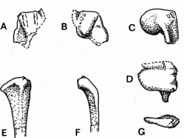
Fig. 1 - Fluviatilavis antunesi sp. nov. Holotype right femur.
Views - A posterior, B anterior, C lateral, D proximal, E distal
(broken areas are indicated by dotted lines, but cracks have-not been shown) [Scale, x 2]
Fluviatilavis gen. nov.
ETYMOLOGY. The name is formed from the Latin fluviatilis (=pertaining to a stream or running water) and
avis (=a bird).
DIAGNOSIS. Wader, a little larger than other Recurvi-rostridae. Femur fairly long and slender, with slight anterior curvature. Distal condyles divergent with broad rotular groove. Medial condyle distally shorter than lateral condyle, more medially prominent and anteropos-teriorly narrow. Popliteal depression triangular, tapering proximally along medial side of shaft with narrow ridge bordering medial edge. Lateral tuberculum proximally sited, level with proximal end of medial ridge. Inter-muscular line occupies about three fifths of the proximal shaft on posterior surface. Trochanteric ridge projects strongly anteriorly, with anterior edge flattened. Proximal end with anterior edge concave between neck and promi-nent trochanter, posterior edge with slight distal hollow. At proximal end elongated attachment scar on lateral side borders trochanter and slants posterodistally across surface.
Type species: Fluviatilavis antunesi
Fluviatilavis antunesi sp. nov.
ETYMOLOGY. The species in named in honour of Professor Dr. Miguel Telles Antunes.
DIAGNOSIS. Only known species of its genus. Cha-racters those of genus.
MATERIAL. Holotype an incomplete right femur; tenta-tively referred specimens a distal end of a right humerus,
14
A
o
B
E
•
" :~ ~f:."
.
~ .:0;. ·t· .'.
'.. '..
t·Jf
,c
~
G
c
o
.r ,F
Fig. 2 - Fluviatilavis antunesisp. nov. Distolateral end of right
humerus. Views - A anconal. B palmar. C lateral. D distal.
Distal end of right radius. Views - E palmar. F medial.
G distal [Scale. about x 2]
A
prominence is at the tip of a triangular lateral flange. In these respects it closely resembles R. avosetta.
The radius shaft is also embedded in matrix, with the flattened medial surface exposed. In anconal view the head is most expanded towards the lateral side, the distal end curving out lateroproximally; and the medial side is more flattened. In medial view some distal curvature is apparent at the tip. The radii of Recent recurvirostrids project slightly proximal to the distal end on the medial side and are thicker at this point. In palmar view the specimen shows a general similarity to recurvirostrids, but again differs on the medial side, the ligamental prominence being more medially and distally sited in the new specimen.
MEASUREMENTS. (in miIIimetres). Holotype femur: greatest length 48.8: length to distal tip of medial condyle 48.3; greatest distal width 9. I; anterior width of rotular groove 4.2; greatest width and approximate length of
popliteal depression 3.3
x
8.5; length of ridge at medialedge of depression 4. I; approximate posterior length of lateral condyle 5.8, of medial condyle 3.8; distal end to lateral tuberculum 10.1; distal tip to commencement of single intermuscular line 20.4; width and thickness at mid-shaft 4.2 x4.3; greatest proximal anteroposterior thickness 7.6; anterior width of trochanteric ridge 2.1. Distal end of humerus; lateral condyle, palmar/anconal thickness 6.5, palmar length 5.3; distal width 3.7; width of tricipital groove 2.5; distal end to tip of ectepicondylar prominence 6.2; palmar tip of lateral condyle to tip of ectepicondylar prominence 3.9.
Radius: length of specimen 72.3; width of medial surface of shaft 2.3; distal width 6.0; greatest distal thickness 3.0.
In posterior view, at the distal end, the lateral condyle projects distally with the posterior edge of the fibular condyle diverging from it laterally. The medial condyle has a medial thrust, and projects less far distally. The intercondylar sulcus is shallow and the ligamental impressions on its posterior surface appears as two small, shallow transverse grooves. Proximal to the lateral condyle the posterior surface of the shaft is a little
pro-minent and rounded, bordered medially bythe popliteal
depression.
The distal edge of the depression slants distolaterally. The depression is deepest near the centre of the shaft and is bordered medially by a narrow ridge similar to that apparent on the specimens of R. avosetta examined. Lines extend proximally from the medial edge of the depression and the lateral side, to join on the shaft at an intermuscular line continuing to the trochanter edge-at the ledge-ateral side of the proximal end. Bordering the line just below the end are two small faint impressions, the impressiones obturatoriae, and there is a shallow transverse groove just below the transverse edge of the proximal surface.
In anterior view the distal end has a broad rotular groove with a medial slant. The condylar edges bordering it do not show any strong anterior projection and appear rather flattened, and similar in this respect to those of other recurvirostrids. The trochanteric ridge projects strongly and is flattened anteriorly to produce a well marked ridge along its medial edge, bordering a concavity. The proximal edge of the trochanter has been damaged but the edge of the slightly curved proximal surface shows a distinct sharp distal curvature between the neck of the missing head and the trochanter, indi-cating that the latter, now damaged, was proximally prominent at this point.
In distal view the medial condyle appears medially broad and anteroposteriorly narrow. In proximal view the distal surface is anteroposteriorly broader than it is lateromedially long. It is smoothly rounded, and the trochanter ridge thickens and curves medially towards its
anterior end. , j
The humerus has an embedded and damaged shaft which appears similar in general shape to that of R. avosetta .The lateral condyle of the distal end is also similar in shape to that of the latter, palmar/anconally elongated, and separated from the medial condyle by only a shallow depression. It bears two tricipital ridges anconally, shows two small pits on the lateral side of the. ectepicondyle tip, and is roughly rectangular in lateral view. Proximal to it on the lateral side the ectepicondylar
REFERENCES
ANTUNES. M. T. and RUSSELL. D. E. (1981) - Le gisement de Si/veirinha (Bas Mondego, Portugal): la plus ancienne faune de Vertebres
eocenes connue en Europe. C. R. Acad. Sci. Paris. t. 293 (21 decernbre 1981). Serie II. pp. 1099-1102.
HARDY. J. W. (1959) - A previously undescribed recurvirostrid from the Eocene of Utah. Auk 76. pp. 106-108.
HARRISON. C. J. O. and WALKER. C. A. (1976) - Birds of the British Upper Eocene. Zoo!. J. Linn. Soc. Lond. 59. pp. 323-351.