O
RIGINALA
RTICLE Revista Brasileira de Fisioterapia©Postural influence of high heels among
adult women: analysis by computerized
photogrammetry
A influência postural do salto alto em mulheres adultas: análise por
biofotogrametria computadorizada
Iunes DH1, Monte-Raso VV1,2,3, Santos CBA1, Castro FA4, Salgado HS4†
Abstract
Introduction: In our society, it is observed an increasing number of models, colors, styles, heights and types of high heels. Objective: To evaluate whether the use of high heel shoes results in postural changes, based on a set of variables measured through computerized photogrammetry. Methods: Twenty individuals who often used high heels (group 1) and 20 individuals who only used high heels sporadically (group 2) were photographed in the frontal and sagittal planes at three conditions: a) without using footwear; b) using stiletto heels; and c) using high platform heels. These photographs were randomized and analyzed by a blinded examiner, by means of photogrammetry. Statistical analysis was performed, using a 2x3 factorial analysis of variance to compare the frequency of high heel use with the type of shoe, at the 5% significance level. Results: Only the head protrusion angle showed a difference between groups 1 and 2 (p<0.01). The effect of the type of shoe observed in the alignment of the right knee, which only showed a difference between stiletto heels and barefoot (p=0.03). For the tibiotarsal angle variable, the effect was also observed for all types of footwear. The other angles evaluated did not present any differences regarding the frequency of high heel use and the types of shoe. Conclusions: The frequency and type of high heel practically did not change the static posture evaluated by photogrammetry.
Key words: posture; high heel; photogrammetry.
Resumo
Introdução: Em nossa sociedade, temos observado uma oferta cada vez maior de modelos, cores, estilos, altura e diversos tipos de salto. Objetivo: Avaliar se o uso de calçados de salto alto influencia nas alterações posturais com base em um conjunto de variáveis mensuradas por meio da fotogrametria computadorizada. Métodos: Vinte indivíduos que utilizam salto alto com freqüência (grupo 1) e 20 indivíduos que utilizam salto alto esporadicamente (grupo 2) foram fotografados no plano frontal anterior e sagital em três momentos: a) sem utilização de calçado, b) utilizando salto agulha e c) utilizando salto plataforma, sendo estas fotografias aleatorizadas e analisadas por um experimentador cego por meio da fotogrametria. A análise estatística foi realizada a partir da análise de variância em esquema fatorial 2x3, ou seja, comparando-se a freqüência do uso de salto com o tipo de calçado, com 5% de significância. Resultados: Apenas o ângulo protrusão da cabeça apresentou diferença quando comparados grupo 1 e 2 (p<0,01). O efeito do tipo de calçado ocorreu na variável alinhamento do joelho direito, sendo que houve diferença apenas entre o sapato agulha e os pés descalços (p=0,03); também para a variável ângulo tibiotársico, o efeito esteve presente em todos os tipos de calçado. Os demais ângulos avaliados não apresentaram diferenças entre a freqüência no uso de salto e os outros tipos de sapato. Conclusões: A freqüência do uso de salto e o tipo de salto praticamente não modificam a postura estática avaliada pela fotogrametria.
Palavras-chave: postura; salto alto; fotogrametria.
Received: 26/11/2007 – Revised: 07/05/2008 – Accepted: 10/09/2008
1 School of Physical Therapy, Universidade José do Rosário Vellano (Unifenas) – Alfenas (MG), Brazil 2School of Physical Therapy, Anhanguera Educacional – Leme (SP), Brazil
3 Department of Biomechanics, Medicine and Rehabilitation of the Locomotor Apparatus, Faculdade de Medicina de Ribeirão Preto da Universidade de São Paulo
(FMRP-USP), Ribeirão Preto (SP), Brazil
4 Physical Therapist
Correspondence to: Vanessa Vilela Monte-Raso, FMRP-USP, Avenida dos Bandeirantes, 3900, Jardim Monte Alegre, CEP 14049-900
Introduction
he foot is the bodily structure that interacts the most with the lower kinetic chain1 and has some of the greatest
struc-tural varieties in the human body2,3. It functions as a
compli-ant mechanism of reception and distribution of body weight, adapting to surface irregularities and acting as a rigid lever that propels the body forward during gait4.
To be able to adapt to surface irregularities, bear the weight of the body and withstand the ground reaction force, the foot is comprised of three plantar arches (internal and external longitudinal arches, and transverse arch). hese act as shock absorbers and distribute the loads on the feet to three regions: calcaneous (60% of the load), midfoot (8% of the load) and forefoot, mainly the 5th and 1st metatarsal head
(32% of the load)5,6.
Footwear gives support to the feet7 and should be worn
to enhance their functions, instead of interfering with the transmission of information from the pressures on the ad-equate support areas or during the movements needed while walking8. Footwear can often change the ideal alignment of
the feet.
In our society, we ind that most women have several pairs of shoes in their personal wardrobe. he footwear industry has an ever-increasing supply of colors, styles and types of heel. An aggravating factor of the current high-heel trend is its increas-ing presence in the lives of children and adolescents who are still in the developmental phase, and therefore in susceptible physical conditions9.
Iunes et al.10 analyzed gait in children who wore 5cm
high-heeled platform shoes through electromyography and found increased muscular activity in the gastrocnemius and anterior tibial muscles. Other changes associated with frequent high-heel use are low back pain, halux valgus, callosity and muscular shortening.
With the advance of science, it is now possible to use the available electronic resources more efectively for postural reeducation with the purpose of improving evaluations, thera-peutic interventions and preventive actions9.
A method called computerized biostereometry or bio-photogrammetry has been used to facilitate the work of health professionals. This method consists in the applica-tion of principles of short distance photogrammetry to the medical field to measure the shapes and dimensions of the human body9.
his method is used in postural evaluations due to the ad-vantages and efectiveness of its clinical application. Among the advantages are low-cost image interpretation system, high precision and reproducibility of results. here is also no contact with the patient or use of visible light, which avoids
radiation exposure9,11. Considering the importance of
appro-priate support for the lower limbs, the aim of this study was to assess, by using computerized photogrammetry, whether the frequency of high heel use has any inluence on postural changes, and whether the type of high heel interferes in the posture.
Methodology
Subjects
We analyzed forty women, chosen at random, who were willing to participate in this research. he participants were divided into two groups: group 1, formed by 20 women (mean age 23.5±2.86 years, mean mass 54.10±7.16 kg, mean height 1.61±0.05m) that wore high-heeled shoes everyday; group 2, formed by 20 women (mean age 22.55±2.68 years, mean mass 51.60±4.54 kg, mean height 1.60±0.03m) that wore high heels occasionally to social functions. he inclusion criterion was women who wear size 35 shoes. Exclusion criteria were neuropsychomotor changes, amputations and sequelae from fractures or rheumatoid arthritis. here were no sample losses during the study. All subjects received information about the project and signed a consent form, agreeing to take part in the research, according to Resolution 196/96 of the National Health Council. he study was approved by the Human Research Ethics Committee of Unifenas, protocol number 100/2005.
Footwear
he participants wore a pair of 6.5cm high-heeled platform sandals and a pair of 8.0cm stilettos, both size 35. Neither pair had been worn prior to the experiment.
Postural analysis
he subjects were photographed in the frontal, anterior frontal and sagittal planes. First, they wore a two-piece swim-suit and no shoes, then one of the previously described pairs of high-heeled shoes for the subsequent postural evaluation. A few anatomical points were marked out, including the occipital protuberance, C4, C7, T7, T12, L3 and L5 spinous processes, greater
trochanter, anterosuperior iliac spine (ASIS), posteroinferior iliac spine (PIIS), tibial tuberosity (TT), head of the ibula, lat-eral malleolus, and head of the 5th toe9.
hese markings were made with white 0.9mm-diameter PIMACO labels and orange-colored plastic rods held in place with double-sided tape9.
For the photo record, a Sony(MAVICA FD 200) digi-tal camera was placed, parallel to the ground, on a level tripod. Images were digitally stored in 1,600x1,200 pixel resolution. The anterior frontal and sagittal planes were photographed.
Subject positioning
For the photo record, subjects were placed in a previ-ously marked position, at a standard distance from the camera, which was also previously marked. In this position, the subject was 15cm from the wall. In order to keep this distance, a 15cm wide, 60cm long and 0.5cm thick rubber marker was placed between the wall and the subjects. The distance between the camera and the subject was 2.4m and the tripod held the camera 1.0m above the ground9. Another
rubber device, measuring 7.5cm in width, was placed be-tween the subjects’ feet to maintain the standard posture, as described by Kendall7. The room was well lit, with a blue,
non-reflexive background, and gave the subject privacy dur-ing the shoot9.
Image analysis
he images were analyzed with the application ALCi-magem-2000 Manipulando Imagens version 1.5 through measures of the angles formed by lines drawn between the previously marked anatomical points. his analysis identiied and quantiied possible asymmetries and vertebral curvature angles.
In the anterior frontal plane, the measured and analyzed angles were between the two ASIS and the TT. hese angles were formed by the line drawn between the right ASIS and the left ASIS and the horizontal line parallel to the ground. Another angle analyzed in this plane corresponded to the alignment of the right knee (rKA) and left knee (lKA). his angle was formed by the intersection of the straight line be-tween points C (ASIS) and D (TT) and the line perpendicular to the ground. he greater the angle is, the greater the lower limb alignment9.
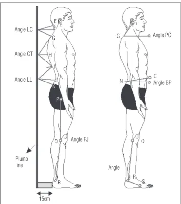
In the sagittal plane, the angles previously described by Iunes9 were analyzed. hey are shown in Figure 1 and described
below.
he head protrusion angle (HP) is formed by the intersec-tion of the straight line between points V (tragus) and G (C7 spinous process) and the line parallel to the ground. he smaller the angle is, the greater the protrusion.
he cervical lordosis angle (CL) is formed by the inter-section of the line between point E (occipital protuberance) and the horizontal extension of point F (C4 spinous process) on the plumb line and the line between point G (C7 spinous process) and the horizontal extension of point F (C4 spinous process) on the plumb line. he larger the angle is, the greater the rectiication, and the smaller the angle, the greater the lordosis.
he thoracic kyphosis angle (TK) is formed by the in-tersection of the line between point G (C7 spinous process) and the horizontal extension of point H (T7 spinous process) on the plumb line and the line between point I (T12 spinous process) and the horizontal extension of point H (T7 spinous process) on the plumb line. he greater the angle is, the greater the rectiication, and the smaller the angle, the greater the kyphosis.
he lumbar lordosis angle (LL) is formed by the intersec-tion of the line between point I (T12 spinous process) and the horizontal extension of point J (L3 spinous process) on the plumb line and the line between point K (L5 spinous process) and the horizontal extension of point J (L3 spinous process) on the plumb line. he larger the angle is, the larger the rectiica-tion, and the smaller the angle, the greater the lordosis.
he pelvic tilt angle (PT) is formed by the intersection of the line between points N (PIIS) and C (ASIS) and the line
Angle PC
Angle BP C
Angle
15cm
Q G
N v Angle LC
Angle CT
Angle LL
Plump line
Angle FJ E
G
H
I
J K
P
R R S
Q
Figure 1. Points and angles evaluated in the right lateral view: C (ASIS),
E (occipital protuberance), F (C4 spinous process), G (C7 spinous
process), H (T7 spinous process), I (T12 spinous process), J (L3 spinous
process), K (L5 spinous process), N (PIIS), P (greater trochanter), Q
(head of the fíbula), R (lateral malleolus), S (distal diaphysis of the 5th
metatarsal of 5th toe), V (tragus), HP (head protrusion), CL (cervical
lordosis), TK (thoracic kyphosis), LL (lumbar lordosis), PT (pelvic tilt), KF (knee flexion), TTA (tibiotarsal angle).
Footwear style
SA(o) 1KA(o) TT(o)
Group 1 Group 2 Group 1 Group 2 Group 1 Group 2
Bare feet 1.92 1.29 175.39 175.3 1.87 1.75
Platform 2.13 1.32 176.21 175.75 1.59 1.66
Stilettos 2.09 1.96 176.28 175.79 1.5 1.69
Table 1. Means of variables SA, lKA and TT with the frequency of high-heel use and the types of shoe, where SA is the symmetry of the ASIS, lKA is the alignment of the left knee, and TT is the symmetry of the tibial tuberosity.
Footwear style
CL(o) TK(o) LL(o)
Group 1 Group 2 Group 1 Group 2 Group 1 Group 2
Bare feet 32.45 35.16 84.88 84.42 56.06 57.35
Platform 33.96 33.99 83.21 83.29 58.54 56.67
Stilettos 33.81 34.93 81.8 83.28 57.6 58.65
Table 2. Means of variables CL, TK and LL with the frequency of
high-heel use and the types of shoe, where CL is cervical lordosis, TK is thoracic kyphosis and LL is lumbar lordosis.
Footwear style
PT(o) KF(o)
Group 1 Group 2 Group 1 Group 2
Bare feet 14.49 14.5 184.93 184.37
Platform 13.44 13.32 186.51 184.69
Stilettos 12.6 13.81 185.24 183.61
Table 3. Means of variables PT and KF with the frequency of high-heel
use and the types of shoe, where PT is pelvic tilt and KF is the position of the knee in the sagittal plane.
Footwear style
HP(o) rKA(o) TTA(o)
Group 1 Group 2 Group 1 Group 2 Mean Group 1 Group 2 Mean
Bare Feet 53.11 50.82 176.7 175.7 176.2ab 110.7 111.5 111.1a
Platform 52.94 51.1 177.2 176.7 177.0bc 129.9 127.6 128.8b
Stiletto 53.39 50.26 177.6 177.6 177.6cd 140.6 136.6 138.6c
Mean 53.15A 50.73B - - -
-Table 4. Means of variables HP, rKA and TTA with the frequency of high-heel use and the types of shoe, where HP is head protrusion, rKA is the
alignment of the right knee and TTA is the tibiotarsal angle.
Different lower case letters (vertical) and capital letters (horizontal) indicate significance (p<0.05) according to Bonferroni’s test12.
parallel to the ground. he larger the angle is, the greater the anterior tilt.
he knee lexion angle (KF) is formed by the intersection of the line between points P (larger trochanter) and Q (lateral knee joint line) and the line between points Q and R (lateral malleolus).
he tibiotarsal angle (TTA) is formed by the intersection of the line between points R (lateral knee joint line) and S (lateral malleolus) and the line between points S and T (head of the 5th toe).
Statistical analysis
For each angle described in the methodology section, three consecutive measures were performed using photo-grammetry, and the arithmetic mean was calculated. he statistical analysis was performed based on the analysis of variance for a completely random model, with 20 repetitions in a 2x3 factorial, i.e. by comparing the frequency of high heel use (regularly or occasionally) with the type of footwear (bare feet, high-heeled platform shoes or stilettos), with a 5% sig-niicance level.
Results
Few modiications in the posture of the examined women were found, regardless of frequency of use and type of high heel. For all the analyzed angles, there was no interaction between frequency of high heel use and the type of footwear (p>0.27, as seen in Tables 1, 2, 3).
However, taking into account the diferences between the group that wore high heels regularly (group 1) and the group that wore high heels occasionally (group 2), only the HP angle that analyzes the positioning of the head showed a difer-ence (p<0.01) in all types of footwear (Table 4). he smaller the measure of this angle, the more head protrusion there is (Figure 1).
Regarding the type of footwear (bare feet, platforms or sti-lettos), there was an efect on the rKA variable that evaluates the lower limb alignment. here was a diference only between
stilettos and bare feet in group 1 (p<0.05) (Table 4). For the TTA variable that evaluates ankle positioning on the sagittal plane, the footwear type had an efect (p<0.01), and there was a difer-ence in all three types of footwear, as expected (Table 4).
Discussion
In general, the exaggerated use of high-heeled footwear causes shortening of the calf musculature, leading frequent high-heel wearers to feel uncomfortable when wearing lat soled shoes. he elevation of the calcaneous leads to modiica-tions in gait pattern and to foot instability6,13,14.
here is an important relationship between heel height and overload on the arches of the foot. High heel use changes body
1. Busquet L. As cadeias musculares. 4a ed. Belo Horizonte: Edições Busquet; 2001. 236p.
2. Cavanagh R, Rodgers MM. The arch index: a useful measure from fooprints. J Biomech. 1987;20(5):547-51.
3. Kulthanan T, Techakampuch S, Bed ND. A study of footprints in athletes and non-athletic people. J Med Assoc Thai. 2004;87(7):788-93.
4. Ledoux WR, Hillstrom HJ. The distributed plantar vertical force of neutrally aligned and pes planus feet. Gait Posture. 2002;15(1):1-9.
mass distribution, reducing the pressure on the calcaneous and shifting it to the forefoot. he weight born by the tip of the foot is in direct proportion to the height of the heel. Continu-ous use of high heels results in overload, which compresses the metatarsals6,15.
Bienfat15 states that there is no good static if the feet are not
planted irmly on the ground. As a result, feet deformities and changes in loading also change static. his raises the issue that if wearing high heels changes the mechanics of the foot, it eventu-ally generates muscular changes to the lower limbs and conse-quently produces ascending compensatory postural changes.
Marques16 reports that a muscle group adapts itself to
certain conditions and that there are variables involved in this mechanism. herefore, it is expected that the frequent high heel use may generate adaptive postural changes.
We suggest that there is a necessity for quantitative studies and clinical investigations to evaluate the relationship between the prolonged use of high heels and orthopedic, joint, and de-generative changes. In the present study, we aimed to quantify the postural modiications produced by the high heel, having photogrammetry as a resource whose reliability has already been tested9,11. Contrary to what has been described by other
authors13 and to our expectations, the frequency of high heel
use and the type of high heel had no correlation with modiica-tions in the static posture.
Regarding the pelvic tilt, the literature states that high heels produce anterior pelvic tilt and an increase in lumbar lordosis3,13,17. However, some authors14,18,19,20,21 have concluded
in their studies that high heels cause posterior pelvic tilt and lumbar rectiication.
Another study14 performed a postural evaluation and
spe-ciic tests for muscular shortening, pelvic mobility and lower limbs on 20 women that wore high and low heels. he sub-jects that frequently wore high heels reported more posterior pelvic tilt.
Bendix et al.18 evaluated 18 women who wore several
types of high heels and found lumbar lordosis by means of an inclinometer. hey concluded that, as the height of the heel increased, the lumbar lordosis and of the posterior pelvic tilt decreased.
Manio et al.21 evaluated seven women without shoes, with
low heels and with high-heeled shoes (85mm), by means of photographs and force platforms, and also found a decrease in anterior pelvic tilt with use of high heels compared to the barefoot women.
In the present study, we did not observe any changes in pelvic position when we evaluated the PB angle, as seen in Table 3, nor did we ind any modiication in lumbar lordosis (evaluated by the LL angle), when we analyzed the relation-ship between frequency of use and type of high heel, as seen in Table 2.
In the literature, wearing high heels is related to knee semilexion, however, a recent study22 evaluated adolescents
with bare feet and high heels by means of photographs and showed that high heels did not change knee positioning. he same result was found in the present study, i.e. women who regularly wore high heels had a similar KF angle to the group of women that wore high heels occasionally. he same rela-tionship was found when diferent types of high heels were compared (Table 3).
No studies were found that correlated head posture, cervi-cal and dorsal regions, nor pelvic and knee asymmetry.
It must be noted that this study included young subjects. It would be useful to investigate a sample of women who have been wearing high heels for several years to verify whether these changes eventually occur.
Conclusions
With the present study, we observed that the frequency of use and type of high heel did not modify static posture in women, according to the photogrammetry evaluation. he only variable that difered between the women who frequently wore high heels and those who wore them occasionally was the head (HP angle). However, this variable did not change when diferent types of heel were worn. Stilettos modiied the rKA in women who did not wear high heels regularly, and ankle po-sitioning on the sagittal plane was the only variable modiied with the use of the diferent types of footwear.
458
5. Kapandji AI. Fisiologia articular: tronco e coluna vertebral. 5a ed. São Paulo: Panamericana; 2000. 253p.
6. Nordin N, Frankel VH. Biomecânica básica do sistema músculo esquelético.
3a ed. Guanabara Koogan; 2003. 401p.
7. Kendall FP, MCcreary EK, Provance PG. Músculos: provas e funções. 5a ed.
São Paulo: Manole; 2007. 454p.
8. Santos A. Postura corporal: um guia para todos. São Paulo: Summus editorial; 2005. 117p.
9. Iunes DH, Castro FA, Salgado HS, Moura IC, Oliveira AS, Bevilaqua-Grossi D. Confiabilidade inter e intra-examinadores e repetibilidade da avaliação postural pela fotogrametria. Rev Bras Fisioter. 2005;9(3):327-34.
10. Iunes DH, Santos CBA, Freitas FP, Gonçalves AR. Análise eletromiográfica da atividade muscular durante a marcha em crianças, utilizando diferentes tipos de calçados. Fisioter Bras. 2005;6(5):328-31.
11. Ribeiro AP, Trombini-Souza F, Iunes DH, Monte-Raso VV.Confiabilidade
inter e examinador da fotopodometria computadorizada e intra-examinador da fotopodoscopia. Rev Bras Fisioter. 2006;10(4):435-9.
12. Neter J, Wasserman W, Kutner MH. Applied linear statistical models. 3a ed. Irwin; 1990.
13. Nasser JP, Mello SIL, Ávila AOV. Análise do impulso em calçados
femininos em diferentes alturas de salto.Anais do VII Congresso Brasileiro
de Biomecânica, 1999:491-93.
14. Albuquerque FMAO, Silva EB. Saltos altos e artralgias nos membros inferiores e coluna lombar. Fisioter Bras. 2003;5(1):18-21.
15. Bienfait M. Os desequilíbrios estáticos: fisiologia, patologia, e tratamento fisioterápico. 3a ed. São Paulo: Summus; 1995.
16. Marques AP. Cadeias musculares: um programa para ensinar avaliação fisioterapêutica global. 2a ed. São Paulo: Manole; 2005. 168p.
17. Snow RE, Willians KR, Holmes Junior GB. The effects of wearing high heeled shoes on pedal pressure in women. Foot Ankle. 1993;13(2): 85-92.
18. Bendix T, Sorenson SS, Klausen K. Lumbar curve, trunk muscles and line of gravity with different heel heightsspine. Spine.1984;9(2):223-7.
19. Opila-Correia KA. Kinematics of high-heeled gait with consideration for age and expierence of wearers. Arch Phys Med Rehabil. 1990;71(11):905-9.
20. De Lauter BJ, Giaconi RM, Questad K, Ko M, Lehmann JF. Footwear and posture: compensatory strategies for heel height. Am J Phys Med Rehabil. 1991;70(5):246-54.
21. Manfio EF, Vilardi Junior NP, Abrunhosa VM, Souza LV, Fernandes BM, Pereira RM. Alterações na marcha descalça e com salto alto. Anais do X Congresso Brasileiro de Biomecânica. 2003;1:87-90.
22. Aguiar Junior AS, Freitas TM. Biomecânica da marcha e da postura com calçado de salto alto. Fisioter Bras. 2004;5(3):183-7.