Acid Proline in the VUV Region
L. H. Coutinho♭♮, M. G. P. Homem♭, R. L. Cavasso-Filho♭, R. R. T. Marinho♯, A. F. Lago ♮ ∗, G. G. B. de Souza♮, and A. Naves de Brito♭♯
♭ Laborat´orio Nacional de Luz S´ıncrotron (LNLS), Box 6192, 13084-971, Campinas-SP Brazil ♯Instituto de F´ısica, Universidade de Bras´ılia, Box 4455, 70910-900, Bras´ılia-DF Brazil
♮ Instituto de Qu´ımica, Universidade Federal do Rio de Janeiro,
Cidade Universit´aria, Ilha do Fund˜ao, 21949-900, Rio de Janeiro-RJ Brazil
Received on 11 January, 2005
Ionic fragmentation of the sublimated amino acid DL-proline has been studied using time-of-flight mass spectrometry and synchrotron radiation. Total ion yield and mass spectra were recorded in the 13 to 21.6 eV energy range. Partial ion yields have been calculated for the produced fragments and the results analyzed in a comparative way. Mass spectrum of proline previously obtained at 21.21 eV using photons from a discharge lamp (He I), was used as reference in the comparison to the synchrotron radiation based spectra. The loss of the COOH fragment represents the most probable dissociation pathway following the photoionization of DL-proline in the valence region. These are the first results of total and partial ion yields spectra for this molecule in its gas phase in the valence region using time-of-flight spectrometry.
I. INTRODUCTION
In recent years, there has been an increasing interest in the spectroscopic study of amino acids due to the fundamental importance of these molecules, which are building blocks of proteins. Knowledge on the valence shell electronic struc-ture of amino acids is necessary for understanding the de-tails of their photo-interaction and reactivity. In addition, va-lence photoionization studies provide a means for understand-ing the photo-stability, as well as the pathways leadunderstand-ing to the photofragmentation products of these molecules follow-ing Vacuum Ultraviolet (VUV) excitation or ionization. Such information is of great importance for several areas, for in-stance, astrophysics and photochemistry, since amino acids have been found in the interstellar medium and meteoritic ma-terials [1–3].
Only a few papers devoted to the experimental study of the amino acids in the VUV regions have been found in the lit-erature. Powis et al. [4] reported on the valence and C 1s core level photoelectron spectra and quantum calculations of gaseous alanine and threonine. Jochims et al. [5] reported an extensive photoionization study in the 6-22 eV energy range, using quadrupole mass spectrometry, of five amino acids: glycine,α-alanine,β-alanine,α-aminoisobutyric acid andα -valine. The Time-Of-Flight (TOF) spectra of the sublimated alpha amino acids L-alanine, L-proline, L-valine and glycine, have been recently reported by Lago et al. [6], from photodis-sociation induced by He I (21.21 eV) photons. This previous results for proline [6], using a He I lamp and a similar spec-trometer to the one used in this work, have also been used here for sake of comparison.
∗Present address: Department of Chemistry, University of North Carolina,
Chapel Hill, NC, 27599-3290, USA
In this paper we report the dissociative photoionization of the amino acid DL-proline in the gas phase, by using Time-Of-Flight Mass Spectrometry (TOF-MS). Photons from a synchrotron radiation source and a discharge lamp (He I) were used as ionizing agents. The resulting photoionization products as well as fragmentation pathways leading to those species have been presented and discussed. Total Ion Yield (TIY) and Partial Ion Yield (PIY) spectra have been recorded in the 13 - 21.6 eV energy range. Mass spectra have been also recorded as a function of photon energy. The resulting decay channels were analyzed in a comparative way. The spectra are, in general, similar to those obtained from electron impact (70 eV) experiments [7]. Within our knowledge, no similar TOF-MS data is available in the literature.
II. EXPERIMENT
FIG. 1: Schematic drawing of the TOF mass spectrometer used for the measurements. The sublimated amino acid enters the ionization region by a needle placed between two plates that accelerate the ions to the flight tube and MicroChannel Plates (MCP) detectors on the right side, and the electrons to the MCP detectors on the left side. The arrival of an electron in the detector initiates a clock that marks the flight-time of the correlated ion.
The vacuum chamber base pressure is usually maintained at about 10−9 mbar. The photons were provided by the
Toroidal Grating Monochromator beamline (TGM) [10] from the LNLS storage ring, located in Campinas-SP, Brazil. This beamline provides linearly polarized radiation from a bend-ing magnet and operates in the range from 12 to 300 eV. The photon intensity was recorded by means of a light sensitive diode. In order to minimize contributions from higher order harmonics in the photon beam, a mixed metallic/gas filter has been added to the beamline. It basically consists in filling a certain region of the beamline with a rare gas (neon), which is isolated from the rest of the vacuum system by two thin metallic foils [11]. Therefore, contributions from higher-order light from the monochromator were negligible in the energy range studied. When using this filter we are able to perform measurements at energies up to 21.6 eV, the absorption edge of Ne. The DL-proline sample was purchased from Sigma-Aldrich in the form of crystalline powder with minimum pu-rity of 99%, and was used without any further purification. Due to the low vapor pressure of this compound, it was nec-essary to use a heated source for sublimation. More details on the sublimation procedure are available in Ref. [6]. The pressure in the chamber was kept in the 10−7mbar range dur-ing the experiments. In order to confirm the absence of de-composition of the samples upon heating, we have performed a Liquid Chromatographic-Mass Spectrometry (LC-MS) ex-periment after the sublimation procedure. The LC-MS results confirmed that no degradation occurred before sublimation of the sample in the temperature range used during our measure-ments.
III. RESULTS
We recorded several PEPICO spectra using synchrotron ra-diation, to analyse both total and partial ion yields. The spec-tral range from 13 to 22 eV was covered, with points being
Relative
Y
ields (arb. units)
arb.arb.
Y
ields (arb
arbarb
Energy (eV)
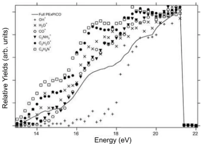
FIG. 2: Total ion yield and partial ion yield of the produced frag-ments after photoionization of DL-proline. Above 21.6 eV the inten-sities drop due to the effect of the harmonic filter.
collected every 0.2 eV. From this set of spectra we were able to construct the graph shown in Fig. 2, where for each photon energy we plot the relative production of the main fragments (PIY) and the TIY. The yields are normalized at their maxi-mum values. The pressure in the experimental chamber and the intensity of the photon beam were used to normalize the data. From these curves we can see that the production of ions increases as the photon energy increases. Besides, a higher slope between 16 and 18 eV can also be observed. From the TIY curve we can distinguish three structures around the ener-gies 14.4 eV, 17.0 eV and 19.2 eV. Above 21.6 eV the intensi-ties drop due to the effect of the harmonic filter. The values of the PIY obtained for DL-proline as function of the photon en-ergy are also shown in table 1. It is important to compare both Fig. 2 and table 1 when analyzing the results. As an example let us take OH+
: in the graph it shows a large increase on its production above 18 eV, but if we look on the table we observe that even at 21.21 eV its production is only about 1% of to-tal. The PIY curves on the graph should not be quantitatively compared one to the other, since their intensities have been normalized at their maximum values, and the table should be used to have a better understanding of the results.
m/z
Intensity (arb. units)
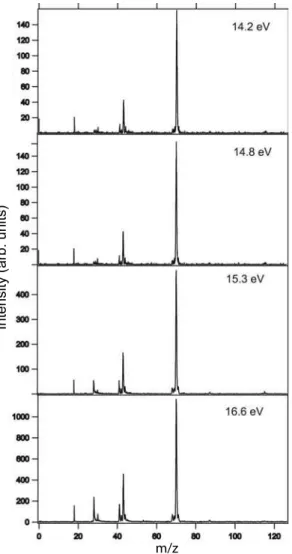
FIG. 3: Mass spectra of DL-proline at 14.2, 14.8, 15.3 and 16.6 eV photon energies.
We also point out the presence of ions formed from rearrange-ment reactions, such as OH+
, H2O+and CO+. However, the presence of water contamination in the peaks at m/z=17 and 18 can not be discarded. The formation of fragments such as OH+
and CO+
may result from the fast dissociation of the COOH+
ion, which was absent from the mass spectra. The CO+
fragment presents a dramatic increase in its production with the increasing of the photon energy when compared to the other fragments, as can be observed both from the table 1 and from Figs. 3 and 4.
The spectrum of L-proline at 21.21 eV (He I) shown in Fig. 5 was taken from Lago et al. [6]. In this figure it is possi-ble to identify a higher number of ionic fragments than in our present results for DL-proline, taken with synchrotron radia-tion. Also the relative productions of the main fragments are different. One hypothesis to explain this divergence is the oc-currence of mass discrimination in the detector used in our spectrometer. The efficiency detection of the microchannel plates is a function of the kinetic energy of the fragments [8]. In this sense, the heavier masses would have smaller detection
m/z
Intensity (arb. units)
FIG. 4: Mass spectra of DL-proline at 18.2, 19.0, 19.3 and 21.21 eV photon energies.
m/z
Intensity (arb. units)
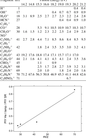
TABLE I: PIY in % for the amino acid DL-proline after photoioniza-tion for several photon energies.
Fragment m/z Photon Energy (eV)
14.2 14.8 15.3 16.6 18.2 19.0 19.3 20.2 21.2
H+ 1 0.4 0.4
OH+
17 0.4 0.7 0.7 0.9 0.9
H2O+
18 3.1 8.9 2.5 2.7 2.7 2.3 2.2 2.4 2.8
HCN+ 27 0.4 0.4 0.9 1.0
C2H3+
CO+
28 5.3 9.1 10.5 10.9 10.7 10.3 10.7
CH2O+ 30 1.6 1.5 1.2 2.3 2.2 2.5 2.4 2.9 2.8
NO+
C2NH3+
41 2.7 2.8 4.4 7.1 8.5 8.6 8.4 8.5 9.5
C3H5+
C2NH4+ 42 1.8 2.4 3.5 3.5 3.0 3.2 4.1
C2H2O+
C2H3O+
43 19.2 17.6 18.8 17.4 17.1 15.7 17.1 17.0
C2H4O+ 44 2.1 1.6 4.1 4.1 4.3 4.1 2.4 3.5 3.6
CHO2+
45 1.1 0.9
C4H6N+
68 2.5 1.7 2.8 2.7 3.9 3.2 3.1
C4H7N+ 69 2.0 1.0 1.8 2.4 2.0 1.6
C4H8N+
70 71.2 67.6 56.3 50.8 46.9 45.3 41.1 44.8 42.6
C2HNO2+ 71 6.7 0.9 0.8 0.7 0.6 0.5 0.4 0.3
PIY He I lamp / PIY SR
70 60 50 40 30 m/z
FIG. 6: The graph shows the ratio between the partial ion produc-tions for the observed fragments using He I lamp and synchrotron radiation (SR) at 21.21 eV. The point at m/z=42 shows the contribu-tion of the peaks at m/z=41, 42 and 43 in the mass spectra in order to minimize the effects due the different mass resolution between the spectra obtained with He I and synchrotron radiation. The peaks at m/z=17, 18 was not included due the different water contamination in spectra.
efficiency. To try to confirm this hypothesis we constructed the graph in Fig. 6, where we compare the relative ionic pro-ductions for two spectra taken at 21.21 eV, one from Ref. [6], using the He I lamp, and the other using synchrotron radia-tion. Care was taken to add together all unresolved peaks in a group of lines thus eliminating spurious fluctuations coming from the different resolution between the synchrotron radia-tion and He I spectra. Furthermore, the peaks H2O+, OH+ and H+
were also removed from analysis since we clearly detect signs of a small water contribution. This contribution changed in the He I and synchrotron radiation spectra. From this graph we can observe that the ratio between the partial productions of the two spectra (the one with the discharge lamp and the one with synchrotron radiation) increases for heavier masses. This could support our hypothesis of mass discrimination stated above.
In summary, time-of-flight mass spectrometry has been em-ployed to the study of the natural amino acid DL-proline us-ing synchrotron radiation as the ionization source. Total ion yield and mass spectra were recorded from 13 to 21.6 eV, us-ing a metallic/gas filter coupled to the beamline in order to reduce the contribution from higher order harmonics contam-ination. Fragmentation products were identified and the rel-ative production among them for a few photon energies was obtained. The loss of the COOH fragment represents the most probable dissociation pathway following the photoionization of DL-proline in the valence region.
IV. ACKNOWLEDGMENTS
The authors wish to acknowledge the staff of the LNLS and support from the Brazilian agencies CAPES, CNPq, FAPERJ, FAP-DF and FAPESP.
[1] A. Brack (Ed.),The Molecular Origins of Life, Cambridge
Uni-versity Press, UK (1998).
[2] L. E. Snyder, Origins Life Evol. Biosphere27, 115 (1997).
[3] M. H. Engel and B. Nagy, Nature (London)296, 837 (1982).
[4] I. Powis, E. E. Rennie, U. Hergenhahn, O. Kugeler, and R.
Bussy-Socrate, J. Phys. Chem. A107, 25 (2003).
[5] H.-W. Jochims, M. Schwell, J-L. Chotin, M. Clemino, F.
Dulieu, H. Baumgartel, and S. Leach, Chem. Phys.298, 279
(2004).
[6] A. F. Lago, L. H. Coutinho, R. R. T. Marinho, A. Naves de
Brito, and G. G. B. de Souza, Chem. Phys.307, 9 (2004).
[7] NIST, Chemistry webbook (http://webbook.nist.gov/chemistry). [8] F. Burmeister, L. H. Coutinho, R. R. T. Marinho, M. G. P.
ter, K. Wiesner, and A. Naves de Brito, Chem. Phys.289, 163 (2003).
[10] P. T. Fonseca, J. G. Pacheco, E. D. Samogin, and A. R. B. de
Castro, Rev. Sci. Instrum.63, 1256 (1992).
Naves de Brito. J. Elect. Spect. Rel. Phen. 144-147, 1125