J. Evid. Based Med. Healthc., pISSN- 2349-2562, eISSN- 2349-2570/ Vol. 3/Issue 31/Apr. 18, 2016 Page 1478
CLINICO-PATHOLOGICAL STUDY OF CERVICAL LYMPHADENOPATHY
Dova Subba Rao1, Mallapraggada Rama Chandra Mohan2, Erabati Santosh Raja3
1Associate Professor, Department of General Surgery, NRI Institute of Medical Sciences. 2Assistant Professor, Department of General Surgery, NRI Institute of Medical Sciences. 3Senior Resident, Department of General Surgery, NRI Institute of Medical Sciences.
ABSTRACT
OBJECTIVES
To know the incidence and aetiological factors of cervical lymphadenopathy. To know the most common group of lymph nodes enlarged. To assess the response to management.
MATERIALS AND METHODS
This study includes 50 patients who attended the Surgical OPD, studied taking detailed clinical history, after physical examination and arriving at clinical diagnosis, confirmation was done by FNAC and biopsy.
RESULTS
Tuberculous lymphadenopathy is the commonest cause of cervical lymphadenopathy with 68% followed by chronic nonspecific lymphadenopathy with 32%. There was no case of sarcoidosis in this series. Disease commonly affected the 2nd and 3rd decades
with 19% and 18% respectively. There is comparatively an increased incidence of tuberculous cervical lymphadenopathy in females than males. The average age of presentation was 30.5 years. There was no definite history of contact with tuberculosis in 82% of cases. In this study series, 44% of the patients belonged to the low income group, 46% belonged to the middle income group. There was only unilateral involvement of node in 72% of cases right side was affected in 32% and left side was affected in 40% of cases. Bilateral involvement was seen in 14% of the cases. The lymph nodes were associated with other groups of lymph nodes in 10% of cases. Chest radiography findings showed the evidence of coexisting active tuberculosis lesions in 8 out of 50 cases (16%) and normal was 42(84%).
CONCLUSION
Knowledge about clinico-demographic perspectives of cervical lymphadenopathy in respect to their cytopathological diagnosis will help to detect/refer the respective cases early for investigations and treatment. Surgical intervention is definitely required in many cases, though most of the cases are medically curable.
KEYWORDS
Cytopathology, Cervical lymphadenopathy, Surgical intervention.
HOW TO CITE THIS ARTICLE: Rao DS, Mohan MRC, Raja ES. Clinico-pathological study of cervical lymphadenopathy. J. Evid. Based Med. Healthc. 2016; 3(31), 1478-1483. DOI: 10.18410/jebmh/2016/335
INTRODUCTION: Neck consists of 300 lymph nodes nearly 1/3 of total lymph nodes of the body. The enlargement of these nodes is significant because of many aetiologic factors. Any infection of the upper respiratory tract can be associated
with cervical adenitis. In adolescents, infectious
mononucleosis may begin with diffuse adenopathy. Chronic granulomatous diseases, particularly cervical lymph node tuberculosis, are endemic in various parts of the world. Sarcoidosis often affects mediastinal and tracheal lymph nodes but cervical adenopathy is also common. Histoplasmosis, Coccidioidomycosis and Actinomycosis can also produce cervical lymphadenopathy. Salivary gland infections can also produce cervical lymphadenopathy, so
also any infection in the oral cavity, ear, nose, throat and scalp can also produce cervical lymphadenopathy. Massive lymphadenopathy in young adults and children is seen in reactive lymphoid lymphoplasia. Malignant metastasis can also be the cause of cervical lymph node enlargement. Lymphomas also present as cervical lymphadenopathy. Among the different infective and inflammatory conditions of cervical lymphadenopathy, tuberculosis is the most commonly found because of the high prevalence of the disease in our country.1 Cervical lymph node involvement is
one of the common extra-pulmonary manifestations of tuberculosis. It is commonly encountered in daily surgical outpatient departments in our country. Tuberculosis is a disease of great antiquity and has even been found in Egyptian mummies. It remains a major disease on a worldwide basis. Fortunately, by effective host defence mechanisms and small number of infecting bacilli, most people overcome the primary infection.2 Better nutrition and
improved social conditions have brought down the disease to low levels in developed countries. It is still common in developing countries like India. Estimates suggest that Financial or Other, Competing Interest: None.
Submission 22-03-2016, Peer Review 31-03-2016, Acceptance 15-04-2016, Published 18-04-2016. Corresponding Author:
Dr. Dova Subba Rao,
Door No. 55-14-109, New P & T Colony, Seetamma Dhara, Visakhapatnam-530013, Andhra Pradesh.
J. Evid. Based Med. Healthc., pISSN- 2349-2562, eISSN- 2349-2570/ Vol. 3/Issue 31/Apr. 18, 2016 Page 1479 worldwide 10 million people develop tuberculosis annually.
The risk is greatly increased in immunocompromised
patients. Tuberculous lymphadenopathy3 commonly affects
adolescents and young adults- Children are also affected. Common age of affected children is 0-5 years. Neck lymph nodes are the commonly affected. Mycobacterium bovis was considered to be the cause of tuberculous lymphadenopathy in the past. But now Mycobacterium tuberculosis is shown to be responsible for most of the tuberculous lymphadenopathy and Mycobacterium bovis in a few cases.
This study was done to know the incidence and aetiological factors of cervical lymphadenopathy; the distribution according to age, sex, urban - rural population, socioeconomic conditions of patients. This study is mainly on
inflammatory and infective causes of cervical
lymphadenopathy. Other causes of cervical
lymphadenopathy are excluded from the study.
MATERIALS AND METHODS: This study includes 50 patients who attended the Surgical O P D during Feb 2014 to June 2015.
Inclusion Criteria: Only inflammatory and infective cases were taken.
Exclusion Criteria: All cases of neck secondary’s and
lymphomas.
Cases were studied taking detailed clinical history, Physical examination and investigations were done. After Physical examination and arriving at clinical diagnosis, confirmation was done by FNAC and Biopsy. Lymph node biopsy was the most important of these.
Name, Age, Sex, Religion, Address, Occupation of the patients were noted. Cases were taken at random and only cases who gave consent for lymph node biopsy were taken for study.
In the history particular emphasis was given to the type of accommodation, the nutritional value of food, history of contact with tuberculosis, any consumption of raw milk. Also history of recurrent pharyngeal infection, scalp infection, ear infections of greater than 3 weeks duration in spite of antibiotics were taken.
After clinical diagnosis was made investigations were done to confirm the diagnosis.
Blood examination for Erythrocyte sedimentation rate (ESR), total white cell count (TC), differential count (DC), haemoglobin percentage (Hb%), Mantoux test was done.by standard method and erythema of more than 12 mm after 48 hours is taken as positive.
Chest x-ray PA view, sputum examination was done. F N A C was done in all cases.
Lymph node biopsy was done in all cases. Macroscopic appearance of the specimen noted down and sent for histopathological examination. Presence of Langhans type of giant cells was taken as the criteria for diagnosing tuberculosis of lymph nodes.
All the specimens were processed by standard procedures like fixing in formalin, slicing by microtome and staining by Gram’s stain and Ziehl-Neelsen stain. All the
slides were examined under 10x, 60x, 100x power using standard microscope. Aspirated material from cold abscess was stained by Gram stain and special stain.
All patients were given anti-tuberculous drugs using DOTS strategy with 2 months intensive therapy and 4 months with continuation phase therapy with drugs isoniazid, rifampicin, ethambutol and pyrazinamide.
Statistical analysis was done by calculating sample percentage value. No correlation studies were done, as this study involves only analysis.
RESULTS: Tuberculous lymphadenopathy is the commonest cause of cervical lymphadenopathy with 68% followed by chronic nonspecific lymphadenopathy with 32%. There was no case of sarcoidosis in this series.
Age (in years) N=50 cases
Number %
0-1 3 6
11-20 19 38
21-30 18 36
31-40 2 4
41-50 4 8
51-60 4 8
>60 --- ---
Gender
Males 24 48
Females 26 52
Table 1: Age distribution and sex distribution
In this series of 50 cases, the disease commonly affected the 2nd and 3rd decades with 19% and 18% respectively.
Next common age group in which tuberculous
lymphadenopathy presented is 5 and 6 decades. 4% of cases affected were in this group in the present study. There is comparatively an increased incidence of tuberculous cervical lymphadenopathy in females than males. The average age of presentation was 30.5 years.
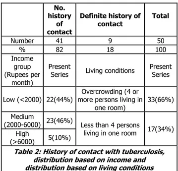
No. history
of contact
Definite history of contact
Total
Number 41 9 50
% 82 18 100
Income group (Rupees per
month)
Present
Series Living conditions
Present Series
Low (<2000) 22(44%)
Overcrowding (4 or more persons living in
one room)
33(66%)
Medium
(2000-6000) 23(46%) Less than 4 persons
living in one room 17(34%)
High
(>6000) 5(10%)
J. Evid. Based Med. Healthc., pISSN- 2349-2562, eISSN- 2349-2570/ Vol. 3/Issue 31/Apr. 18, 2016 Page 1480 There was no definite history of contact with tuberculosis
in 82% of cases. A definite history was obtained in only in 18% of cases. The economic status and living conditions were taken in to consideration to find out the incidence of cervical lymphadenopathy in the studied series. In this study series, 44% of the patients belonged to the low income group, 46% belonged to the middle income group. Only 10% of patients belonged to the higher income group.
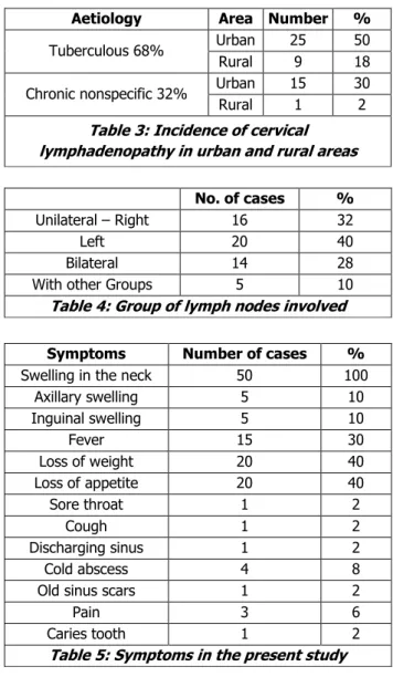
Aetiology Area Number %
Tuberculous 68% Urban 25 50
Rural 9 18
Chronic nonspecific 32% Urban 15 30
Rural 1 2
Table 3: Incidence of cervical lymphadenopathy in urban and rural areas
No. of cases %
Unilateral – Right 16 32
Left 20 40
Bilateral 14 28
With other Groups 5 10
Table 4: Group of lymph nodes involved
Symptoms Number of cases %
Swelling in the neck 50 100
Axillary swelling 5 10
Inguinal swelling 5 10
Fever 15 30
Loss of weight 20 40
Loss of appetite 20 40
Sore throat 1 2
Cough 1 2
Discharging sinus 1 2
Cold abscess 4 8
Old sinus scars 1 2
Pain 3 6
Caries tooth 1 2
Table 5: Symptoms in the present study
The other common presenting symptoms were loss of weight and loss of appetite (40%), fever (30%), axillary and inguinal swellings (10%), cold abscess (8%), pain (6%), sore throat, cough, discharging sinus, old sinus scars, caries tooth (2%).
Groups Present Study
Submandibular& Submental 16%
Upper anterior Deep cervical 28%
Upper Posterior Deep cervical 40%
Lower anterior Deep cervical 28%
Lower posterior Deep cervical 24%
Table 6: Radiography findings
Chest radiography findings showed the evidence of coexisting active tuberculosis lesions in 8 out of 50 cases (16%) and normal was 42(84%).
DISCUSSION: The total number of cases studied is 50 who are attending the surgical out-patient department. From present study, it is seen that tuberculous lymphadenopathy is the commonest cause of cervical lymphadenopathy with 68% followed by chronic nonspecific lymphadenopathy with 32%.
Age Incidence: In this series of 50 cases the disease commonly affected the 2nd and 3rd decades with 19 % and
18% respectively. Next common age group in which tuberculous lymphadenopathy presented is 5 and 6 decades. 4% of cases affected were in this group in the present study. In Wilson's series4 of 100 cases the common age group of
patients was in the 2nd and 3rd decade followed by the 4th
decade with 25%, 32% and 13% respectively.
In B.P. Trivedi's series of 235 cases also the commonest age group of presentation was in the 2nd and 3rd decade with
44% and 35%. Next common age groups affected were 1a
and 4th decade with 10% and 8% respectively. In S P. Pamra
series5 of 322 cases the commonest age group affected were
2nd and 3rd decades with 25% and 35%. Next common age
group were the Ist and 4th decade with 17 % and 11.45 %.In
the present study chronic nonspecific adenopathy affected most commonly the age group of 2nd and 3rd decades with
18% and 8% respectively. In our country the tuberculous lymphadenopathy commonly affects the younger age group. Commonest age group affected is between 11 and 20, 21 and 30 closely followed by 31 and 40 years. Nonspecific lymphadenopathy commonly affects the age group of 11 to 20, 21 to 30 and less commonly 0 to 10. But in western countries the pattern is different. Common age group affected is 0 to 10 years. The causative organism in this age group is atypical mycobacterium. In adults the causative agent is most commonly the Mycobacterium Tuberculosis. Only 5% are due to atypical Mycobacterium. In one study of 343 children with reported lymphadenitis due to atypical mycobacteria 136 were of 3 years or younger age. 194 were younger than 5 years. Only 5 children were younger than 1
year. It cannot be assumed that all cervical
lymphadenopathy in children are caused by atypical mycobacteria. About 5-10 % of childhood lymphadenopathy are due to Mycobacterium Tuberculosis. In another series studied by Hooper,6 tuberculous lymphadenopathy was most
common in the age group of 20 to 40 years. In Prabhakar's series,7 earliest presentation was in a 9-month-old infant and
late age of occurrence was 90 years the average age being 33.6 years. In the present series, the minimum age of presentation was 1 year and the maximum age of presentation was 60 years. The average age of presentation was 30.5 years.
J. Evid. Based Med. Healthc., pISSN- 2349-2562, eISSN- 2349-2570/ Vol. 3/Issue 31/Apr. 18, 2016 Page 1481 in only in 18% of cases. In S. K. Sen's series8 of tuberculous
cervical lymphadenopathy of 386 cases 78.8% cases had no history of contact with tuberculosis, 19.1% had definite history of contact with tuberculosis and a vague history of contact with tuberculosis was obtained in 5.1% of cases.
Sex Incidence: There is comparatively an increased incidence of tuberculous cervical lymphadenopathy in females than males. All the studies in the past as shown a definite increased incidence of cervical lymphadenopathy in females. The incidence was more in S. K. Sen's series- 58.6% and S. D. Pamra series-57.08%.5,8
In the present study though very small the sex incidence was as follows. Males 48% and females 52%. The increased incidence in females may be because of the wide prevalence of malnourishment in females. Other factors influencing the higher incidence in females are overcrowding, lack of education, early marriage, pregnancy, large families, and poor socioeconomic conditions.
Incidence in Different Income Groups and in Different Living Conditions: The economic status and living conditions were taken in to consideration to find out the incidence of cervical lymphadenopathy in the studied series. In this study series 44% of the patients belonged to the low income group, 46% belonged to the middle income group. Only 10% of patients belonged to the higher income group. In S. K. Sen's series,8 65.9% belonged to the
low-income group and 31.6% belonged to the middle-low-income group. Only 2.5% were of the higher income group. Thus, economic status has an important role in the incidence of the disease. The majority of the patients belong to the lower economic status and lesser number of patients are in middle income group. The higher economic status group is the least affected. Here 66% of patients in this study lived in overcrowded conditions i.e. 4 or more than 4 persons lived in one room. In S. K. Sen's series 76.7% lived in
overcrowded conditions.8 In the epidemiology of
tuberculosis overcrowding is an important factor responsible for spread of the disease. The other factors contributing to the higher incidence are population explosion lack of education, large families, poor housing, malnourishment, and unhygienic conditions of living. The distribution of the disease pattern in urban and rural areas was studied in this
series. 50% of the patients had tuberculous
lymphadenopathy and belonged to urban area, 18% of the patients had tuberculosis and belonged to rural area. Chronic lymphadenopathy affected 30% patients and they belonged to urban area, 2% of the patients belonged to rural area. Overcrowding is an important factor for the spread of tuberculosis and its higher incidence in urban areas. Also, in urban areas the people of low socioeconomic group and slum dwellers are mostly affected. There was only unilateral involvement of node in 72% of cases Right side was affected in 32% and left side was affected in 40% of cases. Bilateral involvement was seen in 14% of the cases. The lymph nodes were associated with other groups of lymph nodes in 10% of cases. In S. K. Sen series there was bilateral neck node
involvement in 54.5%, unilateral in 45.5% and neck nodes associated with other group of lymph nodes in 28.5% of cases. The upper anterior deep cervical group of nodes are
the most commonly involved in Wilmont series.9
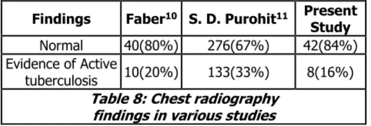
Jugulodigastric nodes were the commonest in this group because tonsils are the common route of entry for the tuberculous bacilli. In the present series upper posterior deep cervical nodes were the commonest (40%) affected followed by upper anterior and lower anterior deep cervical groups (28%). Generalised tuberculosis is very common and may or may not be associated with a known focus in the body. It is characterised by simultaneous enlargement of all the palpable lymph nodes. In Faber's series,10 20% had
associated active lesion on chest x-ray and in S. D. Purohit's series,11 33% of patients had associated active pulmonary
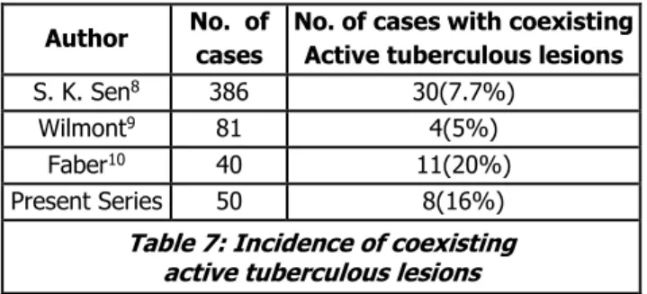
tuberculosis as shown by chest x-ray. In the present series, 16% had associated pulmonary tuberculosis as shown by chest x-ray. The incidence of coexisting tuberculosis in other parts of body is low as shown in table 12. The highest incidence was found in Faber's series10 (20%), lowest in
Wilmont's series9 (5%). In the present study, the incidence
was 16%. Whether the origin of tuberculous
lymphadenopathy is a part of primary complex or haematogenous lesion still remains uncertain. The insidious onset and the absence of constitutional symptoms favour the opinion that the lesion is of primary haematogenous origin. The commonest striking feature reported by all was the insidious onset. In the present study, 68% of cases were of insidious onset. The disease is mainly confined to the cervical group of lymph nodes. Incidence of associated active lesions in other parts of the body was found to be very low. When the primary complex occurs in the lungs, the disease may also be generalised with lesions elsewhere in the body. The behaviour of these nodes closely resembles that of the peripheral adenitis following infection or injury at the drainage site.
Author No. of
cases
No. of cases with coexisting Active tuberculous lesions
S. K. Sen8 386 30(7.7%)
Wilmont9 81 4(5%)
Faber10 40 11(20%)
Present Series 50 8(16%)
Table 7: Incidence of coexisting active tuberculous lesions
Primary Symptoms: As shown, all the 50 patients in the present study had cervical lymph node swelling. The other common presenting symptoms were loss of weight and loss of appetite (40%), fever (30%), axillary and inguinal swellings (10%), cold abscess (8%), pain (6%), sore throat, cough, discharging sinus, old sinus scars, caries tooth (2%).
J. Evid. Based Med. Healthc., pISSN- 2349-2562, eISSN- 2349-2570/ Vol. 3/Issue 31/Apr. 18, 2016 Page 1482 unilateral in 45.5% and neck nodes associated with other
group of lymph nodes in 28.5% of cases.
It is evident that the upper anterior deep cervical group of nodes are the most commonly involved in Bailey (1965), lan Aird (1958) and Wilmont series. Jugulodigastric nodes were the commonest in this group because tonsils are the common route of entry for the tuberculous bacilli.
In the present series, upper posterior deep cervical nodes were the commonest (40%) affected followed by upper anterior and lower anterior deep cervical groups (28%),
Findings Faber10 S. D. Purohit11 Present
Study
Normal 40(80%) 276(67%) 42(84%)
Evidence of Active
tuberculosis 10(20%) 133(33%) 8(16%)
Table 8: Chest radiography findings in various studies
Generalised tuberculosis is very common and may or may not be associated with a known focus in the body. It is characterised by simultaneous enlargement of all the palpable lymph nodes.
In Faber's series 20% had associated active lesion on chest x-ray and in S. D. Purohit's series 33% of patients had associated active pulmonary tuberculosis as shown by chest x-ray. In the present series, 16% had associated pulmonary tuberculosis as shown by chest x-ray.
The incidence of coexisting tuberculosis in other parts of body is low. The highest incidence was found in Faber's series (20%), lowest in Wilmont's series (5%). In the present study the incidence was 16%.8,9
Primary or Secondary: Whether the origin of tuberculous lymphadenopathy is a part of primary complex or haematogenous lesion still remains uncertain. The insidious onset and the absence of constitutional symptoms favour the opinion that the lesion is of primary haematogenous origin. The commonest striking feature reported by all was the insidious onset. In the present study, 68 % of cases were of insidious onset.
The disease is mainly confined to the cervical group of lymph nodes. Incidence of associated active lesions in other parts of the body was found to be very low.
When the primary complex occurs in the lungs, the disease may also be generalised with lesions elsewhere in the body. The behaviour of these nodes closely resembles that of the peripheral adenitis following infection or injury at the drainage site.
Author No. of cases
No. of cases with coexisting active tuberculous lesions
S.K.Sen8 386 30(7.7%)
Wilmont9 81 4(5%)
Faber10 40 11(20%)
Present
Series 50 8(16%)
Table 9: Incidence of coexisting active tuberculous lesions
The prognosis was very good when the patients took regular treatment with antitubercular drugs for the recommended duration of therapy. If the disease is not diagnosed and treated or if there is no patient compliance, prolonged invalidation, dissemination of the disease, complications like cold abscess, sinus formation can occur. Disseminated tuberculosis may cause death eventually. Tubercular cervical lymphadenopathy is very common in our country particularly in people of low socioeconomic group. In the present study, most of the patients responded well to short course chemotherapy with 4 drugs. A few were lost for followup. Surgery was limited in patients with cold abscesses and sinuses along with the antitubercular chemotherapy.
CONCLUSION: Commonest cause of cervical
lymphadenopathy is tuberculosis. (68%) and the next common cause is chronic nonspecific lymphadenopathy (32%).The commonest age group affected in this series are 2nd and 3rd decades. Females (52%) incidence is more in
study. A definite history of contact with tuberculosis was obtained in only 18% in this series. 44% of patients in this series were from low income group and 66 % lived in overcrowded conditions thus proving that tuberculosis is very-common in the low socioeconomic group. In this series, tuberculous lymphadenopathy was found more in the urban population (24%) than in rural population (9%), probably because of the overcrowded living conditions and
atmospheric pollution. Tuberculous cervical
lymphadenopathy is commonly presented as swellings in the neck, other symptoms like fever, loss of weight, loss of appetite and cough are found less commonly in the present study. There were only 16% of patients with associated pulmonary tuberculosis as shown by chest x-ray evidence in the present series. The patients were followed for 6-9 months on monthly basis. Knowledge about clinico-demographic perspectives of cervical lymphadenopathy in respect to their cytopathological diagnosis will help to detect/refer the respective cases early for investigations and treatment. Surgical intervention is definitely required in many cases, though most of the cases are medically curable.
REFERENCES:
1. Minor Madakour M, Kitab E, AL-Otaibi R. AL Swailem.
Text book of Tuberculosis. Page no, 15-26 & 153-160.
2. Ayesha Sarwar, Anwar Ul Haque. Spectrum of
morphological changes in tuberculous lymphadenitis. International Journal of Pathology 2004;2(2):85-89.
3. Leong Anthony SY, Craig L James. Cytological
specimen. Hand book of surgical pathology 1996;1st
edn:205-209.
4. Wilson GR, McLean NR, Chippindale A, et al. The role
of MRI scanning in the diagnosis of cervical lymphadenopathy. Br J Plast Surg 1994;47(3):175-179.
J. Evid. Based Med. Healthc., pISSN- 2349-2562, eISSN- 2349-2570/ Vol. 3/Issue 31/Apr. 18, 2016 Page 1483 6. Hooper AA. TB peripheral lymph nodes. British Journal
of Surgery 1972;89:353-359.
7. Jawahar MS, Sivasubramanian S, Vijayan VK, et al.
Short course chemotherapy for tuberculous
lymphadenitis in children. BMJ 1990;301(6748):359-362.
8. Tripathy SK, Sen RK, Sharma A, et al. Isolated cystic tuberculosis of scapula; case report and review of literature. J Orthop Surg Res 2010;5:72.
9. Newcombe JF. Tuberculous cervical
Lymphadenopathy. Postgraduate Medical Journal 1971;47:713-717.
10.Warren WH, Faber LP. Segmentectomy versus
lobectomy in patients with stage I pulmonary carcinoma. Five-year survival and patterns of
intrathoracic recurrence. J Thorac Cardiovasc
Surg 1994;107:1087–1093.