Half-Barrels Derived from a (β/α)
8
Barrel
β-Glycosidase Undergo an Activation Process
Daniela Beton, Sandro R. Marana*
Departamento de Bioquímica, Instituto de Química, Universidade de São Paulo, São Paulo, Brazil
Abstract
The evolution of (β/α)8barrel proteins is currently thought to have involved the fusion of two (β/α)4half-barrels, thereby conferring stability on the protein structure. After the formation of a whole (β/α)8barrel, this structure could evolve and diverge to form fully active enzymes. Interestingly, we show here that isolated (β/α)4half-barrels derived from the N- and C-termi-nal domains of theβ-glucosidase Sfβgly (Sfβgly-N: residues 1 to 265; Sfβgly-C: residues 266 to 509) undergo an activation process, which renders them catalytically active. The rate constants of the activation process were calculated to be 0.029 and 0.032 h-1for Sfβgly-N and Sfβgly-C, respectively. Moreover, the Sfβgly-N and Sfβgly-C activation processes were simultaneous with modifications in their initial structure, which reduced the exposure of their tryptophan residues. Importantly, this activation was also coincident with an increase in the sizes of Sfβgly-N and Sfβgly-C particles. These novel observations suggest that the change in catalytic activity associated with the transition from a half to whole (β/α)8barrel might also have driven such an evolutionary process.
Introduction
The TIM (or (β/α)8barrel) is the most common fold in enzymes, and enzymes containing this fold function as hydrolases, oxidoreductases, lyases, transferases or isomerases [1,2]. Of those enzymes, the glycoside hydrolases represent the largest group of enzymes that adopt a (β/α)8 barrel fold [1]. Because of its wide distribution, the evolution of this fold has been intensely dis-cussed in the literature [1,3,4]. It has been proposed that the (β/α)8barrel evolved from the duplication and fusion of a half-barrel (β/α)4unit or alternatively resulted from two sequential duplication and fusion events that started from a quarter barrel (β/α)2unit [2,3,5,6,]. Most studies on thein vitroreconstruction of those events have focused on the structure of the initial half-barrels, and again in stability is assumed to have driven (β/α)8barrel evolution [3,2]. Indeed, the fusion of two (β/α)4half-barrels could have contributed to the formation of the active site at the C-terminal top of the barrel, which then could have diverged into different enzymes. Indeed, the (β/α)4half-barrels that have been characterized have not exhibited enzymatic activity when isolated, but when two complementary (β/α)4half-barrels were co-expressed or co-refolded, an active whole (β/α)8barrel was regenerated [6–8]. Nevertheless, as (β/α)8barrels are mainly enzymes, characterization of the enzymatic activity in the transition from pieces to whole barrels is a relevant question that needs to be addressed.
OPEN ACCESS
Citation:Beton D, Marana SR (2015) Half-Barrels Derived from a (β/α)8Barrelβ-Glycosidase Undergo
an Activation Process. PLoS ONE 10(10): e0139673. doi:10.1371/journal.pone.0139673
Editor:Eugene A. Permyakov, Russian Academy of Sciences, Institute for Biological Instrumentation, RUSSIAN FEDERATION
Received:August 14, 2015
Accepted:September 16, 2015
Published:October 2, 2015
Copyright:© 2015 Beton, Marana. This is an open access article distributed under the terms of the
Creative Commons Attribution License, which permits unrestricted use, distribution, and reproduction in any medium, provided the original author and source are credited.
Data Availability Statement:All relevant data are within the paper.
Funding:This work was supported by "Fundação de Amparo à Pesquisa do Estado de São Paulo" (08/ 55914-9) and "Conselho Nacional de
Desenvolvimento Científico e Tecnológico". The funders had no role in study design, data collection and analysis, decision to publish, or preparation of the manuscript.
Herein, we worked with (β/α)4half-barrels derived from the N- and C-terminal halves of theβ-glucosidase Sfβgly (AF052729) [9], a member of Glycoside Hydrolase family 1. We show that those (β/α)4half-barrels undergo an activation process, which renders them catalytically active. Moreover, such activation processes occur simultaneously with modifications in the ini-tial structure of the (β/α)4half-barrels and are coincident with an increase in size. These obser-vations suggest that in addition to the gain in structural stability, the increase in catalytic activity might also have driven the evolution of (β/α)8barrels.
Material and Methods
Assembly of the Expression Vectors for Sf
β
gly-N and Sf
β
gly-C
The cDNAs encoding Sfβgly-N (residues 1 to 265 of Sfβgly) and Sfβgly-C (residues 266 to 509 of Sfβgly) were amplified using a PCR Master Mix kit (Fermentas Life Science; Vilnius, Lithua-nia) and a pAE vector containing an insert for native Sfβgly as template. The reaction cycles (30) were 30 s at 94°C, 60 s at 60°C and 90 s at 72°C. The pair of primers 50-CCGCTCGAGCA GCAGCGCCGCTTC-3-0and 50-CCCAAGCTTTCATTCAGCAGCCATTTCATCC-30was used for Sfβgly-N, whereas the primers 50 -CCGCTCGAGCTTAGGAGACAGGGTGAGTGG-30and 50-CCCAAGCTTTCAATGTCCCTCATCTATAGTCATG-30were used for Sfβgly-C. The amplified products were analyzed by electrophoresis in 1% agarose gels and extracted using a PCR clean-up kit (Promega; Fitchburg, USA). Purified products were digested with the enzymesXhoI andHindIII, for which sites were present in the primers (underlined and double underlined segments, respectively). Next, digested products were cloned into a pAE vector [10], which had been previously digested with the same restriction enzymes. The complete liga-tion reacliga-tion was used to transform XL1 Blue competent cells, which were cultivated on Luria broth agar plates containing 50μg/mL ampicillin. Transformed colonies were cultivated in
5 mL of Luria broth containing 50μg/mL ampicillin for 16 h. Next, they were used for plasmid
extraction using a DNA purification kit (Promega Fitchburg, USA). Plasmids were sequenced using Big Dye termination mix (Applied Biosystems; Waltham, USA).
Expression and Purification of Sf
β
gly-N and Sf
β
gly-C in ArticExpress
(DE3)
A colony of ArticExpress(DE3) freshly transformed with either the vector pAE-Sfβgly-N or pAE-Sfβgly-C was transferred to 10 mL of 2xTY medium (1.6% tryptone, 1% yeast extract and 8.5 mM NaCl) containing 50μg/mL carbenicillin and 10μg/mL gentamicin and cultivated at
washing step was repeated 5 times, and the final pellet, the resin containing the recombinant proteins that were specifically bound, was stored on ice for the next step. Finally, recombinant proteins were eluted in 0.15 mL of 10 mM sodium phosphate buffer, pH 7.0, containing 300 mM imidazole. The buffer of the supernatant containing eluted proteins was exchanged to 50 mM sodium citrate-phosphate buffer, pH 6.0, using a HiTrap desalting column (GE Health-Care; Little Chalfont, UK).
SDS
–
PAGE and Immunoblotting
The protein electrophoresis in denaturing conditions were as described elsewhere [11]. For visualization, proteins were stained using 0.2% (m/v) Coomassie Blue R prepared in 50% meth-anol and 2% acetic acid (v/v). For immuno-detection, proteins were transferred to nitrocellu-lose membranes [12]. Transfer was confirmed by staining the membranes with 0.1% Ponceau-S (m/v) prepared in 10% acetic acid (v/v). Membranes were destained using water and blocked with 5% skimmed milk prepared in 10 mM Tris-HCl, pH 7.4, containing 150 mM NaCl (TBS). For immune detection of the recombinant proteins, the membranes were incubated for 16 h at 4°C with anti-His HRP antibody (Qiagen; hearafter termed anti-His6) prepared in TBS con-taining 5% skimmed milk. Next, free antibodies were washed out by incubation (6 steps of 10 min) in TBS containing 0.1% Tween 20 (m/v). Recombinant proteins specifically recognized by the anti-His6antibody were stained using an Opti-4CN kit (BioRad; Hercules, CA, USA).
β
-Glucosidase Assay
The enzymatic activity ofβ-glucosidase was detected using as a substrate 1 mM methylumbelli-ferylβ-glucoside prepared in 100 mM citrate-sodium phosphate buffer, pH 6.2. The reaction mixture containing substrate and enzyme was incubated at 30°C for defined periods of time. Reactions were stopped with 200 mM glycine-NH4OH, pH 10.5, and the product methylum-belliferone was detected by fluorescence (excitation, 360 nm; emission, 450 nm).
Determination of Rate Constants for the Activation Process
The activation processes of Sfβgly-N and Sfβgly-C were analyzed according to the hysteresis model [13], which results inEq 1for determining the activation rate constant.
lnðv2 vtÞ ¼ lnðv2 v1Þ kt ðequation1Þ
The rates of substrate hydrolysis at different times (vt) were determined by calculating the slope of [P]versustime curve. The valuevtis proportional to the concentration of active enzyme at time t. The valuev2is proportional to the total concentration of enzyme, which was calculated based on the slope at the highest experimental time (48 h).v1is the initial activity of the inactive enzyme, which was assumed to be 0 because the [P]versustime converges to zero at time zero. Thus,vt,v2andv1, which were calculated from the experimental [P]versustime curve, were linearized using a plot of ln (v2-vt)versust, for which the slope corresponds to the activation rate constants (k).
Determination of Circular Dichroism Spectra
Determination of Fluorescence Spectra for Proteins
Protein samples were incubated in a quartz cell (10 mm) at 30°C in a F4500 espectrofluorom-eter (Hitachi; Tokyo, Japan). Samples were excited at 295 nm, and emission spectra were detected from 305 to 400 nm. The scan speed was set to 1.2 nm/min, the integration time to 1 s and the slit opening to 5.0 nm. For fluorescence quenching experiments, spectra were deter-mined in the presence of acrylamide (up to 0.47 M) prepared in 50 mM citrate-sodium phos-phate buffer, pH 6.0. The maximal fluorescence values were recorded at the same wavelength in the absence (F0) or presence (F) of acrylamide. The ratios F0/F at each acrylamide concentra-tion [Q] were plotted as previously described [15] for the determination of the Stern–Volmer constant (Ksv). Quenching experiments were performed with protein samples previously incu-bated at 30°C in 50 mM citrate-sodium phosphate buffer, pH 6.0, for 0 and 48 h.
Dynamic Light Scattering
Protein samples prepared in 50 mM citrate-sodium phosphate buffer, pH 6.0, were passed through 0.22μm filters and transferred to disposable plastic cuvettes (50μL). Samples were
previously incubated at 30°C in 50 mM citrate-sodium phosphate buffer, pH 6.0, for 0, 24 and 48 h. Data were collected at 30°C using a Zetasizer Nano ZS (Malvern; Worcestershire, UK).
Results and Discussion
The cDNA coding the N- and C-terminal halves (residues 1 to 265 and 266 to 509) of the GH1 β-glucosidase Sfβgly [9] (AF052729) was amplified by PCR and cloned into the expression vec-tor pAE. Considering that Sfβgly folds as a (β/α)8barrel, these halves are hereafter termed Sfβgly-N and Sfβgly-C, which are predicted to correspond to (β/α)4half-barrels. Thus, each of these half-barrels, as well as native Sfβgly, were produced as recombinant proteins in ArticEx-press(DE3) bacteria at 12°C for 24 h. Next, they were successfully purified using Ni-NTA aga-rose resin. The homogeneity and identity of Sfβgly-N and Sfβgly-C were confirmed by SDS–
PAGE and western blotting using anti-His6antibody (Fig 1). Approximately 200μg of both
soluble Sfβgly-N and Sfβgly-C was obtained from 3 L induced bacterial media. However, the yields were variable, and freshly transformed bacteria had to be used for each induction. Nota-bly, Sfβgly-N and Sfβgly-C were also expressed in BL21-Gold (DE3) bacteria at 20, 25 and 37°C, but no soluble recombinant protein was obtained from those induced bacteria.
Sfβgly-N and Sfβgly-C were subjected to enzymatic activity assays using methylumbelliferyl β-glucoside as a substrate. Remarkably, both half-barrels exhibited hydrolytic activity toward that substrate, which increased with time and yielded exponential curves (Fig 2). These findings suggest that the concentration of catalytically active Sfβgly-N and Sfβgly-C increased with assay time, indicating that these half-barrels underwent an activation process. Native Sfβgly did not exhibit such behavior in similar activity assays actually generating linear curves as expected. Moreover, control assays prepared with substrate alone, in which the half-barrel sam-ples were replaced by buffer containing 300 mM imidazole,did not showβ-glucosidase activity. Finally, mock“Sfβgly-N and Sfβgly-C samples”produced from extracts of ArticExpress (DE3) bacteria transformed with an“empty”pAE vector were used in theβ-glucosidase assays, and these assays showed no product formation. Accordingly, mock“Sfβgly-N and Sfβgly-C sam-ples”produced from ArticExpress (DE3) bacteria expressing an inactive mutant Sfβgly showed no product formation.
time. Hence, increases in rate, which are proportional to the concentration of active enzyme at each reaction time, were graphically analyzed (Fig 2, inserts), resulting in the rate constants 0.029 and 0.032 h-1for the activation processes of Sfβgly-N and Sfβgly-C, respectively (Table 1). These data indicate that those half-barrels needed 35 and 33 h, respectively, to be converted into an active form.
This observation is in agreement with previous studies showing that half-barrels showed no enzymatic activity just after purification because indeed Sfβgly-N and Sfβgly-C slow activation process was only detected after a long incubation time using a sensitive activity assay based on the formation of a fluorescent product.
Fig 1. Expression and purification of Sfβgly-N and Sfβgly-C.Proteins from the soluble (lane S) and insoluble (lane P) fractions of bacteria (ArticExpress (DE3)) induced to produce Sfβgly-N(A)and Sfβgly-C(B)were analyzed by SDS–PAGE (12% polyacrylamide; Comassie Blue R250 staining), transferred to
a nitrocellulose membrane and immuno-detected using anti-His6antibody (lanes 1 and 2, respectively). Sfβgly-N and Sfβgly-C were purified from the
respective soluble fractions using Ni-NTA agarose and analyzed by SDS–PAGE (lane 3; arrow; Comassie Blue R250 staining).
Fig 2. Enzyme activity assay of Sfβgly-N and Sfβgly-C showing an activation process.Purified samples of Sfβgly-N(A)and Sfβgly-C(B)in 100 mM citrate-phosphate buffer, pH 6.0, were incubated with substrate 1 mM methylumbelliferylβ-glucoside prepared in 100 mM citrate-phosphate buffer, pH 6.0, for 16, 22, 40 and 48 h. The fluorescence of the product methylumbelliferone (excitation, 360 nm; emission, 450 nm) was determined after the addition of 100 mM glycine-NaOH, pH 10.5.Inserts:rates were analyzed according the hysteresis model [10]. The reaction rate at each time (vt) was calculated based on the slope at that specific
It is unclear how isolated Sfβgly-N and Sfβgly-C can catalyze a hydrolytic reaction, as native GH1β-glucosidases depend on two glutamic acid residues, catalytic acid/base and nucleophile, for that activity [16]. However, the observation of an activation process suggests that a modi-fied form of these half-barrels is active.
To understand the putative modifications that Sfβgly-N and Sfβgly-C are subjected to dur-ing the activation process, we physically characterized these half-barrels. Initially, circular dichroism spectra indicated that both half-barrels had a secondary structure (Fig 3). Sfβgly-N is mainly composed ofα-helices (64%) and unstructured regions (33%), whereasβ-strands are a minor component (3%). Conversely, Sfβgly-C is mainly composed of unstructured regions (49%) andβ-strands (47%), andα-helices are a minor component (4%).
Next, the modification of the Sfβgly-N and Sfβgly-C structure along with the activation pro-cess was verified using tryptophan fluorescence quenching experiments (Fig 4). The Stern-Vol-mer constants (Ksv) for acrylamide quenching of Sfβgly-N intrinsic fluorescence decreased from 5.9 to 4.5 after 48 h of incubation in 50 mM citrate-sodium phosphate buffer, pH 6.0. Similarly, for Sfβgly-C, theKsvwas reduced from 6.5 to 3.8 after 48 h. Thus, for both half-bar-rels, 48 h of incubation in 50 mM citrate-sodium phosphate buffer, pH 6.0, which is sufficient for complete Sfβgly-N and Sfβgly-C activation, reduced the access of acrylamide to tryptophan residues [15], suggesting that the activation process involves structure modifications that reduce the exposure of Sfβgly-N and Sfβgly-C tryptophan residues. TheKsvfor native Sfβgly remained unchanged (7.3versus7.5) after a 48 h incubation.
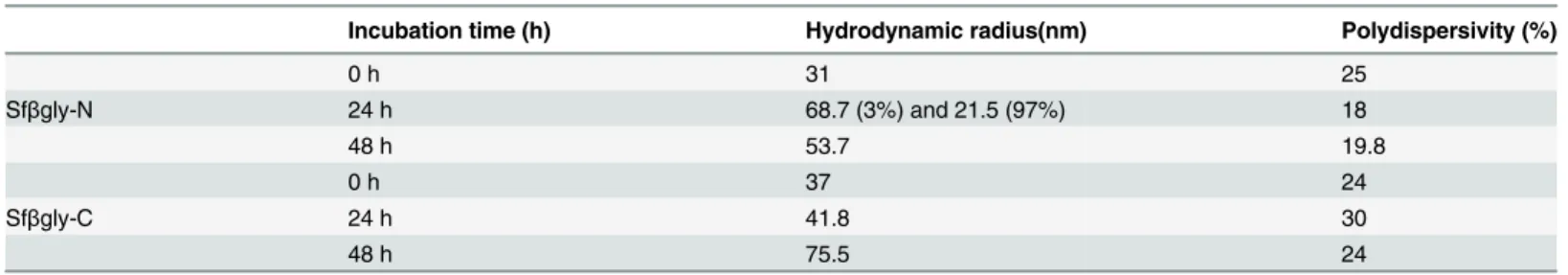
Finally, physical modifications occurring throughout the activation process for Sfβgly-N and Sfβgly-C were also probed by dynamic light scattering. We initially observed that 100% of the Sfβgly-N particles in the sample exhibited a hydrodynamic radius of 30.8 nm. After incuba-tion for 24 h in 50 mM citrate-sodium phosphate buffer, pH 6, 97% of them remained
unchanged (21 nm), whereas 3% increased to 68.7 nm (Table 2). Finally after 48 h, 100% of Sfβgly-N particles in the sample had changed to 53.7 nm. A similar result was obtained for Sfβgly-C. Initially, 100% of particles in the sample had a radius of 37 nm, and after 24 h, they remained unaltered (41 nm), whereas after 48 h, 100% of them exhibited a radius of 75.5 nm (Table 2). The polydispersivity of the samples, which was lower than 25%, indicates that Sfβgly-N and Sfβgly-C were homogeneous throughout the incubation period. Interestingly, samples of Sfβgly-N and Sfβgly-C that were incubated at 4°C for 48 h did not show any modifi-cation in hydrodynamic radius.
Therefore, simultaneously with the enzymatic activation process (Fig 2), Sfβgly-N and Sfβgly-C appear to increase in radius (Table 2), which could indicate an oligomerization pro-cess. We noted that the initial hydrodynamic radius (31 and 37 nm) was not compatible with the expected value (~9 nm) for monomeric Sfβgly-N and Sfβgly-C particles. Perhaps the differ-ence results from the partially unstructured nature of Sfβgly-N and Sfβgly-C, as revealed by the circular dichroism spectra (Fig 3). Alternatively, Sfβgly-N and Sfβgly-C could be present as oligomers at the initial time. Nevertheless, although the real“initial aggregation state”of these particles is not known, it is clear that the initial radius of the Sfβgly-N and Sfβgly-C particles had increased after 48 h.
Table 1. Kinetic parameters for the activation of Sfβgly-N and Sfβgly-C.
Sfβgly-N Sfβgly-C
k(h-1) 0.029±0.006 0.032±0.007
Rate constants (k) for the activation process were calculated based on seven independent assays.
Conclusions
In brief, enzyme activity assays showed that the concentration of active Sfβgly-N and Sfβgly-C increased with time, indicating that these half-barrels underwent an activation process. Physi-cal characterization of Sfβgly-N and Sfβgly-C suggests that they can undergo oligomerization, which involves modification of their structure and culminates in active forms of these half-bar-rels. These observations add interesting data to the discussion of (β/α)8barrel evolution, as it is
currently proposed that the fusion of two (β/α)4half-barrels confers stability on the protein struc-ture because of the removal of hydrophobic surfaces from the aqueous solvent to the protein core [3,17]. In addition the data presented here suggest that the increased catalytic activity in the tran-sition from half to whole (β/α)8barrels might also have driven that evolutionary process.
Acknowledgments
We thank to Dr. Iolanda M. Cuccovia (IQUSP) for the access to the DLS equipment.
Author Contributions
Conceived and designed the experiments: SRM. Performed the experiments: DB. Analyzed the data: DB SRM. Contributed reagents/materials/analysis tools: SRM. Wrote the paper: DB SRM.
Fig 4. Acrylamide quenching of Sfβgly-N and Sfβgly-C fluorescence.Purified samples of Sfβgly-N(A)and Sfβgly-C(B)were used for the determination of fluorescence spectra (excitation, 295 nm; emission, 305 to 450 nm) at different concentrations of acrylamide ([Q], 0 to 0.47 M). The maximum emission was determined at the same wavelength in the absence (F0) and presence (F) of acrylamide. Spectra were determined after incubation for 0 (■) and 48 h (□)
in 50 mM citrate-sodium phosphate buffer, pH 6.0.
doi:10.1371/journal.pone.0139673.g004
Table 2. Characterization of Sfβgly-N and Sfβgly-Cby dynamic light scattering.
Incubation time (h) Hydrodynamic radius(nm) Polydispersivity (%)
0 h 31 25
Sfβgly-N 24 h 68.7 (3%) and 21.5 (97%) 18
48 h 53.7 19.8
0 h 37 24
Sfβgly-C 24 h 41.8 30
48 h 75.5 24
References
1. Nagano N, Orengo CA,Thorthon JM.One fold with many functions: The evolutionary relationships between TIM barrel families based ontheir sequences, structures and functions.J Mol Biol.2002; 321:741–765. PMID:12206759
2. Henn-Sax M, Höcker B, Wilmanns M, Sterner R.Divergent evolution of (β/α)8-barrel enzymes.Biol-Chem. 2001; 382;1315–1320.
3. Lang D,Thomas R,Henn-Sax M, Sterner R,Wilmanns M. Structural evidence for evolution of theβ/α barrel scaffold by gene duplication and fusion.Science 2000; 289:1546–1550. PMID:10968789
4. Gerlt JA,Raushe lFM. Evolution of function in (β/α)8-barrel enzymes. Curr.Op Chem Biol. 2003; 7:252–
264.
5. Gerlt JA,Babbitt PC. Barrels in pieces?.Nat Struct Biol. 2001; 8:5–7. PMID:11135656
6. Höcker B, Beismann-Driemeyer S, Hettwer S, Lustig A,Sterner R.Dissection of a (βα)8-barrel enzyme
into two folded halves.Nat Struct Biol. 2001; 8:32–36. PMID:11135667
7. Soberón X, Fuentes-Gallego P, Saab-Rincón G.In vivofragment complementation of a (βα)8-barrel
protein: generation of variability by recombination.FEBS Lett.2004; 560:167–172. PMID:14988017
8. Akanuma S,Yamagishi A.Experimental evidence for the existence of a stable half-barrel subdomain in the (β/α)8-barrel fold. J Mol Biol. 2008; 382:458–466. doi:10.1016/j.jmb.2008.07.040PMID:18674541
9. Marana SR, Jacobs-Lorena M, Terra WR, Ferreira C.Amino acid residues involved in substrate binding and catalysis in an insect digestiveβ-glycosidase.Biochim Biophys Acta. 2001; 1545:41–52. PMID:
11342030
10. Ramos CR, Abreu PA, Nascimento AL, Ho PL. A high-copy T7Escherichia coliexpression vector for the production of recombinant proteins with a minimal N-terminal His-tagged fusion peptide.Brazilian J Med Biol Res. 2004; 37: 1103–1109.
11. Laemmli UK.Cleavage of structural proteins during the assembly of the head of bacteriophage T4. Nature.1970; 227: 680–685. PMID:5432063
12. Harlow E,Lane D. Antibodies.A Laboratory Manual. Cold Spring Harbor Laboratory, New York; 1998.
13. Purich DL.Enzyme kinetics: catalysis & control. A reference of theory and best-practice methods, Else-vier; 2010.
14. Kelly SM, Jess TJ, Price NC.How to study proteins by circular dichroism.BiochimBiophysActa. 2005; 1751: 119–139.
15. Eftink MR,Ghiron CA. Exposure of tryptophanyl residues in proteins.Quantitative determination by fluo-rescence quenching studies.Biochem.1976; 15:672–680.
16. Kempton JB, Whiters SG. Mechanism ofAgrobacteriumβ-glucosidase: kinetic studies.Biochem.1992; 31: 9961–9969.