219 Radiol Bras. 2012 Jul/Ago;45(4):219–224
Tenosynovitis and synovitis of the first extensor compartment
of the wrist: what sonographers should know
*
Tenossinovite e sinovite do primeiro compartimento extensor do punho: o que o ultrassonografista precisa saber
Carlos Frederico Arend1
Tenosynovitis of the first dorsal compartment of the wrist is a condition surrounded by myths. The aim of this paper is scientifically review relevant aspects about terminology, pathophysiology, diagnosis, and follow-up of this condition.
Keywords: Tenosynovitis; Ultrasonography; Quervain; Review.
A tenossinovite do primeiro compartimento extensor é uma enfermidade frequente, cercada por mitos. O objetivo deste artigo é revisar cientificamente alguns dos aspectos mais pertinentes ao ultrassonografista sobre sua terminologia, fisiopatogenia, diagnóstico e acompanhamento.
Unitermos: Tenossinovite; Ultrassonografia; Quervain; Revisão.
Abstract
Resumo
* Study developed at Radimagem Diagnóstico por Imagem, Porto Alegre, RS, Brazil.
1. MD, Radiologist at Radimagem Diagnóstico por Imagem, Porto Alegre, RS, Brazil.
Mailing Address: Dr. Carlos Frederico Arend. Avenida Cristóvão Colombo, 1691, Floresta. Porto Alegre, RS, Brazil, 90560-001. E-mail: carlos_arend@hotmail.com
Received December 16, 2011. Accepted after revision June 28, 2012.
Arend CF. Tenosynovitis and synovitis of the first extensor compartment of the wrist: what sonographers should know. Radiol Bras. 2012 Jul/Ago;45(4):219–224.
clinical syndrome and not the anatomo-pathological diagnosis.
PHYSIOPATHOGENESIS
To the best of the author’s knowledge, there is no scientific evidence indicating that the condition is caused by occupa-tional or recreaoccupa-tional overuse. Conversely, the analysis of historical series documents its higher prevalence among women over the centuries, even at times when only men effectively participated in the manual, re-petitive work.
Therefore, even though overuse may ex-acerbate symptoms, the cause of tenosyno-vitis is widely unknown, with a probable predominant role of sex-related genetic factors. An illustrative comparison can be made with patients suffering from ischemic heart disease, in whom excessive physical activity exacerbates symptoms, but does not cause the disease. The legal implica-tions of the semantic difference between the verbs cause and exacerbate are con-siderable and must be emphasized.
Genetic factors may equally predispose individuals to anatomical variations in the first compartment such as vertical septum and accessory tendons of the abductor pollicis longus, both considered as risk fac-tors for development of tenosynovitis. Be-such eponym is controversial, since the
original article simultaneously describes the cases of three physicians, Quervain, Sandos and Kocher, the latter having for-merly been boss of Quervain and a Nobel prize winner in 1909 for his studies on the thyroid gland(1). The pioneerism in these reports is also questionable, since a very similar clinical entity had been described about 20 years before by Henry Gray on his classical anatomy book, where the term washerwoman’s sprain was coined(2).
The term tenosynovitis is also somewhat inappropriate, since the main histological marker for the condition is the muco-polysaccharide deposition in the tendon sheath, indicating degenerative process(3,4). More precisely, the external reticular layer of the sheath is histologically normal, while the internal layer demonstrates myxoid or mucinous degeneration, and the central layer demonstrates vascular proliferation, and macrophage infiltration. Despite this remark, the authors routinely use the term tenosynovitis to describe the tendon sheath thickening in order to facilitate the com-munication with the referring physician. Fluid distension of the tendon sheath, with-out associated parietal thickening or ten-don abnormalities, is described as synovi-tis. In the present paper, the term tenos-ynovitis is liberally utilized to describe the INTRODUCTION
Tenosynovitis of the first extensor com-partment of the wrist is a common condi-tion, surrounded by myths. In the recent years, there is a growing interest in the uti-lization of ultrasonography either as a di-agnostic tool or as a forensic tool in medi-cal-legal litigations involving the disease. The present article is aimed at scientifically reviewing some of the most relevant as-pects regarding terminology, physiopatho-genesis, diagnosis and follow-up of tenos-ynovitis of the first extensor compartment of the wrist.
TERMINOLOGY
sides predisposing to the development of anatomical variations, genetic factors may also be involved in the physiopathogenesis while modulating individual’s sensitivity to pain and psychological profile.
CLINICAL DIAGNOSIS
The diagnosis of tenosynovitis of the first extensor compartment of the wrist has been historically based on the finding of pain over the styloid process of the radius that is exacerbated with thumb and wrist movement. Crepitation and triggering may also be rarely observed. However, most re-cently, a worrisome question on the sub-jectivity and reproducibility of the clinical diagnosis in medical-legal litigations has emerged, and helped to popularize the use of ultrasound in the assessment of the pa-tients. In this context, it is widely known that there may be discrepancies between the clinical and sonographic diagnoses. The choice between clinical assessment and imaging evaluation as the best diag-nostic method is controversial, with fervorous supporters in both sides. A rel-evant factor contributing to such contro-versy is the lack of a gold standard, so the comparison between different methods is complex and imperfect. In the authors’
opinion the evaluation by the referring physician is sovereign to determine the treatment, but not to establish the anatomi-cal diagnosis. Regardless such uproar, it is interesting to recognize the correct termi-nology of clinical tests frequently used in the clinical assessment of these patients, which could be utilized as a strong argu-ment to neutralize those who heavily criti-cize the virtues of the imaging modality. In fact, no other area of semiology has a misconception so deeply rooted as the Finkelstein’s test. Over the years, several variations of the test have been described without taking historical background into consideration, which has lead to innumer-able redundant, ambiguous and equivocal designations. In order to solve such mis-understandings, one should get back to 1930, when Finkelstein published his clas-sical article describing the original tech-nique for assessing patients with tenosyno-vitis of the first extensor compartment(5) (Figure 1A): “On grasping the patient’s thumb and quickly abducting the hand ulnarward, the pain over the styloid tip is excruciating. This is probably the most pathognomonic objective sign.”
At that time, other physicians shared the Filkelstein’s interest in the assessment of individuals with tenosynovitis of the first
extensor compartment, including Eichoff, who had published his work in 1927(6). The Eichoff’s report, however, had poor glo-bal penetration, possibly because it was written in a non-English language. Ironi-cally, it was Finkelstein himself who first translated into English and published the description of the clinical test created by Eichoff, duly referencing his peer(5) (Fig-ure 1B): “Eichoff explains… a simple ex-periment… if one places the thumb within the hand and holds it tightly with the other fingers, and then bends the hand severely in ulnar abduction, an intense pain is ex-perienced on the styloid process of the ra-dius, exactly at the place where the tendon sheath takes its course.”
For unknown reasons, the historical cor-rection has faded away over time. In the recent years, with few exceptions(7,8), a number of textbooks and scientific papers have been unfair to Eichoff, as they men-tioned the maneuver of grasping the thumb in the palm of the hand as Finkelstein’s test.
SONOGRAPHIC DIAGNOSIS
In the authors’ routine, ultrasound ex-amination is performed while the patient comfortably rests the ulnar aspect of his hand on the examination table, utilizing
Figure 1. Tests utilized for clinical diagnosis of tenosynovitis of the first extensor compartment. A: Finkelstein’s test, which consists inholding the patient’s thumb while the hand is forced into ulnar deviation. B: Eichoff’s test, which consists in grasping the thumb in the palm of the hand while the wrist is ulnar deviated. Both tests are positive in the presence of pain over the radial styloid process during the ulnar deviation of the wrist. The tests names were not confused or equivocally written, for surprise of those who equivocally describe the maneuver of grasping the thumb in the palm of the hand as Finkelstein’s test.
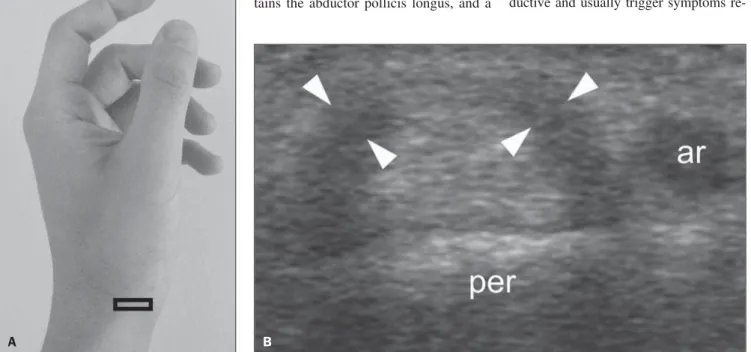
only one focal zone of the equipment ad-justed for the depth of the first extensor compartment. The investigation often re-quires generous amount of conductive gel on the skin surface in order to bring the region of interest into the focal zone. The transducer is placed transversely, adjacent to the radial tubercle, over the second tensor compartment. From the second ex-tensor compartment, the transducer is moved to transversely scan the first exten-sor compartment, adjacent to the radial sty-loid process. The extensor pollicis brevis (EPB) and abductor pollicis longus (APL) occupy the first extensor compartment. A useful tip to memorize the names of the extensor tendons passing over the dorsal wrist consists in recognizing that their sur-names alternate between longus and brevis as they advance towards the ulnar direc-tion from the first to the third compartment: abductor pollicis longus, extensor pollicis brevis, extensor carpi radialis longus, ex-tensor carpi radialis brevis and extensor pollicis longus. In the first compartment, the abductor pollicis longus is thicker than the extensor pollicis brevis, which should not be confused with disease. The synovial
sheath is depicted as a thin structure that surrounds the echogenic tendon fibers.
On ultrasound, the typical finding of tenosynovitis is the thickening and hypoechogenicity of the tendon sheath at the level of the radial styloid process (Fig-ure 2). We rely on the subjectivity rather than on a mathematical definition of sheath thickening, which reinforces the relevance of the experience of the examiner, particu-larly because tendon sheath usually mea-sures less than 1 mm thick. Contralateral comparison may be useful to identify subtle lesions. In chronic cases, bone erosion may be associated (Figure 3). More rarely, the tendon sheath has normal thickness, but there is fluid distension of the compartment caused by synovitis (Figure 4). The exami-nation does not end with the diagnosis, since predisposing factors such as vertical septum and accessory tendons must be ac-tively investigated as a cause of synovitis or secondary tenosynovitis. The role of these anatomical variations as risk factor for development of disease is intuitive, in the first case, with space reduction, and in the second case, with increase in the com-partment contents.
Vertical septum may be present in up to half of cases. It divides the first com-partment into a ventral larger area, that con-tains the abductor pollicis longus, and a
dorsal smaller space, that contains the ex-tensor pollicis brevis(9). Subcompartimen-talization and incomplete tenosynovitis is defined as vertical septum restricting the process to the sheath that surrounds only one of the tendons (Figure 5).
Accessory tendons of the abductor pol-licis longus are other recognized predis-posing factor for the development of teno-synovitis of the first extensor compartment. Its detection is rather intuitive, distally to the radial styloid process where the sheath thickening is not so noticeable (Figure 6).
FOLLOW-UP
Although the usefulness of ultrasonog-raphy in the diagnosis of tenosynovitis of the first extensor compartment is unqutionable, its role in the follow-up is not es-tablished. In the follow-up of more than 200 patients over the last ten years, we have never observed resolution of the tendon sheath thickening after conservative treat-ment, thus we do not recommend sono-graphic follow-up to record the disappear-ance of the process after symptoms remis-sion. Curiously, the authors’ personal ex-perience demonstrates that follow-up examination in patients who became as-ymptomatic after treatment are counterpro-ductive and usually trigger symptoms
re-Figure 2. Tenosynovitis of the first extensor compartment. A: Transducer positioning. B: Corresponding image demonstrating thickening and hypoechogenicity of the tendon sheath (arrowheads), at the level of the radial styloid process (per), adjacent to the radial artery (ar). The involvement is most prevalent in women at a (at least) 10:1 ratio. The particular predisposition of tenosynovitis to affect puerperal women is also well established, primarily by endocrine influence on water retention, although mechanical factors resulting from the newborn care activities cannot be neglected.
lapse as the patient is informed about the persistence of the anatomic injury (Figure 7). Sheath thickening is not an inflamma-tory or infectious process (like sinusitis and pneumonia), but rather a degenerative phe-nomenon (like osteoarthritis). Thus, the treatment is not aimed at correcting an al-ready-established anatomic injury, but
rather at improving symptoms and slow-ing the progression of the disease. The au-thors’ experience in the follow-up after surgical treatment is more positive, with some cases demonstrating complete sheath thickening regression (Figure 8). Synovi-tis is morphologically less refractory and fluid distension of the sheath usually dis-appears after conservative management.
CONCLUSION
Tenosynovitis of the first extensor com-partment of the wrist is a common condi-tion, with controversial terminology and poorly known physiopathology. In this
group of patients, ultrasonography can dif-ferentiate tenosynovitis from synovitis, the first one being an eminently inflammatory condition, and the later, predominantly degenerative. Additionally, ultrasonogra-phy can identify anatomical elements such as vertical septum and accessory tendons of the abductor pollicis longus, both po-tentially responsible for symptoms refrac-toriness, which may guide the adoption of a more aggressive management.
REFERENCES
1. de Quervain F. Ueber das Wesen und die Behand-lung des stenosierenden Tendovaginitis am Pro-cessus styloideus radii. Munch Med Wochenschr. 1912;59:5–6.
Figure 4. Synovitis of the first extensor compartment. Transverse scan dem-onstrating fluid distension of the tendon sheath (asterisks) which presents normal parietal thickness at the level of the radial styloid process. Also, note radial artery (ar), abductor pollicis longus (al), and extensor pollicis brevis (ec). Synovitis of the first extensor compartment is rare, but should be recog-nized, since its response to the conservative management tends to be more favorable, while its surgical indication is more controversial as compared with the classical presentation of tenosynovitis.
Figure 3. Tenosynovitis of the first extensor compartment in association with bone erosion. Transverse scan demonstrating thickening and hypoechogenicity of the tendon sheath (arrowheads) and a bone erosion (arrow) at the level of the radial styloid. Additionally, note the radial artery (ar).
Figure 5. Tenosynovitis of the first extensor compartment, incomplete form. A: Transducer positioning. B: Corresponding image, at the level of the radial styloid process (per), demonstrating the tenosynovitis (arrowhead) restricted to the extensor pollicis brevis (ec). Note the abductor pollicis longus (al) with normal appearance. The description of the incomplete form of tenosynovitis of the first extensor compartment is relevant for management because it restricts the diffusion of locally injected substances; and from the surgical point of view it requires the release of the septum in order to avoid refractory postoperative symptoms.
B
A B
Figure 7. Follow-up of tenosynovitis of the first extensor compartment in a patient submitted to conservative treatment. A: Baseline transverse scan demon-strating thickening and hypoechogenicity of the sheath of the first extensor compartment (arrowhead) at the level of the radial styloid process (per). Also, note the radial artery (ar). B: Follow-up image acquired from a patient responsive to conservative treatment. In this specific case, the patient was asymptomatic and satisfied with the treatment as he entered the examination room, but symptoms relapsed after being informed about the persistence of the anatomical findings.
Figure 8. Follow-up of tenosynovitis of the first extensor compartment in a patient submitted to surgical treatment. A: Baseline transverse scan demonstrating thickening and hypoechogenicity of the first compartment sheath (arrowhead) at the level of the radial styloid process (per). B: Follow-up image acquired one year after surgical management, documenting complete resolution of the anatomical injury. Note the normal appearance of the extensor pollicis brevis (ec) and the abductor pollicis longus (al) after tendon sheath release. Also, note the radial artery (ar).
A B
Figure 6. Tenosynovitis of the first extensor compartment with accessory tendons of the abductor pollicis longus. A: Transverse scan at the level of the radial styloid process (per), demonstrating tenosynovitis (arrowheads) affecting both the extensor pollicis brevis (ec) and the abductor pollicis longus (al), in an apparently trivial case. B: However, distal transverse evaluation demonstrates accessory tendons of the abductor pollicis longus. The identification of such anatomical variation that is seen in up to 75% of autopsies requires screening distally to the radial styloid process, where one can observe divergent tendons towards their insertion points. Also, note the radial artery (ar).
2. Gray H. Anatomy, descriptive and surgical. Phila-delphia, PA: Lea Brothers & Co.; 1893.
3. Clarke MT, Lyall HA, Grant JW, et al. The histo-pathology of de Quervain’s disease. J Hand Surg Br. 1998;23:732–4.
4. Read HS, Hooper G, Davie R. Histological appear-ances in post-partum de Quervain’s disease. J Hand Surg Br. 2000;25:70–2.
5. Finkelstein H. Stenosing tenovaginitis at the radial styloid process. J Bone Joint Surg. 1930;12:509– 40.
6. Eichoff E. Zur Pathogenese der Tendovaginitis stenosans. Bruns’ Beitrage Z Klin Chir. 1927; CXXXIX:746.
7. Arend CF. Master – Ultrassonografia musculoes-quelética. 2ª ed. Rio de Janeiro, RJ: Revinter; 2012.
8. Dawson C, Mudgal CS. Staged description of the Finkelstein test. J Hand Surg Am. 2010;35:1513– 5.