ABSTRACT
http://dx.doi.org/10.1590/1678-7757201302295
Marginal adaptation of class V composite
restorations submitted to thermal and mechanical
cycling
123, Luis Roberto Marcondes MARTINS
1- DDS, MSc, PhD, Department of Dentistry, Federal University of Ceará, Sobral, CE, Brazil. 2- DDS, MSc, PhD, Department of Dentistry, Federal University of Sergipe, Aracaju, SE, Brazil. 3- DDS, MSc, PhD, Private Clinic, Fortaleza, CE, Brazil.
4- DDS, MSc, PhD, Department of Restorative Dentistry, State University of Campinas, Piracicaba, SP, Brazil.
! André Luis Faria-e-Silva - Universidade Federal de Sergipe - Centro de Ciências Biológicas e da Saúde - Departamento de Odontologia Rua Cláudio Batista, s/n – Sanatório - 49060-100 - Aracaju - SE - Brazil -e-mail: [email protected]
"#!$%&'(&)"*!+4';&'(;""*!+4(&'(;
O
bjective: This study evaluated the effect of the margin location and an adhesive system on the marginal adaptation of composite restorations. Material and Methods: Class V cavities were prepared in bovine teeth with the gingival margin on the dentin and the incisal margin on the enamel. The cavities were restored with a micro-hybrid composite ! " #"$%"&"" The marginal adaptation was analyzed using scanning electronic microscopy (SEM, 500 $' ) "&' &)*! evaluation, the samples were submitted to thermal cycling (2,000 cycles of 5°C±2°C followed by 55°C±2°C – T1) and mechanical cycling (100,000 cycles of 50 kN and 2 Hz – T2). Replicas of samples were rebuilt after each cycling and analyzed under SEM. The data were submitted to Mann-Whitney, Wilcoxon and Friedman testing (α/**346)SB presented higher gaps in the dentin than the enamel, while there was no difference between the substrate for the CL. In the dentin, the CL showed better marginal sealing than )""')& 7& the baseline, thermal and mechanical cycling for any experimental condition. Conclusions: The outcomes of the present study showed that the adhesive system and margin location have an important effect on the marginal adaptation of composite restorations.
Key words: Dental restoration failure. Composite resins. Dentin-bonding-agents.
INTRODUCTION
Despite the improvements of restorative material in recent decades, the marginal integrity of restorations remains a challenge for dentistry. Poor marginal adaptation may produce marginal discoloration, postoperative sensibility, and secondary caries21. These are the most frequent reasons to replace or repair an adhesive restoration3,24. The marginal failure of composite resin restorations is related mainly to the quality of bonding to the dental structures2 and to stress generated on the restoration21.
Traditionally, the bonding to the dental tissue is obtained by etching the substrate using phosphoric
acid, followed by rinsing and applying an adhesive agent25. Later, simpler adhesives were introduced with the development of self-etching primers/ adhesives, eliminating the previous conditioning, rinsing, and drying steps that were critical for the adhesion protocol. However, it has been demonstrated that this simplification did not improve the bonding performance7,25.Moreover, the substrate where the adhesive was applied can ="' systems25,28.
intermolecular distance between the monomers and consequential shrinkage16.Bonding the composite resin to the cavity walls impairs the material deformation and generates shrinkage stress on the bonding interfaces18,26. If stress exceeds the bond strength between the dental substrate and the adhesive system, a contraction gap will be formed, jeopardizing the restoration’s longevity17,21.
In addition to stress shrinkage, the occlusal loads and alterations of the temperature of the oral behavior produce stress on the restoration and can also compromise the marginal sealing14,27. Clinical evaluations of restorations are very complicated because of ethical reasons, and they are time-consuming and expensive. In vitro studies
simulating oral conditions have been performed in order to permit an estimation of the restoration longevity. Thus, the aim of this study was to evaluate the effect of the substrate and adhesive system on the marginal integrity of composite restorations submitted to thermal and mechanical cycling. The null hypotheses were that the following have no effect on the marginal adaptation of composite restorations: (I) the localization of the restoration margin (dentin or enamel), (II) the adhesive system (etch-and-rinse or self-etching), and (III) thermal and mechanical cycling.
METHODOLOGY
One week after extraction, 40 sound bovine incisors were cleaned and examined under a light microscope (Eclipse E 600; Nikon, Shinagawa-ku, Tokyo, Japan) in order to exclude those with cracks. The teeth were stored in distilled water at 5°C for less than one month before the restorative procedure. Standard-shaped Class V cavities (3x3 mm, and 2 mm of depth) were prepared using a #169L carbide bur (KG Sorensen Ind. Com. Ltda. – Barueri, SP, Brazil) on the buccal surface. Each preparation was designed so that the incisal margin was in the enamel and the gingival margin was in the dentin. Within these dimensions, the C-factor [ratio between the bonded area (33 m2) and the
free surface (9 mm2)] of the cavity was 3.7. The cavities were prepared with a water-cooled high-speed turbine using a standard cavity preparation device. The turbine is attached to this device that permits the controlled movement of the bur on the x, y and z axes. A new bur was used for each of the ""
The cavities were restored using a two-step etch-and-rinse [Single Bond 2 (SB)], or a two-" /* ) # '" manufacturers of the adhesive systems used are described in Figure 1.
The cavities were randomly restored using one of the following adhesive protocols (SB or CL). For the SB groups, a 35% phosphoric acid gel (3M Scotchbond Etchant, 3M ESPE, St. Paul, MN, USA) was applied to the entire cavity for 15 sec. The acid was rinsed off with water for 15 sec and the excess water was removed with a small damp cotton pellet. The SB adhesive system was applied according to the manufacturer’s instructions to all cavity walls, which were checked for a shiny surface. The adhesive layer was thinned with a directed low-pressure air stream and light-cured for 20 sec. For the CE groups, the self-etching primer was applied to the cavities, left undisturbed for 20 sec and evaporated with an air-syringe. The adhesive was then applied, spread gently with an air-syringe and light-cured for 20 sec.
The cavities were restored with a micro-hybrid composite resin (Filtek Z-250, 3M ESPE, St. Paul, MN, USA), filled in one (bulk) increment of 2 mm and light-cured for 20 sec. The light-curing procedures were performed with LED Radii-Cal (SDI, Bayswater, Victoria, Australia) devices. The output of the light-curing unit was periodically checked using a handheld radiometer (Model 100, Demetron Kerr, Orange, CA) and was determined to be near 600 mW/cm2. All restored cavities were stored in distilled water at 37°C for 24 h and "&=$7''$\ Lex Pop-on®, 3M ESPE, St. Paul, MN, USA) under a water spray. The disks were used in descending
Adhesive $)"* Manufacturer ?*@
Single Bond 2 2-step,
etch-and-rinse
3M ESPE, St. Paul, MN, USA
Bis-GMA, HEMA, DUDMA, polyalkenoic acid copolymer, CQ, DHEPT, water, ethanol, silica
2-step, self-etch
Kuraray, Osaka, Japan
Primer: 10- MDP, HEMA, hydrophilic dimethacrylate, photo-initiator, water
Bonding agent: Bis-GMA, HEMA, 10-MDP, CQ, DHEPT, colloidal silica
Figure 1-
^ # ''# & 7%$
Impressions of restorations were taken using a polyvinyl siloxane impression material (Express, 3M ESPE, St. Paul, MN, USA) and replicas were done with epoxy resin (Epoxide, Buehler Ltd, Lake Bluff, IL). Replicas were mounted on aluminum stubs, gold sputter-coated (SCD 050, Baltec, Vaduz, Liechtenstein) and examined by scanning electron microscopy (SEM; JSM-5600LV, JEOL, Tokyo, Japan). The enamel and gingival margins were divided into 3 regions each for SEM analysis. The margins were analyzed under SEM at 500x ' )'$'' ' gap of each region was recorded (T0).
! # '" & submitted to the thermo-cycling procedure. The designed number of cycles for thermal stress
was 5000 cycles using a thermo-cycling machine (MCT2; Instrumentos de Precisão Ltda, São Paulo, SP, Brazil). Each cycle consisted of immersion of samples in water at 5±2°C followed by 55±2°C with a dwell time of 2 min for each bath. The transfer time between baths was 15 sec. New impressions were taken immediately after the thermo-cycling procedure and the replicas were evaluated under SEM (T1).
Afterward, the samples were placed into resin cylinders through their root portions. This procedure allowed the adaptation of the samples to a cyclic mechanical loading device (ERIOS Representações e Comércio Ltda, São Paulo, SP, Brazil). A vertical load of 50 kN was applied on the samples’ incisal edges. With a frequency of 2 Hz, 100000 cycles of loading were performed. The samples remained in distillated water at 37°C during the mechanical cycling. Replicas of the samples were rebuilt immediately after the mechanical cycling and analyzed under SEM (T2). All impressions were performed immediately following the cycling.
The Mann-Whitney test was used to compare the adhesive systems in each level of the substrate. The Wilcoxon test was used to analyze the factor location of margin in each level of the adhesive system. The repeated measures Friedman test was used to compare the time of evaluation in each $"')
Location of margins Adhesive Time of evaluation p-value
T0 T1 T2
Dentin SB 4.73 6.27 5.73 0.794
CL 0.00 1.53 2.73 0.197
Enamel SB 0.00 0.00 0.00 1.000
CL 1.07 0.27 1.13 0.066
Table 2- Medians of gap measurements (in μm) within each time of evaluation
Figure 2- Intact margin in enamel obtained with Single Bond 2 after thermo-cycling (T1). E – Enamel; C – Composite
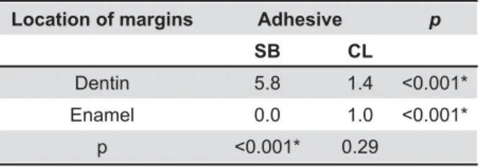
Figure 3- Photomicrography obtained at T0 showing the presence of gap in the dentinal margin of a restoration performed with Single Bond 2. D – dentin; C – Composite Location of margins Adhesive p
SB
Dentin 5.8 1.4 !"""#
Enamel 0.0 1.0 !"""#
p !"""# 0.29
Table 1- Medians of gap measurements (in μm)
all analyses was established at 5%.
RESULTS
The results of the gap measurements for the adhesive and substrate factors are displayed in Table 1. The SB presented higher values of gap widths than the CL when the margin of restoration was located in the dentin. The opposite was observed for the evaluation of margins in the enamel, where the SB showed a better marginal adaptation than the CL. The SB presented a better performance in the margins in the enamel than in the dentin. There was no difference between the locations of the margins for the gap measurement when the CL was used. The comparison between the times of evaluation is presented in Table 2. The widths of the gap measured in the baseline, after thermal and mechanical cycling were statistically similar for all experimental conditions. Illustrative micrographs obtained of the marginal integrity of restorations are shown in Figures 2 and 3.
DISCUSSION
A proper marginal sealing is essential to improve the longevity of composite resin restorations10,12,21. Class V cavities were chosen in this study because they remain a challenge for restorative procedures. Thus, most of the clinical studies evaluating the performance of an adhesive system use class V cavities. The C-factor of these cavities impairs the '"=& "%'^ shrinkage, increasing the stress over the boding interface10,23. Moreover, these cavities frequently present gingival margins in the dentin, consisting of an additional challenge to obtain a proper marginal sealing23. However, in the present study, differing from clinical situations, the cavities were & ' '" ) 7\ & ^ and to increase the effects of stress shrinkage and, consequently, the challenge over the bonding interfaces.
In composite restorations, stresses submitted on the restoration can disrupt the bonding and lead to the formation of gaps. Thus, a proper bond of an adhesive to the dental tissue contributes to avoid marginal microleakage7,14. In the present study, the ' = gap formation only for the SB, while this adhesive presented the best marginal adaptation to the enamel margins. Conversely, the CL presented similar behavior in both the margin in the enamel )# %" was partially accepted.
B o n d i n g t o e n a m e l i s p r e d i c t a b l e a n d stable because of this substrate’s high mineral
content25. In contrast with the enamel, dentin is a more heterogeneous substrate, consisting of %$%"# 7#&) conditioning of the dentin widens the opening of the dentinal tubules, exposes a layer of mineral depleted
7#&25.
The presence of organic content and water impairs proper bonding. Furthermore, the presence of solvents and hydrophilic components in the adhesive layer of the SB can additionally compromise the adhesive’s proper polymerization5,11, mainly in the presence of dentinal wetness, contributing to a reduction of the bonding performance9. These aspects can explain the inferior results of the SB when the margins were located in the dentin.
On the other hand, the CL presents a hydrophobic adhesive that is applied on the etched dentin by a self-etching primer. The absence of solvents and the more hydrophobic characteristic of this adhesive layer contribute to form a more homogeneous and stable bonding1,19. This explains the lowest gaps observed in the margin in the dentin when the CL was used, compared with the SB. The opposite was observed in the margins in the enamel. Thus, the second null hypothesis was rejected. The poorer performance of the CL on the enamel margins when compared to the SB is possibly related to the relatively low acidity of its self-etching primer. CL’s self-etching primer contains the acidic monomer 10-MDP and presents a pH level of approximately 2 (milder acid)15. Self-etching adhesives with relatively high pH levels are unable to produce ' & % the enamel13. In contrast, phosphoric acid used previously to the application of the SB is able to % ' )# ' 7 bonding to the dental substrate contributes to maintaining the margin sealing.
study. One possible explanation may reside in the water absorption from the samples during the cycling tests. Thus, the hygroscopic expansion of the composite can partially compensate a possible gap increase generated by the stresses27.
Laboratorial studies simulating clinical conditions are usually performed trying to predict the restoration behavior. The present study used thermal and mechanical cycling in order to promote stress on the restorations. Despite the absence of statistical differences between the moments of evaluation (before and after cycling), the outcomes of this study must be carefully evaluated. Clinically, there are other variables and different results can be observed. Furthermore, the current study used bovine teeth as a bonding substrate to evaluate the leakage of the adhesive restorations. The use of bovine teeth as a substitute for human teeth is a controversial matter. However, Reis, et al.20 (2004) analysed the bond strength and the enamel and dentinal morphology of possible substitutes for human teeth in bonding tests. The values of the bond strengths obtained with bovine and human teeth are similar, either for the enamel or the dentine. In addition, the morphology presented by these two substrates was also similar.
CONCLUSION
Within the limitations of the current study, the following conclusions can be drawn:
Single Bond 2 showed higher means of gaps in the dentin margins, while the location of margins = "'
"' 7 ' sealing than the Single Bond when the margins in the dentin were observed. In contrast, the Single Bond presented the best performance in the enamel margins.
The thermal and mechanical cycling utilized did not alter the gap measurements.
REFERENCES
1- Andrade e Silva SM, Carrilho MR, Marquezini L Junior, Garcia FC, Manso AP, Alves MC, et al. Effect of an additional hydrophilic versus hydrophobic coat in the quality of dentinal sealing provided by two-step etch-and-rinse adhesives. J Appl Oral Sci. 2009;17:184-9. 2- Atoui JA, Chinelatti MA, Palma-Dibb RG, Corona SA. Micro-leakage in conservative cavities varying the preparation method and surface treatment. J Appl Oral Sci. 2010;18:421-5. 3- Bernardo M, Luis H, Martin MD, Leroux BG, Rue T, Leitão J, et al. Survival and reasons for failure of amalgam versus composite posterior restorations placed in a randomized clinical trial. J Am Dent Assoc. 2007;138:775-83.
4- Borges AF, Santos JS, Ramos CM, Ishikiriama SK, Shinohara MS. Effect of thermo-mechanical load cycling on silorane-based composite restorations. Dent Mater J. 2012;31:1054-9.
5- Cadenaro M, Breschi L, Rueggeberg FA, Suchko M, Grodin E, Agee K, et al. Effects of residual ethanol on the rate and degree of $"'_`**{|36}~ 6- Cenci MS, Pereira-Cenci T, Donassollo TA, Sommer L, Strapasson !#_'='' % of restorative materials. J Appl Oral Sci. 2008;16:106-10. 7- De Munck J, Van Landuyt K, Peumans M, Poivitein A, Lambrechts P, Braem M, et al. A critical review of the durability of adhesion to tooth tissue: methods and results. J Dent Res. 2005;84:118-32. 8- Ehrenberg D, Weiner GI, Weiner S. Long-term effects of storage and thermal cycling on the marginal adaptation of provisional resin crowns: a pilot study. J Prosthet Dent. 2006;95:230-6.
9- El-Askary FM, Nassif MS, Andrade AM, Reis A, Loguercio AD. Effect of surface area and air-drying distance on shear bond strength of etch-and-rinse adhesive. Braz Oral Res. 2012;26:418-23.
10- Eliguzeloglu Dalkilic E, Omurlu H. Two-year clinical evaluation of three adhesive systems in non-carious cervical lesions. J Appl Oral Sci. 2012;20:192-9.
11- Faria-e-Silva AL, Lima AF, Moraes RR, Piva E, Martins LR. Degree of conversion of etch-and-rinse and self-etch adhesives light-cured using QTH or LED. Oper Dent. 2010;35:649-54. 12- Heintze SD. Systematic reviews: I. The correlation between laboratory tests on marginal quality and bond strength. II. The correlation between marginal quality and clinical outcome. J Adhes Dent. 2007;9:77-106.
13- Ibrahim IM, Elkassas DW, Yousry MM. Effect of EDTA and phosphoric acid pretreatment on the bonding effectiveness of self-etch adhesive to ground enamel. Eur J Dent. 2010;4:418-28. 14- Kenshima S, Grande RH, Singer JM, Ballester RY. Effect of '% \ '" restorations. J Appl Oral Sci. 2004;12:307-11.
15- Li N, Nikaido T, Takagaki T, Sadr A, Makishi P, Chen J, et al. The role of function in bonding to enamel: acid-base resistant zone and bonding performance. J Dent. 2010;38:722-30.
16- Nagem H Filho, Nagem HD, Francisconi PA, Franco EB, Mondelli RF, Coutinho KQ. Volumetric polymerization shrinkage of contemporary composite resins. J Appl Oral Sci. 2007;15:448-52. 17- Papadogiannis D, Kakaboura A, Palaghias G, Eliades G. Setting characteristics and cavity adaptation of low-shrinking resin composites. Dent Mater. 2009; 25:1509-16.
18- Pereira RA, Araujo PA, Castañeda-Espinosa JC, Mondelli RF. Comparative analysis of the shrinkage stress of composite resins. J Appl Oral Sci. 2008;16:30-4.
19- Reis A, Leite TM, Matte K, Michels R, Amaral RC, Geraldeli S, et al. Improving clinical retention of one-step self-etching adhesive system with an additional hydrophobic adhesive layer. J Am Dent Assoc. 2009;140:877-85.
20- Reis AF, Giannini M, Kavaguchi A, Soares CJ, Line SR. Comparison of micro-tensile bond strength to enamel and dentin of human, bovine, and porcelain teeth. J Adhes Dent. 2004;6:117-121.
21- Rodrigues SA Jr, Pin LF, Machado G, Della Bona A, Demarco FF. =' of class II composite restorations. J Appl Oral Sci. 2010;18:37-43. 22- Sampaio PC, Almeida AA Jr, Francisconi LF, Casas-Apayco LC, # #' glass-ionomer liner on dentin adhesive interface of Class I cavity walls after thermo-cycling. Oper Dent. 2011;36:403-12. 23- Santiago SL, Passos VF, Vieira AH, Navarro MF, Lauris JR, Franco EB. Two-year clinical performance of resinous restorative system in non-carious cervical lesions. Braz Dent J. 2010;21:229-34.
24- Sarrett DC. Prediction of clinical outcomes of a restoration based on in vivo marginal quality evaluation. J Adhes Dent. 2007;9:117-20.
26- Van Ende A, De Munck J, Mine A, Lambrechts P, Van Meerbeek B. Does a low-shrinking composite induce less stress at the adhesive interface? Dent Mater. 2010;26:215-22.
27- Verluis A, Tanbirojn D, Lee MS, Tu LS, Delong R. Can hygroscopic expansion compensate polymerization shrinkage? Part 1. Deformation of restored teeth. Dent Mater. 2011;27:126-33.
28- Villela-Rosa AC, Gonçalves M, Orsi IA, Miani PK. Shear bond strength of self-etch and total-etch bonding systems at different dentin depths. Braz Oral Res. 2011;25:109-15.