375
MRI in premature neonates
Radiol Bras. 2010 Nov/Dez;43(6):375–378 Original Article • Artigo Original
Reliability of qualitative assessment of brain magnetic
resonance imaging in extremely premature infants*
Confiabilidade da análise qualitativa da ressonância magnética do encéfalo em prematuros extremos
Andre Dietz Furtado1, Marcus Vinicius Rocha Pinto2, Cláudio de Carvalho Rangel3, Luiz Celso Hygino da Cruz Jr4, José Maria A. Lopes5, Manoel de Carvalho5, Jofre Antônio Oliveira Cabral5, Romeu Côrtes Domingues6, Emerson Leandro Gasparetto7
OBJECTIVE: The present study was aimed at evaluating the reliability of the qualitative visual assessment of brain abnormalities using conventional brain MRI in extremely preterm infants. MATERIALS AND METHODS: A cohort of 45 consecutive infants with gestational age of 30 weeks or less (median of 27 weeks, ranging from 25 to 30 weeks) was enrolled in this study. Two independent, experienced neuroradiologists blindly reviewed MRI studies of the infants’ brain for diffuse and excessive high-signal intensity (DEHSI), dilated lateral ventricles, intracranial hemorrhage, areas of abnormal signal in the basal ganglia and cortex, cyst-like areas, ventricular deformities, enlargement of subarachnoid spaces, early leukoencephalomalacia, and cortical abnormalities. RESULTS: Forty-one patients (91.1%) presented abnormalities at MRI. The most common findings were DEHSI in the white matter (75.6%) and ventricular dilatation (42.2%). The interobserver agreement was high (κκκκκ > 0.60) for most of the abnormal MRI findings. The kappa statistic values were moderate for enlargement of the subarachnoid space (κκκκκ = 0.52) and was only low for DEHSI in the white matter (κκκκκ = 0.39). CONCLUSION: Conventional MRI seems to be a reliable method for evaluating the most common brain abnormalities in extremely premature infants; however, the presence of DEHSI in the white matter demonstrated to be is a less reliable finding.
Keywords: Preterm; Brain; Magnetic resonance imaging; Imaging; Hypoxia.
OBJETIVO: O objetivo deste estudo foi avaliar a confiabilidade da análise visual qualitativa dos achados de imagem de ressonância magnética (RM) em recém-nascidos prematuros extremos. MATERIAIS E MÉTODOS: Uma coorte de 45 recém-nascidos de idade gestacional de 30 semanas ou menos foram inseridos neste estudo. Dois neurorradiologistas, cegos quanto aos dados clínicos, avaliaram de forma independente as RMs de crânio em relação aos seguintes achados: presença de hipersinal difuso e excessivo (DEHSI), dilatação dos ventrículos laterais, hemorragia intracraniana, áreas de sinal anormal em núcleos da base e córtex, áreas de aspecto cístico, deformidades ventriculares, dilatação do espaço subaracnóideo, leucoencefalomalácia precoce e anormalidades corticais. RESULTADOS: Quarenta e um pacientes (91,1%) apresentaram exame de RM anormal. Os achados mais comuns foram DEHSI (75,6%) e dilatação dos ventrículos (42,2%). A concordância interobservadores entre os dois experientes neurorradiologistas foi alta (κκκκκ > 0,60) para a maioria das alterações detectadas pela RM. O valor de kappa foi moderado (κκκκκ = 0,52) para alargamento do espaço subaracnoide e fraco (κκκκκ = 0,39) para DEHSI na substância branca. CONCLUSÃO: A avaliação qualitativa da maioria dos achados de imagem por RM de neonatos prematuros extremos foi considerada confiável, entre-tanto, a presença de DEHSI na substância branca demonstrou um grau de confiabilidade menor.
Unitermos: Prematuros; Cérebro; Imagem por ressonância magnética; Imagem; Hipóxia.
Abstract
Resumo
* Study developed at CDPI – Clínica de Diagnóstico Por Ima-gem, Rio de Janeiro, RJ, Brazil.
1. Clinical Fellow in Neuroradiology, Children’s Hospital of Pitts-burgh, PittsPitts-burgh, PA, USA.
2. Graduate Student of Medicine, Federal University of Rio de Janeiro (UFRJ), Rio de Janeiro, RJ, Brazil.
3. Radiologist, CDPI – Clínica de Diagnóstico Por Imagem and Clínica Multi-Imagem, Rio de Janeiro, RJ, Brazil.
4. Radiologist, CDPI – Clínica de Diagnóstico Por Imagem and Clínica Multi-Imagem, Fellow PhD degree, Federal University of Rio de Janeiro (UFRJ), Rio de Janeiro, RJ, Brazil.
5. Neonatologist, Clínica Perinatal Laranjeiras, Rio de Janeiro, RJ, Brazil.
INTRODUCTION
Over the last decades, there have been significant advances in perinatal and neo-natal care, which have dramatically im-proved survival rates for infants with very low birth weights. Nevertheless, there has been a increasing concern regarding the later neurodevelopmental challenges faced by surviving infants(1). The developing Furtado AD, Pinto MVR, Rangel CC, Cruz Jr LCH, Lopes JMA, Carvalho M, Cabral JAO, Domingues RC, Gasparetto EL. Reliability of qualitative assessment of brain magnetic resonance imaging in extremely premature infants. Radiol Bras. 2010; 43(6):375–378.
0100-3984 © Colégio Brasileiro de Radiologia e Diagnóstico por Imagem
6. Radiologist and Medical Director, CDPI – Clínica de Diag-nóstico Por Imagem and Clínica Multi-Imagem, Rio de Janeiro, RJ, Brazil.
7. Radiologist, CDPI – Clínica de Diagnóstico Por Imagem and Clínica Multi-Imagem, Associated Professor at Department of Radiology of Federal University of Rio de Janeiro (UFRJ), Rio de Janeiro, RJ, Brazil.
Mailing Address: Dr. Emerson L. Gasparetto. Avenida das Amé-ricas, 4666, sala 325, Barra da Tijuca. Rio de Janeiro, RJ, Brazil, 22640-102. Email: [email protected]
376
Furtado AD et al.
Radiol Bras. 2010 Nov/Dez;43(6):375–378 brain is highly vulnerable to injury from a
variety of ischemic, inflammatory, infec-tive, and neurotoxic factors(2). Extremely preterm infants are at higher risk of brain hemorrhage, white matter (WM) lesions, and poor cerebral development(3). The cen-tral nervous system (CNS) damage in-creases the probability of neurological and developmental disabilities in this group of patients(4). Conventional neuroimaging is usually employed to assess the presence and the extent of brain injury as well as to predict neurodevelopmental outcomes.
The two major neuroimaging modali-ties used in the evaluation of the premature infant’s brain are cranial ultrasonography (US) and magnetic resonance imaging (MRI)(5–8). Cranial US is reliable in the evaluation of hemorrhagic lesions, hydro-cephalus, and cystic changes(5,6). Such tech-nique, however, is typically obtained through the anterior fontanel, which has a limited field of view. Moreover, cranial US is not accurate enough to evaluate diffuse or subtle brain injuries, specially in the WM(5–8). MRI of the brain is more sensi-tive and specific than cranial US to detect hemorrhage, ischemia, and WM lesions(5, 6,9–11). It also provides better
characteriza-tion of the most common brain abnormali-ties in preterm infants(5,6,12).
The aim of the present study was to evaluate the reliability of the qualitative visual assessment of brain abnormalities using conventional brain MRI in a cohort of 45 extremely preterm infants.
MATERIALS AND METHODS
Patient population
The Institutional Review Board of our Hospital approved the study and term of free and informed consent to perform MRI under sedation at term-equivalent age was obtained from the parents. A cohort of 45 consecutive infants with gestational age of 30 weeks or less (median of 27 weeks, ranging from 25 to 30 weeks) was enrolled in this prospective study. The gestational age was calculated from the date of the last menstrual period and confirmed with data from early US scans. The patients had a median birth weight of 890 g (ranging from 385 g to 1225 g). Critically ill patients were not considered for the study, at least until
the neonatologist in charge of these patients confirmed that they could be submitted to MRI study at our imaging center affiliated to the hospital with no risk for the patient.
Imaging protocol
All the patients underwent MRI in a 1.5 T scanner (Magnetom Avanto – Siemens Medical Systems; Erlangen, Germany) us-ing a head coil. The followus-ing sequences were obtained: T1-weighted sagittal three-dimensional gradient-echo (repetition time (TR)/echo time (TE) = 1770/3.9 ms, field of view (FOV) = 190 × 190 mm, matrix = 256 × 256, slice thickness = 0.7 mm), T2-weighted axial fast spin-echo (FSE) (TR/ TE = 5610/159 ms, FOV = 180 × 180 mm, matrix = 256 × 256, slice thickness = 4 mm), and T2-weighted axial gradient-echo (TR/TE = 786/35 ms, FOV = 180 × 180 mm, matrix = 256 × 256, slice thickness = 4 mm, flip angle = 30°).
Imaging analysis
All the MRI studies were independently and blindly reviewed by two neuroradi-ologists (five and six years of experience). In cases of disagreement, a third neuroradi-ologist reviewed the images and final de-cisions were defined by consensus.
The following MRI findings were evaluated: 1) diffuse and excessive high-signal intensity (DEHSI), defined as areas of abnormal, diffuse high-signal on T2-weighted FSE images within the periven-tricular and/or subcortical WM (signal in-tensity similar to the CSF)(7); 2) dilated lat-eral ventricles, if the transverse ventricular diameter was >10 mm, measured at the level of the atria(13–15), presence of intrac-ranial hemorrhage defined as abnormal areas with signal characteristics compat-ible with blood products, classified accord-ing to its location in 3) intraparenchymal hemorrhage (IPH), 4) intraventricular hem-orrhage (IVH), and 5) germinal matrix hemorrhage (GMH); 6) areas of abnormal signal in the basal ganglia and cortex; 7) cystic-like areas; 8) ventricular deformities; 9) enlargement of subarachnoid spaces overlying the cortical convexities (> 3 mm); 10) enlargement of the interhemispheric fissure (> 3 mm); 11) early foci of leuko-encephalomalacia; 12) and gyri abnormali-ties(7).
Statistical analysis
Interobserver agreement in the MR im-ages analysis was assessed with calculation of the kappa (κ) index, and the following ranges for agreement were used: 0.00, poor; 0.00 to 0.20, slight; 0.21 to 0.40, fair; 0.41 to 0.6, moderate; 0.61 to 0.8, substan-tial; and 0.81 to 1.0, almost perfect(16). The p value of less than 0.05 was considered as statistically significant.
RESULTS
Prevalence of brain MRI abnormalities in extreme preterm infants
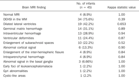
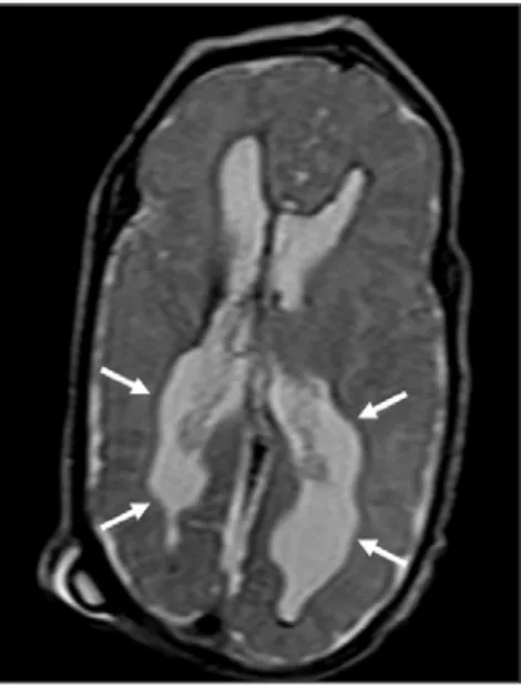
Among the 45 studied cases, only four patients (8.9%) had normal MR images (Table 1). The remaining 41 patients (91.1%) presented abnormalities on the MRI. The most common findings were DEHSI in the WM (75.6%) (Figure 1), lat-eral ventricles dilatation (42.2%) (Figure 2), GMH (31.1%) (Figure 3), IVH (28.9%) (Figure 4), ventricular deformities (24.4%) (Figure 5), and enlargement of the sub-arachnoid spaces (22.2%).
Reliability of qualitative visual assessment of conventional brain MRI
All the κ values obtained in the analy-sis of the interobserver variability were sta-tistically significant. The interobserver agreement was high (κ > 0.60) for most of the abnormal MRI findings (Table 1). The
κ statistic value was moderate for enlarge-ment of the subarachnoid spaces overlying the cortical convexities (κ = 0.52) and was only fair for DEHSI in the WM (κ = 0.39).
DISCUSSION
377
MRI in premature neonates
Radiol Bras. 2010 Nov/Dez;43(6):375–378
ventional brain MRI performed at term-equivalent age in a cohort of 45 extremely preterm infants. The interobserver agree-ment was high (κ > 0.60) for most of the MRI findings, with the exception of the DEHSI in the WM. In our study, we ob-served a relatively low κ statistic value for DEHSI in the WM (κ = 0.39). Even though previous papers have defined the MR sig-nal characteristics of DEHSI in the WM, it is still highly dependent on the radiologist’s experience(1,7,22,23). A thorough knowledge of the normal patterns of myelinization is essential when considering the possibility of DEHSI in the WM. Additionally, in some cases, CNS lesions were very severe and occurred early in the fetal develop-ment, making the evaluation of normal myelinization patterns even more difficult. This finding of relatively low interobserver agreement for WM DEHSI in our study may have relevant clinical implications because WM abnormalities have been re-ported as the main brain MRI abnormality related to the long- and short-term progno-sis of extremely preterm infants; and clini-cal therapeutic decisions are frequently based on whether WM abnormalities are present or not(5,20).
We acknowledge some limitations to our study. Critically ill patients were not considered for the study to avoid additional risk for them. MRI was performed at the term-equivalent age, but not at birth, which
Table 1 Brain MRI findings at term-equivalent age in 45 extremely preterm infants.
Brain MRI finding
Normal MRI DEHSI in the WM
Dilated lateral ventricles Germinal matrix hemorrhage
Intraventricular hemorrhage Ventricular deformities
Enlargement of subarachnoid spaces Abnormal cortical signal
Enlargement of the inter-hemispheric fissure Intraparenchymal hemorrhage
Abnormal signal in the basal ganglia Early foci of leukoencephalomalacia
Gyri abnormalities Cystic-like areas
No. of infants (n = 45)
4 (8.9%) 34 (75.6%)
19 (42.2%) 14 (31.1%)
13 (28.9%) 11 (24.4%)
10 (22.2%) 6 (13.3%)
4 (8.9%) 4 (8.9%)
3 (6.66%) 1 (2.2%)
1 (2.2%) 1 (2.2%
Kappa statistic value
1.00 0.39
0.653 0.89
0.94 0.87
0.52 0.90
0.84 0.84
1.00 1.00
1.00 1.00
MRI, magnetic resonance imaging; DEHSI, diffuse and excessive high signal intensity; WM, white matter.
with 45 extremely preterm infants is simi-lar to previous studies(7,22).
Cranial US is less sensitive to demon-strate most of these WM abnormalities. Moreover, the significance of the US find-ing of WM echogenicity is controver-sial(1,5–9,20,21). Inder et al.(21) have compared serial cranial US and brain MRI at term in a cohort of 96 extremely preterm infants. These authors emphasized the significant limitations of US for the detection of non-cystic WM injuries. Additionally, cranial US failed to demonstrate subtle WM inju-ries between birth and term in a group of 32 preterm infants as compared with brain
MRI(5). In conclusion, US seems to have poorer sensitivity and specificity for the evaluation of WM abnormalities in ex-tremely preterm infants as compared with MR imaging, and does not correlate with the clinical outcome(5,21). Brain MRI is, thus, considered as the main imaging mo-dality to predict neurodevelopment out-come in extremely preterm infants. It has been suggested that the severity of conven-tional MRI abnormalities is directly related to adverse long-term neurodevelopmental outcomes(20).
In our study, we assessed the reliability of the subjective visual assessment of
con-Figure 1. Coronal T2WI (5610/159) image dem-onstrates diffuse and excessive high-signal inten-sity, evidenced as areas of abnormal high-signal intensity within the periventricular white matter.
Figure 2. Axial T2WI (5610/159) image demon-strates lateral ventricles dilation measured at the level of ventricular atria (> 10 mm) (*).
378
Furtado AD et al.
Radiol Bras. 2010 Nov/Dez;43(6):375–378 is similar to other studies(6,7,12,20–23). Finally,
as the aim of our study was to assess the reliability of brain MRI abnormalities, we did not correlate the MRI findings with the long-term outcome, which has already been studied(20).
In conclusion, conventional MRI is usu-ally employed to assess brain abnormalities in extremely preterm infants. The most com-mon brain MRI findings at term-equivalent age in extremely preterm infants were DEHSI in the WM, ventricular dilation, GMH, and IVH, which have been associated with adverse neurodevelopmental outcome. According to the present study, conventional MRI seems to be a reliable method for evalu-ating the most common brain abnormalities in extremely premature infants; however, it seems that the presence of DEHSI in the WM is a less reliable finding.
REFERENCES
1. Inder TE, Wells SJ, Mogridge NB, et al. Defin-ing the nature of the cerebral abnormalities in the premature infant: a qualitative magnetic reso-nance imaging study. J Pediatr. 2003;143:171–9. 2. Kuban KC, Leviton A. Cerebral palsy. N Engl J
Med. 1994;330:188–95.
3. Vohr BR, Allen M. Extreme prematurity – the continuing dilemma. N Engl J Med. 2005;352: 71–2.
4. Wood NS, Marlow N, Costeloe K, et al. Neuro-logic and developmental disability after
ex-tremely preterm birth. EPICure Study Group. N Engl J Med. 2000;343:378–84.
5. Maalouf EF, Duggan PJ, Counsell SJ, et al. Com-parison of findings on cranial ultrasound and magnetic resonance imaging in preterm infants. Pediatrics. 2001;107:719–27.
6. Inder TE, Warfield SK, Wang H, et al. Abnormal cerebral structure is present at term in premature infants. Pediatrics. 2005;115:286–94. 7. Maalouf EF, Duggan PJ, Rutherford MA, et al.
Magnetic resonance imaging of the brain in a cohort of extremely preterm infants. J Pediatr. 1999;135:351–7.
8. Hope PL, Gould SJ, Howard S, et al. Precision of ultrasound diagnosis of pathologically verified lesions in the brains of very preterm infants. Dev Med Child Neurol. 1988;30:457–71.
9. Blankenberg FG, Norbash AM, Lane B, et al. Neonatal intracranial ischemia and hemorrhage: diagnosis with US, CT, and MR imaging. Radi-ology. 1996;199:253–9.
10. Blankenberg FG, Loh NN, Bracci P, et al. Sonography, CT, and MR imaging: a prospective comparison of neonates with suspected intracra-nial ischemia and hemorrhage. AJNR Am J Neuroradiol. 2000;21:213–8.
11. Barkovich AJ, Westmark K, Partridge C, et al. Perinatal asphyxia: MR findings in the first 10 days. AJNR Am J Neuroradiol. 1995;16:427–38. 12. Battin MR, Maalouf EF, Counsell SJ, et al. Mag-netic resonance imaging of the brain in very preterm infants: visualization of the germinal matrix, early myelination, and cortical folding. Pediatrics. 1998;101:957–62.
13. Farrell TA, Hertzberg BS, Kliewer MA, et al. Fetal lateral ventricles: reassessment of normal values for atrial diameter at US. Radiology. 1994;193: 409–11.
14. McArdle CB, Richardson CJ, Nicholas DA, et al. Developmental features of the neonatal brain: MR imaging. Part I. Gray-white matter differentiation and myelination. Radiology. 1987;162(1 Pt 1): 223–9.
15. McArdle CB, Richardson CJ, Nicholas DA, et al. Developmental features of the neonatal brain: MR imaging. Part II. Ventricular size and extracere-bral space. Radiology. 1987;162(1 Pt 1):230–4. 16. Landis JR, Koch GG. The measurement of ob-server agreement for categorical data. Biometrics. 1977;33:159–74.
17. Saigal S, Feeny D, Rosenbaum P, et al. Self-per-ceived health status and health-related quality of life of extremely low-birth-weight infants at ado-lescence. JAMA. 1996;276:453–9.
18. Hack M, Flannery DJ, Schluchter M, et al. Out-comes in young adulthood for very-low-birth-weight infants. N Engl J Med. 2002;346:149–57. 19. Marlow N, Wolke D, Bracewell MA, et al. Neu-rologic and developmental disability at six years of age after extremely preterm birth. N Engl J Med. 2005;352:9–19.
20. Woodward LJ, Anderson PJ, Austin NC, et al. Neonatal MRI to predict neurodevelopmental outcomes in preterm infants. N Engl J Med. 2006; 355:685–94.
21. Inder TE, Anderson NJ, Spencer C, et al. White matter injury in the premature infant: a compari-son between serial cranial compari-sonographic and MR findings at term. AJNR Am J Neuroradiol. 2003; 24:805–9.
22. Arthur R. Magnetic resonance imaging in preterm infants. Pediatr Radiol. 2006;36:593–607.
23. Valkama AM, Pääkkö EL, Vainionpää LK, et al. Magnetic resonance imaging at term and neuro-motor outcome in preterm infants. Acta Paediatr. 2000;89:348–55.
Figure 5. Axial T2WI (5610/159) image demon-strates ventricular deformities evidenced as irregular ventricular walls (arrows).
Figure 4. (A) Axial T2WI (5610/159) and (B) axial gradient-echo image (1770/3.9; flip angle = 30°) demonstrate low-signal intensity material layering in the occipital horn of the left lateral ventricle (arrows). Note marked blooming of the susceptibility effect from blood products on the gradient-echo image.