Dement Neuropsychol 2016 September;10(3):185-197
185
Holanda Júnior and Almondes Sleep and executive functions
Original Article
Sleep and executive functions in older adults
A systematic review
Francisco Wilson Nogueira Holanda Júnior1, Katie Moraes de Almondes2
ABSTRACT. Introduction: A recent increase in studies suggests a role of age-related sleep changes in executive functions (EF). However, this relationship remains unclear and mixed results have emerged. Objective: To investigate how age-related sleep changes may play an important role in the extent to which healthy older adults exhibit decline in
EF. Methods: A systematic strategy was employed to identify the available literature on age-related sleep changes and
EF. Results: Of the 465 studies identified, 26 were included. Results suggest that multiple sleep parameters differ in the way they benefit or impair EF. Parameters such as greater wake after sleep onset and lower sleep efficiency, in addition to circadian fragmentation of sleep, showed more consistent results and are potentially correlated with worsening in EF measures. However, other results seem inconclusive. Conclusion: These findings were discussed based on the prefrontal circuitry vulnerability model, in which sleep has been identified as a beneficial factor for prefrontal cortex functioning and hence for EF, which relies mostly on this brain area and its related networks.
Key words: executive functions, sleep, older adults, prefrontal cortex.
SONO E FUNÇÕES EXECUTIVAS EM IDOSOS: REVISÃO SISTEMÁTICA
RESUMO. Introdução: Um aumento recente de estudos sugere um papel das mudanças do sono relacionadas ao envelhecimento nas funções executivas (FE). Entretanto, essa relação ainda não é clara e resultados mistos têm emergido.
Objetivo: Investigar como as mudanças do sono relacionadas à idade podem desempenhar um papel importante na
medida em que idosos saudáveis apresentam um declínio nas FE. Métodos: Uma estratégia de revisão sistemática foi empregada para identificar a literatura disponível sobre mudanças no sono relacionadas ao envelhecimento e FE.
Resultados: Dos 465 estudos identificados, 26 foram incluídos. Os resultados sugerem que múltiplos parâmetros do
sono diferem na forma como beneficiam ou comprometem as FE. Parâmetros como maiores despertares após o início do sono e baixa eficiência do sono, além da fragmentação circadiana do sono, mostraram resultados mais consistentes e potencialmente se correlacionaram com a piora nas medidas de FE. Contudo, outros resultados parecem inconclusivos.
Conclusão: Esses achados foram discutidos baseadamente no modelo de vulnerabilidade pré-frontal, no qual o sono
tem sido apontado como um fator benéfico para o funcionamento do córtex pré-frontal e, consequentemente, para as FE que têm como substrato principal essa área cerebral e regiões relacionadas.
Palavras-chave: funções executivas, sono, idoso, córtex pré-frontal.
INTRODUCTION
T
he process of normal aging afects the sleep-wake regulatory system in ultra-dian, circadian and homeostatic levels. Changes in ultradian rhythm of sleep and sleep parameters include decreases in slow wave sleep (SWS, stage N3), which appear to begin as early as midlife, as well as decrease in total sleep time (TST) and increased wakeafter sleep onset (WASO), the latter resulting in poor sleep eiciency.1-3 he percentage of SWS linearly decreases at a rate of approxi-mately 2 per cent per decade up to 60 years and then stabilizes through the mid-90s.4,5
Older adults spend an increasing percent-age of sleep in stpercent-ages N1 and N2, resulting in less restorative sleep.6 Crowley, Trinder, Kim, Carrington & Colrain found that sleep
This study was conducted at the Postgraduate Program in Psychology, Federal University of Rio Grande do Norte, Natal RN, Brazil.
1Master’s Student on the Postgraduate Program in Psychology, Federal University of Rio Grande do Norte. 2Associate Professor at the Department of Psychology and
on the Postgraduate Program in Psychology, Federal University of Rio Grande do Norte, Natal RN, Brazil.
Francisco Wilson Nogueira Holanda Júnior. Postgraduate Program in Psychology / Federal University of Rio Grande do Norte / Lagoa Nova – 59078-970 Mailbox 1622 – Natal RN – Brazil. E-mail: [email protected]
Disclosure: The authors report no conflicts of interest.
Received July 11, 2016. Accepted in final form August 16, 2016.
Dement Neuropsychol 2016 September;10(3):185-197
186 Sleep and executive functions Holanda Júnior and Almondes spindle number, density and duration, as well as K-com-plex number and density, are lower in younger than in older adults.7 A slight decrease in percentage of rapid eye movement (REM) sleep may occur, decaying by less than 1% per decade or may remain relatively unchanged.8
Regarding circadian organization of sleep in elderly, there is commonly a phase advance in the circadian sleep cycle: a propensity toward an earlier sleep onset, together with an earlier morning wake pattern.9 In other words, older people become sleepy in the evening and tend to sleep quite early, hence waking up earlier in the morning.10 his advance in sleep phase is complicated by social pressure to stay up later in the evening, despite their altered internal rhythm.
here are also changes in homeostatic organization of sleep: it has been hypothesized that in the elderly, homeostatic sleep pressure undergoes a decrease and may be a cause of reduced TST, SWS and sleep ei-ciency.11,12 his theory is evidenced by an increased amount of nocturnal awakenings and diminished day-time sleepiness in elderly compared to young adults.13 Despite this reduced daytime sleepiness, the coexistence of a high frequency of daytime napping seems paradoxi-cal. Up to 60% of older adults takes a nap,14 most likely resulting from lifestyle factors, such as opportunities to nap due to schedule lexibility as a result of retirement, maladaptive habits or even medication side efects. Although considered non-pathological, the ultradian, circadian and homeostatic age-related changes men-tioned above may lead to complaints in the elderly and, when combined with lifestyle factors, lead to poor sleep quality or sleep deprivation.
Healthy aging also involves changes in cognitive functioning. hereby a certain amount of cognitive decline is a normal part of aging, varying considerably across individuals and cognitive domains, with some cognitive functions appearing more susceptible than others to the efects of aging.15 Declines in cognition tend to be most marked in executive functions (EF).16,17 EF is an umbrella term denoting a branch of top-down processes involved in the coordination and control of goal-directed behavior.18-20 Diamond21 describes three core executive functions: (1) inhibitory control (the ability to restrain one’s habitual responses to override strong internal predispositions or external draw); (2) working memory (the ability to hold information in mind to support the completion of tasks); and (3) cog-nitive lexibility (the ability to ponder multiple sources and forms of information at one time and adaptively switch between them when engaging in a task). From these core functions, other EF functions are derived, such as planning, reasoning, problem-solving. However,
other EF are also considered in other models, such as the ability to sustain attention, resistance to interfer-ence, utilization of feedback, multitasking, and verbal luency.19 Adequate executive functioning is necessary for selecting and monitoring actions that facilitate the fulillment of chosen goals and supports older adults to perform complex activities in everyday life.22
Changes in executive functioning related to healthy aging can be explained at least partially by structural and functional changes in the central nervous system. Although the loss of neurons in the elderly is moder-ate,23 studies have shown that loss of neuronal and den-dritic architecture, rather than loss of neurons, underlies neocortical volume loss with increasing healthy age.24 Changes in frontal-striatal circuits are the most likely signiicant cause of reduced EF in non-mild cognitive impairment (MCI) and nondemented older adults.25 Frontal-striatal systems are preferentially vulnerable to white matter change, atrophy, and certain forms of neurotransmitter depletion.25-27 Frontal-striatal and frontoparietal circuitry play an important role in execu-tive functioning.28,29
Given that both sleep components and EF undergo alterations in healthy aging, associations between these factors have been considered, as well as the impact of sleep changes (i.e. ultradian, circadian and homeostatic) in prefrontal circuits that underlie EF. Horne & Harri-son irst hypothesized that the waking function of the prefrontal cortex (PFC) and the frontal predominance of EEG delta activity in sleep may be linked.30-32 Accord-ing to this model and more recent indAccord-ings, sleep loss selectively afects the eiciency of PFC circuitry, produc-ing changes in brain metabolism and afectproduc-ing executive functioning.33-35 Since then, some studies have argued that cognitive functioning related to the PFC is particu-larly vulnerable to sleep loss or sleep deprivation.36,37
Assuming the prefrontal circuit vulnerability to sleep loss hypothesis and the parallel decreases in sleep and EF in older adults, the present article provides a sys-tematic review of evidence of interaction between these two functions in healthy aging, that is, a growing body of literature suggesting that changes in sleep may play an important role in the extent to which healthy older adults exhibit decreases in executive functioning.
METHOD
Dement Neuropsychol 2016 September;10(3):185-197
187
Holanda Júnior and Almondes Sleep and executive functions terms were systematically applied across these three
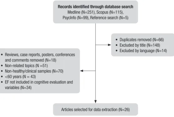
databases. he numbers of relevant hits from these databases are summarized in Figure 1. he retrieved articles were referenced in EndNote (Version 17.0) and duplicates removed. his search strategy was augmented with hand searches of reference lists of included studies. Searches were conducted on 10th May 2016.
Study selection. Inclusion and exclusion criteria were outlined prior to the search. Inclusion criteria included: 1) articles published in the last 25 years (1990-2016); 2) sample comprising healthy older adults (60 years or older, without dementia, mild cognitive impairment, sleep disorders or loss of functionality); 3) empirical research; and 4) focus on sleep variables (e.g. ultra-dian, circadian and homeostatic), healthy aging and cognitive functioning, addressing EF or at least one EF domain evaluated. he exclusion criteria established were: 1) case studies, letters to the editor, and confer-ence abstracts; 2) non-human experimental model studies; 3) non-healthy/clinical samples of older adults, such as diagnosed with sleep disorders, dementia and cognitive impairment; 4) EF not included in cognitive evaluation and variables; 5) non-English language; and 6) review articles.
he 465 articles initially retrieved from the preced-ing detailed searches were screened by their titles and abstracts, and articles not meeting the inclusion and/or exclusion criteria described earlier were removed. his gave a total of 26 articles eligible for review.
RESULTS
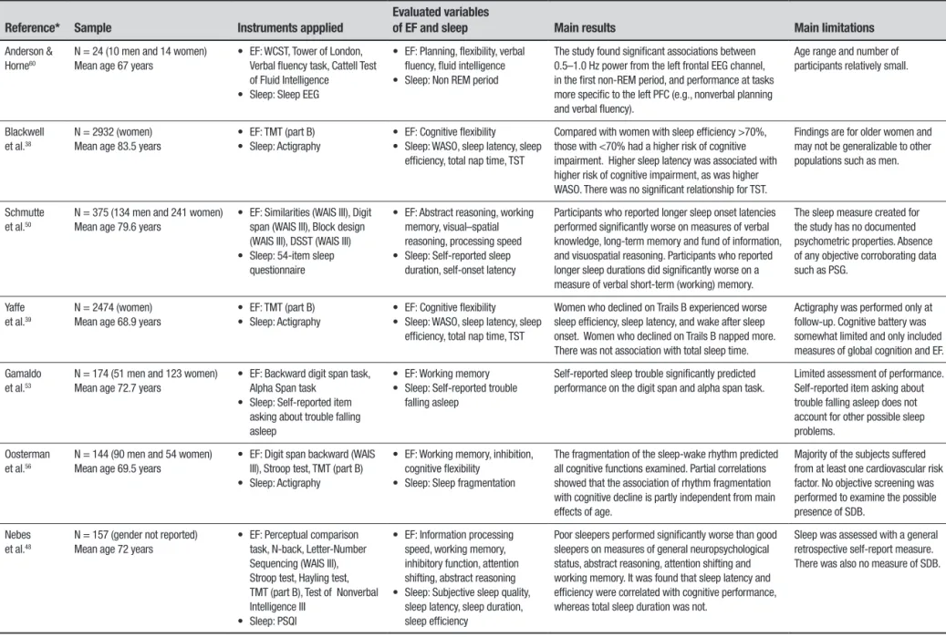
We included 26 articles in the review. A summary and analysis of these articles are given in Table 1. In general, the studies varied widely in their research design, partic-ipants and sleep and EF variables. A large portion of the studies were conducted by researchers in the United States (n=16). his was followed by the United Kingdom (n=2), France (n=2), the Netherlands (n=2), Canada (n=1), Ireland (n=1), Japan (n=1), and Switzerland (n=1). In relation to study populations, all the articles in this review were based on non-clinical elderly samples, most of them including home/community-dwelling subjects. he main sleep variables included in articles were WASO, sleep latency, sleep eiciency, TST, sleep quality and other less frequent variables such as sleep deprivation and sleep stages. Sleep circadian and homeo-static inluences were also included in some studies.
A total of 10 studies (n=9094) evaluated sleep using objective measures only. Seven studies (n=6383) were based on objective and subjective measures. A total of 9 studies (n=2261) evaluated sleep using subjective measures only. Actigraphy was the most used objective measure by studies (n=10), followed by polysomnogra-phy (PSG) (n=4) and other sleep detection devices (n=3). he most common subjective measure was the Pitts-burgh Sleep Quality Index (PSQI). Others included the Epworth sleepiness scale (ESS) (n=4), sleep diary (n=4) and other sleep questionnaires (n=3). A large variety of tests and instruments were used to assess EF. he Trail Making Test (TMT) was used in more than half of the
• Duplicates removed (N=66) • Excluded by title (N=148) • Excluded by language (N=14) • Reviews, case reports, posters, conferences
and comments removed (N=18) • Non-related topics (N =51) • Non-healthy/clinical samples (N=70) • <60 years (N = 43)
• EF not included in cognitive evaluation and variables (N=34)
Articles selected for data extraction (N=26) Records identified through database search
Medline (N=251), Scopus (N=115), PsycInfo (N=99), Reference search (N=5)
Dement Neuropsyc
hol 2016 September;10(3):185-197
188
Sleep and e
xecutive functions
Holanda Júnior and
Almondes
Table 1. Overview of studies assessing the impact and the relation of sleep and executive functioning.
Reference* Sample Instruments appplied
Evaluated variables
of EF and sleep Main results Main limitations
Anderson & Horne60
N = 24 (10 men and 14 women) Mean age 67 years
• EF: WCST, Tower of London, Verbal fluency task, Cattell Test of Fluid Intelligence
• Sleep: Sleep EEG
• EF: Planning, flexibility, verbal fluency, fluid intelligence • Sleep: Non REM period
The study found significant associations between 0.5–1.0 Hz power from the left frontal EEG channel, in the first non-REM period, and performance at tasks more specific to the left PFC (e.g., nonverbal planning and verbal fluency).
Age range and number of participants relatively small.
Blackwell et al.38
N = 2932 (women) Mean age 83.5 years
• EF: TMT (part B) • Sleep: Actigraphy
• EF: Cognitive flexibility • Sleep: WASO, sleep latency, sleep
efficiency, total nap time, TST
Compared with women with sleep efficiency >70%, those with <70% had a higher risk of cognitive impairment. Higher sleep latency was associated with higher risk of cognitive impairment, as was higher WASO. There was no significant relationship for TST.
Findings are for older women and may not be generalizable to other populations such as men.
Schmutte et al.50
N = 375 (134 men and 241 women) Mean age 79.6 years
• EF: Similarities (WAIS III), Digit span (WAIS III), Block design (WAIS III), DSST (WAIS III) • Sleep: 54-item sleep
questionnaire
• EF: Abstract reasoning, working memory, visual–spatial reasoning, processing speed • Sleep: Self-reported sleep
duration, self-onset latency
Participants who reported longer sleep onset latencies performed significantly worse on measures of verbal knowledge, long-term memory and fund of information, and visuospatial reasoning. Participants who reported longer sleep durations did significantly worse on a measure of verbal short-term (working) memory.
The sleep measure created for the study has no documented psychometric properties. Absence of any objective corroborating data such as PSG.
Yaffe et al.39
N = 2474 (women) Mean age 68.9 years
• EF: TMT (part B) • Sleep: Actigraphy
• EF: Cognitive flexibility • Sleep: WASO, sleep latency, sleep
efficiency, total nap time, TST
Women who declined on Trails B experienced worse sleep efficiency, sleep latency, and wake after sleep onset. Women who declined on Trails B napped more. There was not association with total sleep time.
Actigraphy was performed only at follow-up. Cognitive battery was somewhat limited and only included measures of global cognition and EF.
Gamaldo et al.53
N = 174 (51 men and 123 women) Mean age 72.7 years
• EF: Backward digit span task, Alpha Span task
• Sleep: Self-reported item asking about trouble falling asleep
• EF: Working memory • Sleep: Self-reported trouble
falling asleep
Self-reported sleep trouble significantly predicted performance on the digit span and alpha span task.
Limited assessment of performance. Self-reported item asking about trouble falling asleep does not account for other possible sleep problems.
Oosterman et al.56
N = 144 (90 men and 54 women) Mean age 69.5 years
• EF: Digit span backward (WAIS III), Stroop test, TMT (part B) • Sleep: Actigraphy
• EF: Working memory, inhibition, cognitive flexibility
• Sleep: Sleep fragmentation
The fragmentation of the sleep-wake rhythm predicted all cognitive functions examined. Partial correlations showed that the association of rhythm fragmentation with cognitive decline is partly independent from main effects of age.
Majority of the subjects suffered from at least one cardiovascular risk factor. No objective screening was performed to examine the possible presence of SDB.
Nebes et al.48
N = 157 (gender not reported) Mean age 72 years
• EF: Perceptual comparison task, N-back, Letter-Number Sequencing (WAIS III), Stroop test, Hayling test, TMT (part B), Test of Nonverbal Intelligence III
• Sleep: PSQI
• EF: Information processing speed, working memory, inhibitory function, attention shifting, abstract reasoning • Sleep: Subjective sleep quality,
sleep latency, sleep duration, sleep efficiency
Poor sleepers performed significantly worse than good sleepers on measures of general neuropsychological status, abstract reasoning, attention shifting and working memory. It was found that sleep latency and efficiency were correlated with cognitive performance, whereas total sleep duration was not.
Sleep was assessed with a general retrospective self-report measure. There was also no measure of SDB.
Dement Neuropsyc
hol 2016 September;10(3):185-197
189
Holanda
Júnior
and
Almondes
Sleep and e
xecutive functions
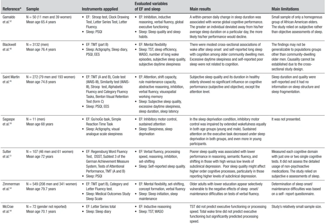
Table 1. Continuation.
Reference* Sample Instruments appplied
Evaluated variables
of EF and sleep Main results Main limitations
Gamaldo et al.52
N = 50 (11 men and 39 women) Mean age 65.4 years
• EF: Stroop test, Clock Drawing Test, Letter Series Test, Letter Fluency.
• Sleep: PSQI
• EF: Inhibition, inductive reasoning, verbal fluency, global executive functioning • Sleep: Sleep quality and sleep
habits.
A within-person daily change in sleep duration was associated with worse global cognitive performance. The greater an individual deviated away from his/her average sleep duration on a particular day, the more likely his/her performance would decline.
Small sample of only a homogenous group of African American elders. The study relied on subjective rather than objective assessments of sleep.
Blackwell et al.40
N = 3132 (men) Mean age 76.4 years
• EF: TMT (part B)
• Sleep: Actigraphy, Sleep diary, PSQI, EES
• EF: Mental flexibility • Sleep: TST, sleep efficiency,
WASO, number of long wake episodes, subjective sleep quality, subjective daytime sleepiness
There were modest cross-sectional associations of wake after sleep onset and self-reported long sleep with cognition among older community dwelling men. Excessive daytime sleepiness and self-reported poor sleep were not related to cognition.
The findings may not be generalizable to populations groups other than community-dwelling older men. Causality cannot be established due to the cross-sectional study design.
Saint Martin et al.45
N = 272 (79 men and 193 women) Mean age 74.8 years
• EF: TMT (A and B), Code test III), Similarity test (WAIS-III), Stroop test, Alphabetic Fluency and Category Fluency Tasks, Benton Visual Retention Test (form C)
• Sleep: PSQI, EES
• EF: Attention, shift capacity, rule maintenance capacity, abstractive reasoning, inhibition, verbal fluency, visuospatial working memory
• Sleep: Subjective sleep quality, excessive daytime sleepiness, sleep duration, sleep latency
Subjective sleep quality and its duration in healthy elderly showed no significant influence on cognitive performance (subjective and objective), except the attention level.
Sleep duration and quality were self-reported and it had no information on sleep structure and sleep fragmentation.
Sagaspe et al.59
N = 11 (men) Mean age 68 years
• EF: Go/noGo task, Simple Reaction Time Task • Sleep: Actigraphy, visual
analogue scale sleepiness
• EF: Inhibitory motor control, sustained attention • Sleep: Sleepiness, sleep
deprivation
In the sleep deprivation condition, inhibitory motor control was impaired by extended wakefulness equally in both age groups (young and male). Sustained attention on the executive task decreased under sleep deprivation in both groups, and even more in young participants.
It was not presented.
Sutter et al.51
N = 107 (46 men and 61 women) Mean age 72 years
• EF: Regensburg Word Fluency Test, DSST, Subtest 3 of the German Achievement Measure System, Tests of Attentional Performance, TMT (A and B) • Sleep: PSQI
• EF: Verbal fluency, processing speed, reasoning, inhibition, set-shifting
• Sleep: Self-reported sleep quality.
Poorer sleep quality was associated with lower performance in reasoning, semantic fluency, and shifting in those with high versus low levels of subclinical depression. Poor sleep quality might affect higher order cognitive processes, particularly in those reporting higher levels of subclinical depression.
Measured each cognitive domain with just one or two single cognitive tests. It did not assess the detailed usage of non-psychoactive medications. The study relied on subjective e assessments of sleep.
Zimmerman et al.54
N = 549 (208 men and 341 women) Mean age 79.7 years
• EF: TMT (part B), Category and Letter Fluency test.
• Sleep: Medical Outcomes Study Sleep Scale
• EF: Mental flexibility, set-shifting, concept formation, verbal fluency • Sleep: Sleep initiation, sleep
maintenance
Older adults with lower education appear selectively vulnerable to the negative effects of sleep onset/ maintenance difficulties on tests of verbal fluency.
Determination of sleep onset/ maintenance difficulties was based on a self- report questionnaire.
McCrae et al.44
N = 72 (gender not reported) Mean age 70.1 years
• EF: Letter Series total • Sleep: Sleep diary
• EF: Inductive reasoning • Sleep: TST, WASO
TST did not predict executive functioning or processing speed. Total wake time did not predict executive functioning but significantly predicted processing speed.
Study’s relatively small sample size.
Dement Neuropsyc
hol 2016 September;10(3):185-197
190
Sleep and e
xecutive functions
Holanda Júnior and
Almondes
Table 1. Continuation.
Reference* Sample Instruments appplied
Evaluated variables
of EF and sleep Main results Main limitations
Lim et al.57
N = 700 (172 men and 528 women) Mean age 82.4 years
• EF: Digit span test, Digit ordering test • Sleep: Actgraphy
• EF: Working memory
• Sleep: Fragmentation of rest and activity
Greater fragmentation of rest and activity were associated with lower levels of cognitive performance, with preferential involvement of perceptual speed, semantic memory, working memory, and visuospatial abilities.
Primarily women aged 80 and over.
Miyata et al.43
N = 78 (16 men and 62 women) Mean age 72.2
• EF: Number (n)-back test • Sleep: Actigraphy, PSQI, EES
• EF: Working memory • Sleep: TST, WASO, sleep
efficiency, sleep latency, daytime sleepiness
Short sleep duration decreased short-term memory capacity. Participants with sleep efficiency <85% showed a significant decrease on short-term memory and working memory test accuracy compared with those with sleep efficiency >85%.
The study included a high percentage of female participants.
Wilckens et al.41
N = 45 (13 men and 32 women) Mean age 62.8 years
• EF: Sternberg working-memory task, N-back task, Stroop task, Flanker task, National Adult Reading Test (NART), Categorical and Lexical Fluency tasks
• Sleep: Sleep detection device
• EF: Working memory, inhibition, verbal fluency and proficiency • Sleep: WASO, TST
In the older group, higher sleep continuity was associated with better inhibitory control, memory recall, and verbal fluency. TST was not associated with cognitive performance in any domains for the older group.
Participants were not excluded based on any sleep measures or sleep disorders. The study used an accelerometer-based sleep detection device, whereas PSG is considered the “gold standard” for sleep measurement.
Wilckens et al. 42
N = 53 (gender not reported) Mean age 62.6
• EF: TMT (A and B), DSST (WAIS-III), Stroop Task, N-Back, task-switching
• Sleep: A sleep detection device
• EF: Attention, cognitive flexibility, working memory, inhibition • Sleep: WASO, TST
Better global switching performance was associated with longer and more continuous sleep. Young and older adults may benefit similarly from lower wake time after sleep onset and longer total sleep time in overall performance.
The study used an accelerometer-based sleep detection device, whereas PSG is considered the “gold standard” for sleep measurement.
McHugh et al.55
N = 505 (gender not reported) Mean age 73.4 years
• EF: Digit span backward, CAMCOG similarities, TMT (A and B)
• Sleep: PSQI
• EF: Divided attention, working attention, psychomotor speed • Sleep: Time to bed, time do rise
Early and late sleepers were significantly slower on attention, learning and praxis tasks than those whose bedtime did not differ significantly to the robust norm. Wake-times were not associated with cognitive functioning in this cohort.
Self-report measures of sleep. There were no guidelines to categorise morningness-eveningness behavior of a less extreme type among otherwise healthy older adults.
Groeger et al.62
N= 31 (6 men and 25 women) Mean age 70.8 years
• EF: DSST, Sustained attention to Response Task, Choice Reaction Time Test, Lexical Decision Time, Serial Reaction Task, Continuous Tracking Task, Pursuit Tracking Task,Verbal n-Back and Spatial n-Back, Goal Neglect Task, Paced Visual Serial Addition Task, Verbal Fluency Task
• Sleep: PSG, PSQI
• EF: Sustained attention, divided attention, processing speed, decision, sequence & motor control, working memory, verbal fluency.
• Sleep: Slow wave sleep disruption
Slow wave sleep disruption resulted in less positive affect, slower or impaired information processing and sustained attention, less precise motor control, and erroneous implementation, rather than inhibition, of well-practiced actions. At baseline, younger participants performed better than older participants across many cognitive domains, with largest effects on executive function, response time, sustained attention, and motor control.
It was not presented.
Dement Neuropsyc
hol 2016 September;10(3):185-197
191
Holanda
Júnior
and
Almondes
Sleep and e
xecutive functions
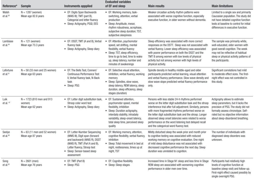
Table 1. Continuation.
Reference* Sample Instruments appplied
Evaluated variables
of EF and sleep Main results Main limitations
Walsh et al.58
N = 1287 (women) Mean age 82.8 years
• EF: Digits Span Backwards (WAIS III); TMT (part B), Categorial and letter fluency • Sleep: Actigraphy, PSQI, EES
• EF: Working memory, task-switching, attention, verbal production
• Sleep: Amplitude, mesor, rhythm robustness, acrophase, subjective sleep duration; TST, subjective sleepiness
Weaker circadian activity rhythm patterns were associated with worse cognitive function, especially executive function, in older women without dementia.
Limited to a single sex and primarily Caucasian population. The study did not have detailed cognitive function tests at baseline to control for initial differences in executive function.
Lambiase et al.49
N = 121 (women) Mean age 73.3 years
• EF: DSST, TMT (A and B), Verbal fluency task
• Sleep: Actigraphy, Sleep diary
• EF: Attention, psychomotor speed, set shifting, mental flexibility, verbal fluency • Sleep: TST, sleep efficiency,
time to go to bed, time to wake up, sleep latency, number and minutes of awakenings
Sleep efficiency was associated with more correct responses on the DSTT. Sleep was not associated with verbal fluency. Lower sleep efficiency was associated with poorer performance on both the DSST and the TMT B among women with low levels of physical activity but not among women with high levels of physical activity.
The sample was primarily white, well-educated, older women with good overall cognition. The week may not be reflective of habitual sleep or physical activity patterns of the participants.
Lafortune et al.61
N = 58 (33 men and 25 women) Mean age 63 years
• EF: The Bells Test, Conners’ Continuous Performance Test II, Verbal fluency task, N-Back task
• Sleep: PSG
• EF: Selective visual attention, inhibition, verbal fluency, working memory
• Sleep: Spindles, slow wave, sleep latency, REM latency, sleep duration, sleep efficiency, sleep stages (duration)
Spindle density in healthy middle-aged and older participants predicted verbal learning, visual attention and verbal fluency performance. Slow wave density and slow wave slope predicted verbal fluency performance only.
Significant correlations had mild to moderate effect sizes. The first-night effect was not controlled in this study.
Luik et al. 47
N = 1723 (810 men and 913 women)
Mean age 62 years
• EF: Letter digit substitution task, Stroop color word test • Sleep: Actigraphy, Sleep diary
• EF: Sustained attention, psychomotor speed, mental flexibility, inhibition • Sleep: Duration actigraphy,
interdaily stability, intradaily variability, sleep-onset latency, total sleep time, perceived sleep quality
Persons with less stable 24-h rhythms performed worse on the letter digit substitution task and the stroop interference trial after full adjustment. Similarly, persons with more fragmented rhythms performed worse on the letter digit substitution task and the stroop. Longer observed sleep onset latencies were related to worse performance on the word listening test delayed recall and the categorical word fluency test.
Actigraphy allows to estimate sleep parameters, but it lacks the precision of PSG. The study did not formally assess chronotype. Self-rated but no objective information about sleep-disordered breathing.
Seelye et al.46
N = 63 (11 men and 52 women) Mean age 87 years
• EF: Letter-Number Sequencing (WMS III), Digit span (forward and backward) (WAIS R), DSST (WAIS R), TMT (Part A and B), Letter fluency, Stroop test • Sleep: Sensor-based sleep
assessment
• EF: Working memory, attention, cognitive flexibility, verbal fluency, inhibition
• Sleep: Total movement in bed at night, restlessness, times up at night, TST
Mildly disturbed sleep the week prior and month prior to cognitive testing was associated with reduced working memory on cognitive evaluation. One night of mild sleep disturbance was not associated with decreased cognitive performance the next day. Sleep duration was unrelated to cognition.
The number of individuals with diagnosed sleep disorders was unknown.
Song et al.63
N = 2601 (men) Mean age 76 years
• EF: TMT (Part B) • Sleep: PSG
• EF: Cognitive flexibility • Sleep: Sleep stages
Increased time in Stage N1 sleep and less time in Stage REM sleep are associated with worsening cognitive performance in older men over time.
Participants had relatively high levels of cognitive function at baseline (sleep visit) and follow-up. First-night effect caused possibly by single overnight PSG.
Dement Neuropsychol 2016 September;10(3):185-197
192 Sleep and executive functions Holanda Júnior and Almondes studies (n=14). Other more frequent measures were Ver-bal Fluency Tasks (n=11), the Stroop test (n=7), Digit Span Test (n=7), n-back Task (n=6), and Digit Symbol
Substitution Test (DSST) (n=6). Other less frequent tests used are given in Table 1.
Wake after sleep onset (WASO). WASO means the total amount of time awake after falling sleep and it is considered a better relection of sleep fragmentation. here is evidence that WASO could be related to execu-tive functioning and global cognition. Using actigraphy, Blackwell et al.38 found that higher WASO was associ-ated with higher risk of cognitive impairment, using the TMT as a measure of executive functioning and the Mini-Mental State Examination (MMSE) for global cognition. In a longitudinal community-based study by Yafe et al.39 elderly women who showed decline on the TMT (part B) experienced worse WASO. Using the same measures, another study by Blackwell et al.40 found a modest association. Recently, in two studies Wilckens et al.41,42 found that in older adults, higher sleep continuity (i.e. lower WASO) was associated with better inhibitory control and that individuals with less WASO were more likely to engage preparatory strate-gies to reduce switch costs and boost task-switching performance. hese indings suggest that a higher WASO negatively impacts executive functioning. Inversely, older adults may beneit from lower WASO in performing complex tasks.
In another study, however, although participants with WASO longer than 30 min tended to have lower accuracy on the n-back test (attention and working
memory measure) than those with WASO <5 min or 5–30 min, these diferences were not signiicant.43 McCrae et al.44 found that WASO did not predict execu-tive functioning (measured by a reasoning test) but signiicantly predicted processing speed. his seems to indicate that in some cases, sleep fragmentation primar-ily afects basic processes, such as processing speed and alertness, rather than more complex ones.
Total sleep time (TST). In some studies, TST was examined as a continuous variable while in other studies it was dichotomized into short and long sleep duration. he results of its impact on EF and other cognitive domains are controversial. Many studies found no association between objectively measured total sleep duration and executive functioning.38-40,42,46,47 his lack of relation-ship was also detected in subjectively measured total sleep duration.44,45,48 Miyata et al.43 found that TST was correlated with the 0-back test from the n-back test, a
simple measure of attention and short-term memory,
but not for the 1-back test, which relects working memory capacity.
Nevertheless, in the study by Wilckens et al.41 a bet-ter performance on task-switching, a model paradigm of executive functioning that involves cognitive lexibility and the ability to shift attention between one task and another, was associated with longer TST. here have been cases in which subjective and objective measures diverged in associations: Lambiase et al.49 reported that total sleep duration measured using a sleep diary, a subjective instrument, was associated with cognitive lexibility; on the other hand, in the same study, actig-raphy measures did not show this association. Taken together, these studies of sleep duration and executive functioning in older adults have produced mixed results, although most of the evidence mentioned above indi-cates no relationship between them and may suggest that it is interruption of sleep, such as higher WASO, rather than quantity, that most afects EF.
Sleep latency. In relation to sleep latency, deined as the length of time that it takes to accomplish the transi-tion from wakefulness to sleep, the reviewed articles reported mixed results. When examining objective reports, Blackwell et al.,38 Yafe et al.39 and Blackwell et al.40 showed that higher sleep latency was associ-ated with worse executive functioning based on perfor-mance on the TMT (part B). Schmutte et al.50 reported that longer self-reported sleep latency was signii-cantly and inversely related to verbal-based cognitive measures, abstract reasoning, and longer latencies associated with poorer cognitive functioning. Nebes et al.48 also showed that a longer self-reported time to fall asleep was associated with poorer abstract reasoning. Others reported lack of association between sleep latency and EF through subjective45 and objective43 measures. It may be that longer sleep latency disturbs sleep quality and quantity and thus exacerbates the negative impacts on EF.
objec-Dement Neuropsychol 2016 September;10(3):185-197
193
Holanda Júnior and Almondes Sleep and executive functions tive sleep eiciency decreased attention and working
memory worsened.43 Lambiase et al.49 also reported that lower actigraphy-assessed sleep eiciency was associated with poorer performance on executive function tasks of attention, set-shifting and cognitive lexibility. On subjective measures, sleep eiciency was correlated with measures of abstract reasoning and working memory.48 Nevertheless, using the same measures as Blackwell et al.38 and Yafe et al.,39 Black-well et al.40 failed to ind these associations. According to the authors, this less consistent inding observed for sleep eiciency may relate to greater measurement error of this variable. Overall, the studies indicated that older adults with lower percentage sleep eiciency exhibited worse executive functioning.
Sleep quality. Sleep quality does not refer only to the number of hours in bed. It is the quality of these hours and how other factors afect sleep that are important, such as fragmentation, sleep deprivation, perception of restorative sleep, daytime sleepiness, having trouble falling sleep and waking up in the morning or staying alert during the daytime. Most of the studies used the PSQI as a general measure of sleep quality. he results in the literature regarding the efect of this parameter on EF tend to be mixed. In older women, good (PSQI < 6) and poor (PSQI ≥ 6) sleepers difered signiicantly on tests of working memory, attention, set shifting, and abstract problem solving,48 suggesting an important role of sleep quality in executive functioning. Sutter et al.51 sought to clarify the relationship between sleep quality and cognitive performance in healthy older adults, and to evaluate the moderating role of subclin-ical depression in this relationship. he study found that self-reported sleep quality in healthy older adults seemed to be selectively related to higher order execu-tive functions in those participants with high versus low levels of subclinical depression.
In other lines of evidence, these efects were not found. Gamaldo et al.52 used inhibition, inductive rea-soning, verbal luency and global executive functioning together with other cognitive domains, to create a global composite score and found that sleep quality measured by PSQI was not a signiicant predictor of performance across cognitive domains. Although Blackwell et al.40 reported that almost half of the men (44%) had self-reported poor sleep quality, deined as PSQI > 5, no signiicant association between this subjective measure and executive functioning was conirmed. Also, exces-sive daytime sleepiness did not correlate with cognitive outcomes. Martin et al.45 observed that good and poor sleepers did not difer on any of the cognitive
func-tion measures, including those evaluating EF domains, except on the TMT (part A), where this latter result may relect attention impairment. Luik et al.47 also found no relationship between lower reported sleep quality, as measured by a sleep diary and global cognitive func-tioning or performance on speciic cognitive tasks, such as inhibition. Taken together, these contrary indings do not seem to conirm the relationship between sleep quality and EF.
Some articles reviewed also used other variables related to sleep quality. Gamaldo et al.53 examined the relationship between elders’ cognitive performance and self-reported trouble falling asleep and found that this complaint signiicantly predicted performance on the digit span and alpha span task (measures of working memory and attention). his demonstrates that a self-report of sleep diiculty may be a predictor of cognitive performance. Zimmermann et al.54 reported that sleep diiculties (i.e. sleep initiation and sleep maintenance) appear to selectively impact the verbal luency process in older adults with lower education measured by category luency and letter luency tests, suggesting that those with higher education are better able to engage active cognitive compensation against sleep diiculties than individuals with lower education.
Circadian, homeostatic and ultradian factors. Another study found an association between time to bed and cognitive functioning, independent of sleep duration. Older adults who went to bed much later showed a poor performance on the TMT (A and B) and on the drawing test (prefrontally-mediated tasks) compared to older adults who went to bed within the robust norm window.55 hus, it appears that severe deviations in time to bed represent a marker of circadian misalign-ment and a possible marker of cognitive impairmisalign-ment. he study by Oosterman et al.56 reported that fragmen-tation of the sleep-wake rhythm predicted neuropsy-chological functioning such as mental speed, verbal memory and EF (cognitive lexibility, interference and working memory) in home-dwelling elderly people. Partial correlations showed that the association of rhythm fragmentation with cognitive decline is partly independent of the main efects of age. herefore, part of age-related cognitive decline could independently be associated with sleep and its circadian organization. Another study found that greater fragmentation of rest and activity measured by actigraphy was associated with lower levels of cognitive performance, including working memory.57
Dement Neuropsychol 2016 September;10(3):185-197
194 Sleep and executive functions Holanda Júnior and Almondes weaker circadian activity rhythm patterns (i.e. ampli-tude, mesor, rhythm robustness, and acrophase) were associated with worse cognitive function, especially executive functioning measured by the TMT (part B).58 Disrupted circadian activity rhythms could be an early indicator of future executive function decline. In another study, actigraphy recordings were used to quantify 24-h rhythms by calculating the stability and fragmentation of the rhythm over a period of days. Both aspects of the 24-h activity rhythm and sleep parameters were related to global cognitive functioning, but speciically, distur-bances in the 24-h activity rhythm were mostly related to tasks that draw on perceptual speed and executive functioning (mental lexibility and inhibition).47 Perhaps his association could relect a direct efect of disturbed rhythms on perceptual speed and executive functioning. Sagaspe et al.59 evaluated inhibitory motor control and sustained attention under controlled high or low sleep pressure conditions in young and older males. Under the sleep deprivation condition, inhibitory motor control was equally impaired by extended wakefulness in both age groups. Although sustained attention also decreased under sleep deprivation conditions in both groups, this efect was more pronounced for young par-ticipants. his might indicate that aging is a protective factor against the efects of extended wakefulness on simple tasks (i.e. sustained attention) due to an attenu-ation of sleep pressure with durattenu-ation of time awake. In other words, homeostatic sleep pressure would be lower in the older people, allowing them to be less vulnerable to sustained attentional failure after a night of sleep deprivation.
Lastly, some of the studies reviewed focused on sleep stages and sleep EGG measures. Anderson & Horne60 examined associations between neuropsychological per-formance, such as planning, lexibility, verbal luency, and luid intelligence, and sleep EEG characteristics within the prefrontal cortex in 24 healthy 61-75-year-olds. hey found signiicant associations between 0.5– 1.0 Hz power from the left frontal EEG channel, in the irst non-REM period, and performance on tasks more speciic to the left PFC (e.g., nonverbal planning and verbal luency). hese results pointed to a sleep EEG correlate of neuropsychological performance center-ing on the PFC. Lafortune et al.61 found that spindle density in healthy middle-aged and older participants predicted verbal learning, visual attention and verbal luency performance. Slow wave density and slow wave slope predicted verbal luency performance only. hese results suggest that spindle density is a marker of cogni-tive functioning in older adults and may relect neuro-anatomic integrity.
Groeger et al.62 assessed the efects of reducing SWS on daytime functioning and whether these efects difer across groups of healthy young, middle-aged, and older individuals. Evaluating domains such as sustained and divided attention processing speed, decision, sequence and motor control, working memory, and verbal lu-ency, the study showed that SWS disruption resulted in slower or impaired information processing and sustained attention, less precise motor control, and erroneous implementation, rather than inhibition, of well-practiced actions. Younger participants performed better at baseline than older participants across many cognitive domains, with largest efects on executive function, response time, sustained attention, and motor control. SWS can possibly be considered a potential mediator of age-related decline in performances. Song et al.63 investigated the relationship between sleep stage distributions and subsequent decline in cognitive func-tion in older men over time, using the TMT (part B) as a measure of executive functioning and the Modiied Mini-Mental State Examination (3MS) for global mea-surement of cognitive function. Increased time in stage N1sleep (considered a “light” sleep and marker of poorer sleep quality) and less time in stage REM sleep were associated with worsening general cognitive and execu-tive performance in older men, suggesting a beneicial role of REM sleep in cognition.
DISCUSSION
he aim of the present review was to systematically understand the evidence of interaction between sleep and executive functions in healthy aging, considering a growing body of literature suggesting that changes in sleep parameters and in its ultradian, circadian and homeostatic organization may play an impor-tant role in the executive functioning. In the elderly population, however, the evidence for this association reviewed tends to be mixed and involves various incon-sistencies. Multiple sleep parameters appear to difer, with some beneiting and others impairing executive functioning.
Dement Neuropsychol 2016 September;10(3):185-197
195
Holanda Júnior and Almondes Sleep and executive functions spent in SWS.41,42 SWS and slow wave activity (SWA)
have been identiied as a beneicial factor for prefron-tal cortex functioning and hence for executive func-tioning, which relies mostly on this brain area and its connections.64,65 Supporting lines of evidences include: (1) slow waves show frontal predominance both under baseline conditions and in response to sleep loss;66,67 (2) delta activity that is high during SWS is associated with cognitive performance;68 (3) selective SWS disrup-tion is associated with poor cognitive performance, and even impaired executive functioning;62 (4) age-related medial prefrontal cortex gray matter atrophy has recently been shown to be associated with reduced SWA in older adults. Lower SWS was associated with reduced functional connectivity within PFC-hippocam-pal networks, suggesting that neural synchrony during sleep strengthens connections between the PFC and functionally related brain regions;69 (5) greater relative slow wave activity was associated with higher dorsolat-eral prefrontal metabolism.65 In addition, there is also migration of cortical activity patterns from the posterior (decreased at occipital and temporal channels) to ante-rior (increased at central and frontal channels) area after sleep deprivation.70
hus, this growing body of evidence supports a potential role of certain aspects of sleep cerebral activ-ity in beneiting prefrontal areas and its connections. his highlights the PFC vulnerability model which holds that sleep deprivation and poor sleep afect PFC circuitry, producing a mild, sub-clinical level of impair-ment.32,33,71,72 he cognitive processes supported by PFC-associated networks (i.e. frontal-striatal and frontopa-rietal), especially EF, seem to be the most sensitive to individual diferences in sleep.34,72 his relationship may be strengthened considering that in normal aging both sleep and executive functioning change, which may pro-duce a synergistic efect on the performance of executive tasks.
High levels of sleep fragmentation (recurrent awak-enings and/or stage shifts) may result in complaints of non-restorative sleep even when an apparently normal total sleep time is present. his may be a factor explain-ing why most studies reviewed here found inconsistent or mixed associations between TST and EF: interrup-tion of sleep, such as higher sleep fragmentainterrup-tion and lower sleep eiciency appears to afect EF more than sleep quantity. It seems that continuous and consoli-dated sleep, which allows adequate progression through sleep stages and NREM-REM cycling, is important for executive functioning.
Contrary to the PFC vulnerability model, other lines of evidence suggest that impaired sleep continuity and
deprivation may cause sleepiness, which in turn may result in reduced vigilance and attentional failures. his reduced vigilance may negatively impact executive functioning. herefore sleep could afect EF mediated by basic process impairment, such as vigilance, alert-ness and attention processes.73 In this case, we can only cautiously infer that older adults with poor sleep quality and sleep deprivation may be less alert during the day, which directly worsens their performance on executive functioning tasks. On the other hand, another potential factor is that it can be assumed that impairment in the executive performance of elderly caused by sleep loss only becomes apparent when the PFC areas of the brain are activated under excessively high demand.51
Age-related changes in sleep parameters and ultra-dian sleep factors do not act solely on executive func-tioning. he review also showed that the changes in circadian organization of wake-sleep cycle (i.e. time to bed as marker of sleep phase, rhythm fragmentation, and weaker circadian activity) and the homeostatic fac-tors also appear to play an important role. he aging process appears to generate vulnerability to the impact of sleep and circadian rhythm disturbances on execu-tive performance.47 By afecting both cognition and the rest-activity rhythm, age-related changes in brain structures may account for an association between these variables. Regarding homeostatic pressure, some results have indicated that an increase in errors on an execu-tive task under extended wakefulness can be attributed mainly to the efect of sleep pressure with duration of time awake.59
Taken together, all of the relationships discussed above have important implications for clinicians and other health professionals, where psychoeducational and cognitive-behavioral interventions focusing on sleep hygiene and sleep parameters could improve global sleep and consequently may beneit cognitive functioning.74 Sleep quality in the elderly population is often poor not because of speciic pathologies, but due to lifestyle fac-tors, such as daytime napping, alcohol consumption, medication side efects, that detract from adequate nocturnal sleep. In these cases, psychoeducational and cognitive-behavioral interventions can help by reducing the efects of insuicient sleep on waking performance. In addition, higher physical activity levels can protect against the negative efects of poor sleep on executive functioning, and also promote other well-documented beneits of a physically active lifestyle.49
Dement Neuropsychol 2016 September;10(3):185-197
196 Sleep and executive functions Holanda Júnior and Almondes abilities. Few studies focus solely on the relationship between sleep and executive functioning. Concerning limitations of the literature reviewed, some studies did not exclude participants based on measures of sleep dis-orders, for example examining the possible presence of sleep disordered breathing, a condition that impairs cog-nition in older adults. In addition, many studies relied on subjective rather than objective assessments of sleep. For example, self-reported items about trouble falling asleep or time of sleep duration do not account for other possible sleep problems and are highly dependent on participant’s perception and beliefs about sleep, which may lead to diferential misclassiication and selective drop-out. For example, polysomnography is considered the “gold standard” for sleep measurement and may shed light on brain activity during sleep and on its role in cognitive functioning. Covariables such as depression, anxiety, and medical illness should also be accounted for when both sleep and EF are investigated. Another important consideration is that most of the studies used relatively few EF tests and tasks. Further studies should employ a more comprehensive range of EF tasks and components to establish a better association between sleep and executive functioning.
In conclusion, healthy aging is marked by changes in both sleep and executive functioning. Although evidence of the role of sleep and its ultradian, circa-dian and homeostatic organization in EF has grown, the association between them is partially inconclu-sive and studies using valid and reliable measures for sleep and EF variables are clearly needed. However, sleep parameters such as wake after sleep onset and sleep eiciency, in addition to circadian fragmenta-tion of sleep, showed less mixed results and are poten-tially correlated with EF measures. his relationship has important implications for clinicians and other health professionals in that psychoeducational and cognitive-behavioral interventions focusing on sleep hygiene and sleep parameters can improve global sleep and consequently may beneit cognitive functioning.
Author contribution. All authors have contributed signii-cantly and are in agreement with the content of the manuscript. FWNHJ and KMA were responsible for study conception and design. FWNHJ was respon-sible for writing the manuscript. FWNHJ and KMA were responsible for the critical revision. Both authors approved the inal manuscript.
REFERENCES
1. Ancoli-Israel S, Ayalon L, Salzman C. Sleep in the elderly: normal variations and common sleep disorders. Harv Rev Psychiatry. 2008; 16:279-286.
2. Buysse DJ, Monk TH, Carrier J, Begley A. Circadian patterns of sleep, sleepiness, and performance in older and younger adults. Sleep. 2005;28:1365-1376.
3. O’Donnell D, Silva EJ, Munch M, Ronda JM, Wang W, Duffy JF. Compar-ison of subjective and objective assessments of sleep in healthy older subjects without sleep complaints. J Sleep Res. 2009;18:254-263. 4. Cajochen C, Münch M, Knoblauch V, Blatter K, Wirz-Justice A.
Age-related changes in the circadian and homeostatic regulation of human sleep. Chronobiol Int. 2006;23:461-474.
5. Ohayon MM, Carskadon MA, Guilleminault C, Vitiello MV. Meta-analysis of quantitative sleep parameters from childhood to old age in healthy individuals: developing normative sleep values across the human lifespan. Sleep. 2004;27:1255-1273.
6. Roepke SK, Ancoli-Israel S. Sleep disorders in the elderly. Indian J Med Res. 2010;131:302-310.
7. Crowley K, Trinder J, Kim Y, Carrington M, Colrain IM. The effects of normal aging on sleep spindle and K-complex production. Clin Neuro-physiol. 2002;113:1615-1622.
8. Floyd JA, Janisse JJ, Jenuwine ES, Ager JW. Changes in REM-sleep percentage over the adult lifespan. Sleep 2006;30:829-836. 9. Duffy JF, Zitting KM, Chinoy ED. Aging and Circadian Rhythms. Sleep
Med Clin. 2015;10:423-434.
10. Wolkove N, Elkholy O, Baltzan, M, Palayew M. Sleep and aging: 1. Sleep disorders commonly found in older people. Can Med Assoc J. 2007;176:1299-1304.
11. Duffy JF, Willson HJ, Wang W, Czeisler CA. Healthy older adults better tolerate sleep deprivation than young adults. J Am Geriatr Soc. 2009;57:1245-1251.
12. Wigren HK, Rytkonen KM, Porkka-Heiskanen T. Basal forebrain lactase release and promotion of cortical arousal during prolonged waking is attenuated in aging. J Neurosci. 2009;29:11698-11707.
13. Dijk DJ, Groeger JA, Stanley N, Deacon S. Age-related reduction in daytime sleep propensity and nocturnal slow wave sleep. Sleep. 2010;33:211-223.
14. Dautovich ND, McCrae CS, Rowe M. Subjective and objective napping and sleep in older adults: are evening naps “bad” for nighttime sleep? J Am Geriatr Soc. 2008; 56:1681-1686.
15. Spreng RN, Wojtowicz M, Grady CL. Reliable differences in brain activity between young and old adults: a quantitative meta-analysis across multiple cognitive domains. Neurosci Biobehav Rev. 2010;34:1178-1794. 16. Colcombe SJ, Kramer AF, Erickson KI, Scalf P. The Implications of
Cortical Recruitment and Brain Morphology for Individual Differences in Inhibitory Function in Aging Humans. Psychol Aging 2005;20:363-375. 17. Turner GR, Spreng RN. Executive functions and neurocognitive aging:
dissociable patterns of brain activity. Neurobiol Aging. 2012;33:826. e1–826.e13.
18. Cartwright KB. Insights From Cognitive Neuroscience: The Importance of Executive Function for Early Reading Development and Education. Early Educ Dev. 2012;23:24-36.
19. Chan RCK, Shumb D, Toulopoulou T, Chen EYH. Assessment of execu-tive functions: Review of instruments and identification of critical issues. Arch Clin Neuropsychol. 2008;23:201-216.
20. Titz C, Karbach J. Working memory and executive functions: effects of training on academic achievement. Psychol Res. 2014;78:852-868. 21. Diamond A. Executive Functions. Annu Rev Psychol. 2013;64:135-168. 22. Johnson JK, Lui LY, Yaffe K. Executive function, more than global
cognition, predicts functional decline and mortality in elderly women. J Gerontol A Biol Sci Med Sci. 2007;62:1134-1141.
23. Morterá P, Herculano-Houzel S. Age-related neuronal loss in the rat brain starts at the end of adolescence. Front Neuroanat. 2012;6:1-9. 24. Freeman SH, Kandel R, Cruz L, et al. Preservation of neuronal number
despite age-related cortical brain atrophy in elderly subjects without Alzheimer disease. J Neuropathol Exp Neurol. 2008;67:1205-1212. 25. Buckner RL. Memory and Executive Function Review in Aging and AD:
Dement Neuropsychol 2016 September;10(3):185-197
197
Holanda Júnior and Almondes Sleep and executive functions
26. Bloss EB, Janssen WG, Ohm DT, et al. Evidence for Reduced Experi-ence-Dependent Dendritic Spine Plasticity in the Aging Prefrontal Cortex. J Neurosci. 2011;31:7831-7839.
27. Magnusson KR, Brim BL, Das SR. Selective vulnerabilities of N-methyl-D-aspartate (NMDA) receptors during brain aging. Front Aging Neurosci. 2010;2:1-15.
28. Alvarez JA, Emory E. Executive Function and the Frontal Lobes: A Meta Analytic Review. Neuropsychol Rev. 2006;16:17-42.
29. Jacob SN, Nieder A. Complementary roles for primate frontal and pari-etal cortex in guarding working memory from distractor stimuli. Neuron. 2014;83:226-237.
30. Horne JA. Human sleep, sleep loss and behaviour. Implications for the prefrontal cortex and psychiatric disorder. Br J Psychiatry. 1993; 162:413-419.
31. Harrison Y, Horne JA. Sleep loss impairs short and novel language tasks having a prefrontal focus. J Sleep Res. 1998;7:95-100.
32. Harrison Y, Horne JA, Rothwell A. Prefrontal neuropsychological effects of sleep deprivation in young adults--a model for healthy aging? Sleep. 2000;23:1067-1073.
33. Harrison Y, Jones, K, Waterhouse J. The influence of time awake and circadian rhythm upon performance on a frontal lobe task. Neuropsy-chologia. 2007;45:1966-1972.
34. Killgore WD, Kahn-Greene ET, Lipizzi EL, Newman RA, Kamimori GH, Balkin TJ. Sleep deprivation reduces perceived emotional intelligence and constructive thinking skills. Sleep Med. 2008;9:517-526. 35. Schmidt C, Peigneux P, Cajochen C. Age-related changes in sleep and
circadian rhythms: impact on cognitive performance and underlying neuroanatomical networks. Front Neurol. 2012;3:5-15.
36. Beebe DW, Gozal D. Obstructive sleep apnea and the prefrontal cortex: Towards a comprehensive model linking nocturnal upper airway obstruction to daytime cognitive and behavioral deficits. J Sleep Res. 2002;11:1-16.
37. Almondes KM, Holanda Júnior FWN, Alves NT. Sleep deprivation and implications for recognition and perception of facial emotions. Sleep Biol Rhythms. 2015;14:13-22.
38. Blackwell T, Yaffe K, Ancoli-Israel S, et al. Poor sleep is associated with impaired cognitive function in older women: The study of osteoporotic fractures. J Gerontol A Biol Sci Med Sci 2006;6:405-410.
39. Yaffe K, Blackwell T, Barnes DE, Ancoli-Israel S, Stone KL. Preclinical Cognitive Decline and Subsequent Sleep Disturbance in Older Women. Neurology. 2007;69:237-422.
40. Blackwell T, Yaffe K, Ancoli-Israel S, et al. Association of sleep character-istics and cognition in older community-dwelling men: The MrOS sleep study. Sleep. 2011;34:1347-1356.
41. Wilckens K A, Woo SG, Erickson KI, Wheeler ME. Sleep continuity and total sleep time are associated with task-switching and preparation in young and older adults. J Sleep Res. 2014;23:508-516.
42. Wilckens KA, Woo SG, Kirk AR, Erickson KI, Wheeler ME. The Role of Sleep Continuity and Total Sleep Time in Executive Function Across the Adult Lifespan. Psychol Aging. 2014;29:658-665.
43. Miyata S, Noda A, Iwaoto K, Kawano N, Okuda M, Ozaki N. Poor sleep quality impairs cognitive performance in older adults. J Sleep Res. 2013; 22:535-541.
44. McCrae CS, Vatthauer KE, Dzierzewski JM, Marsiske M. Habitual sleep, reasoning, and processing speed in older adults with sleep complaints. Cogn Ther Res. 2012;36:156-164.
45. Saint Martin M, Sforza E, Barthélémy JC, Thomas-Anterion C, Roche E. Does subjective sleep affect cognitive function in healthy elderly subjects? The Proof cohort. Sleep Med. 2012;13:1146-1152. 46. Seelye A, Mattek N, Howieson D, Riley T, Wild K, Kaye J. The Impact
of Sleep on Neuropsychological Performance in Cognitively Intact Older Adults Using a Novel In-Home Sensor-Based Sleep Assessment Approach. Clin Neuropsychol. 2015;29:53-66.
47. Luik AI, Zuurbier LA, Hofman A, Van Someren EJ, Ikram MA, Tiemeier H. Associations of the 24-hour activity rhythm and sleep with cognition: A population-based study of middle-aged and elderly persons. Sleep Med. 2015;16:850-855.
48. Nebes RD, Buysse DJ, Halligan EM, Houck PR, Monk TH. Self-reported sleep quality predicts poor cognitive performance in healthy older adults. J Gerontol B Psychol Sci Soc Sci. 2009;64:180-187.
49. Lambiase MJ, Gabriel KP, Kuller LH, Matthews KA. Sleep and Executive Function in Older Women: The Moderating Effect of Physical Activity. J Gerontol A Biol Sci Med Sci. 2014;69:1170-1176.
50. Schmutte T, Harris S, Levin R, Zweig R, Katz M, Lipton R. The
rela-tion between cognitive funcrela-tioning and self-reported sleep complaints in nondemented older adults: results from the Bronx aging study. Behav Sleep Med. 2007;5:39-56.
51. Sutter C, Zöllig J, Allemand M, Martin M. Sleep Quality and Cognitive Function in Healthy Old Age: The Moderating Role of Subclinical Depres-sion. Neuropsychology. 2012;26:768-775.
52. Gamaldo AA, Allaire JC, Whitfield KE. Exploring the within-person coupling of sleep and cognition in older African Americans. Psychol Aging. 2010;25:851-857.
53. Gamaldo AA, Allaire JC, Whitfield KE. The Relationship Between Reported Problems Falling Asleep and Cognition Among African Amer-ican Elderly. Res Aging. 2008;30:752-767.
54. Zimmerman ME, Bigal ME, Katz MJ, Brickman AM, Lipton RB. Sleep Onset/Maintenance Difficulties and Cognitive Function in Nondemented Older Adults: the Role of Cognitive Reserve. J Int Neuropsychol Soc. 2012;18:461-470.
55. McHugh JE, Walsh L, Lawlor BA. Time to bed is associated with cogni-tive outcome: an analysis of sleep-times and wake-times in community-dwelling older adults. Biol Rhythm Res. 2014;45:103-114.
56. Oosterman JM, Van Someren EJW, Vogels RLC, Van Harten B, Scherder EJA. Fragmentation of the rest-activity rhythm correlates with age-related cognitive deficits. J Sleep Res. 2009;18:129-135.
57. Lim AS, Yu L, Costa MD, et al. Increased fragmentation of rest-activity patterns is associated with a characteristic pattern of cognitive impair-ment in older individuals. Sleep. 2012;35:633-640.
58. Walsh CM, Blackwell T, Tranah GJ, et al. Weaker circadian activity rhythms are associated with poorer executive function in older women. Sleep 2014;37:2009-2016.
59. Sagaspe P, Taillard J, Amiéva H, et al. Influence of Age, Circadian and Homeostatic Processes on Inhibitory Motor Control: A Go/Nogo Task Study. Plos One. 2012;7(6): e39410.
60. Anderson C, Horne JA. Prefrontal cortex: links between low frequency delta EEG in sleep and neuropsychological performance in healthy, older people. Psychophysiology. 2003;40:349-357.
61. Lafortune M, Gagnon JF, Martin N, et al. Sleep spindles and rapid eye movement sleep as predictors of next morning cognitive performance in healthy middle-aged and older participants. J Sleep Res. 2014; 23:159-167.
62. Groeger JA, Stanley N, Deacon S, Dijk DJ. Dissociating effects of global SWS disruption and healthy aging on waking performance and daytime sleepiness. Sleep. 2014;37:1127-1142.
63. Song Y, Blackwell T, Yaffe K, Ancoli-Israel S, Redline S, Stone KL; Osteo-porotic Fractures in Men (MrOS) Study Group. Relationships between sleep stages and changes in cognitive function in older men: the MrOS Sleep Study. Sleep. 2015;38:411-421.
64. Wilckens KA, Erickson KI, Wheeler ME. Age-related decline in controlled retrieval: the role of the PFC and sleep. Neural Plast. 2012;2012: 1-14.
65. Wilckens KA, Aizenstein HJ, Nofzinger EA, et al. The role of non-rapid eye movement slow wave activity in prefrontal metabolism across young and middle-aged adults. J Sleep Res. 2016; 25:296-306.
66. Cajochen C, Foy R, Dijk DJ. Frontal predominance of a relative increase in sleep delta and theta EEG activity after sleep loss in humans. Sleep Res Online. 1999;2:65-69.
67. Blatter K, Cajochen C. Circadian rhythms in cognitive performance: methodological constraints, protocols, theoretical underpinnings. Physio Behav. 2007;90:196-208.
68. Scullin MK. Sleep, Memory, and Aging: The Link Between Slow-Wave Sleep and Episodic Memory Changes From Younger to Older Adults. Pychol Aging. 2012;28:105-114.
69. Mander BA, Rao V, Lu B, et al. Prefrontal atrophy, disrupted NREM slow waves and impaired hippocampal-dependent memory in aging. Nat Neurosci.2013;16:357-364
70. Boonstra T, Stins J, Daffertshofer A, Beek P. Effects of sleep depriva-tion on neuronal funcdepriva-tioning: An integrative review. Cell Mol Life Sci. 2007;64:934-946.
71. Harrison Y, Horne JA. The impact of sleep deprivation on decision making: A review. J Exp Psychol Appl. 2000;6:236-249.
72. Muzur A, Pace-Schott E, Hobson A. The prefrontal cortex in sleep. Trends Cogn Sci. 2002;6:475-481.
73. Lim J, Dinges DF. A meta-analysis of the impact of short-term sleep deprivation on cognitive variables. Psychol Bull. 2010;136:375-389. 74. Pace-Schott EF, Spencer RMC. Age-related changes in the cognitive