Licenciado sob uma Licença Creative Commons DO): http://dx.doi.org. . / - . . .AO
[T]
Cervical-scapular muscles strength and severity of
temporomandibular disorder in women with mechanical neck pain
[)]
Força dos músculos cérvico-escapulares e a severidade da disfunção
temporomandibular em mulheres com cervicalgia mecânica
[A]
Fernanda Pasinato[a], Juliana Bordin[a], Clarissa C. Santos-Couto-Paz[b], Juliana Alves Souza[c],
Eliane C. R. Corrêa[c]*
[a] Universidade Federal do Pampa Unipampa , Uruguaiana, RS, Brazil [b] Universidade de Brasília UnB , DF, Brazil
[c] Universidade Federal de Santa Maria UFSM , Santa Maria, RS, Brazil
[R]
Abstract
Introduction: Changes in cervical muscle function have been observed in patients with neck pain NP and
TMD. (owever, the relationship between TMD severity and neck muscle strength in the presence/absence of NP is unknown. Objective: To determine the prevalence of TMD in women with and without mechanical NP
and assess the cervical-scapular muscle strength and its association with TMD severity. Methods: Fifteen
vo-lunteers without neck pain CG and women with mechanical neck pain NPG took part and were selected by the Neck Disability )ndex. The diagnosis and severity of TMD were determined by the Research Diagnostic Criteria for TMD and Temporomandibular )ndex T) , respectively. The strength of the upper trapezius muscle, and cervical lexor and extensor muscles was measured by digital hand dynamometer. Results: . % of
wo-men with NP and . % without NP were diagnosed with TMD p = . . The NPG showed lower strength of the cervical lexor p = . and extensor p= . muscles, and higher T) p = . than in the CG. )t was
*FP: PhD, e-mail: fepas. [email protected] JB: Grad, e-mail: [email protected]
CCSCP: PhD, e-mail: [email protected] JAS: MSc, e-mail: [email protected]
also veri ied moderate negative correlation between T) and the strength of dominant p = . , r = - . and non-dominant p = . , r = - . upper trapezius, and cervical lexors p = . , r = - . in the NPG. Conclusion: There was no difference in the prevalence of TMD in women with and without NP. (owever,
women with NP have lower cervical muscle strength – compared to those without NP – which was associa-ted with greater severity of TMD. Thus, in women with NP associaassocia-ted with TMD, it is advisable to assess and address the severity of this dysfunction and identify the cervical-scapular muscles compromise.
Keywords: Temporomandibular Joint Disorders. Neck Pain. Muscle Strength Dynamometer. Muscle Strength.
Resumo
Introdução: Modiϔicações da função muscular cervical têm sido veriϔicadas em pacientes com cervicalgia e DTM.Entretanto, ainda não é conhecida a relação entre a severidade da DTM e a força muscular cervicalna presença/ausência de cervicalgia. Objetivo:Veriϔicar a prevalência de DTM em mulheres com e sem cervical-gia mecânica, avaliar a força dos músculos cérvico-escapulares e sua associação com a severidade da DTM.
Métodos: Participaram 15 voluntárias sem dor cervical (GC) e 14 mulheres com cervicalgia mecânica (GCM), selecionadas por meio do Índice de Disfunção Relacionada ao Pescoço. O diagnóstico e a gravidade da DTM foram determinados pelos Critérios diagnósticos para pesquisa em desordens temporomandibulares e Índice Temporomandibular (IT), respectivamente. A força dos músculos trapézio superior, ϔlexores e extensores cer-vicais foi aferida por dinamometria digital manual. Resultados: 64,5% das mulheres com cervicalgia e 33,3% das sem dor cervical apresentaram diagnóstico de DTM (p = 0,095). O GCM apresentou menor força dos mús-culos ϔlexores (p = 0,044) e extensores cervicais (p = 0,006) e maior IT (p = 0,038) que o GC. Também foi veri-ϔicada correlação negativa moderada entre o IT e força dos músculos trapézio superior dominante (p = 0,046, r = -0,547), não dominante (p = 0,007, r = -0,695) e ϔlexores cervicais (p = 0,023, r = -0,606) no GCM. Conclusão:
Não houve diferença na prevalência de DTM entre mulheres com e sem cervicalgia. Entretanto, mulheres com cervicalgia apresentaram menor força muscular cervical (comparadas às sem cervicalgia) que esteve associa-da a maior severiassocia-dade associa-da DTM. Assim, em mulheres com cervicalgia associaassocia-da à DTM, é recomendável avaliar e abordar a severidade desta disfunção e o comprometimento dos músculos cérvico-escapulares.
Palavras-chave:Desordens da Articulação Temporomandibular. Cervicalgia. Dinamômetro de Força Muscular. Força Muscular.
Introduction
Mechanical neck pain is a non-speci ic disorder of the cervical spine, characterized by pain and dis-comfort exacerbated by neck movement, present in periods of remission and exacerbation . Cervical pain is the fourth leading cause of disability in adults, after lower back pain, depression and arthralgia . Epidemiologic studies estimate an annual prevalence of to % in general population , and a system-atic review reports the average frequency of . %
. Annually, . % of economically active popu-lation experience some kind of limitation resulting from cervical pain , affecting the quality of life and causing signi icant socioeconomic impact .
)t has been shown that individuals with chronic neck pain exhibit structural and functional changes including weakness and cervical lexor and extensor
muscles imbalance - , a decrease in the cervical range of motion, as well as proprioceptive de icits and compromise of postural control . Those changes are thought to be assigned to the re lex inhibition associated with pain, causing damage to the muscu-lar function and favoring the chroni ication of mechanical neck pain , . Although the biome-chanical and neuroanatomic relationship between the cervical spine and the stomatognathic system has been veri ied in clinical and experimental studies - , the link between the dysfunctions affecting these systems remains in dispute.
imbalances and tensions . Furthermore, the ster-nocleidomastoid muscle SCM acts as a synergist in the support and stabilization of the head and the cervical region. )ts coactivation during teeth clench-ing varies from to % of the maximal voluntary contraction , suggesting a functional relationship between the stomatognathic and cervical systems. Thus, compensations between these systems may be a necessary mechanism for achieving stability and ef iciency during the masticatory function , .
)n addition to the biomechanical relationship, evi-dence exists , that cervical and stomatognathic systems interact mutually in the nociceptive and pro-prioceptive domains. The association between orofacial and cranio-cervical symptoms can be explained by the convergence of second-order nociceptive neurons, which receive cervical and trigeminal afferents in the brainstem sensory nuclear complex. So, nociception resulting from a cervical dysfunction may increase the central sensitization of the trigeminal central nucleus, leading to diffusion of pain to the craniofacial area and vice versa , .
Temporomandibular disorder is a generic term re-ferring to painful and/or dysfunctional conditions of the mastication muscles, temporomandibular joints and structures related, with the cervical spine among them . A comorbity between neck pain and TMD has been observed , , and there is evidence that TMD might be related to the decrease in cervical muscles resistance , . (owever, the role of TMD severity, as well as its association with the isometric strength of cervical-scapular muscles, in the presence and/or absence of neck pain, has not been described in the literature.
Furthermore, a few research addressed the classi ication of TMD regarding its severity. The Temporomandibular )ndex allows for this classi i-cation, as provides scores regarding the clinical aspects relevant for the diagnosis of TMD such as: limitation of mandibular movement range, muscle and joint pain, as well as the presence of joint noise. Clinically, the utiliza-tion of this index makes possible the determinautiliza-tion of the dysfunction severity and the veri ication of therapeutic interventions results .
Therapeutic approaches, targeted to the cranio-cervical-mandibular functional unit, have been rec-ommended for patients with primary dysfunctions, both in the cervical spine and the stomatognathic system , . Additional studies are required, how-ever, that analyze the relationship among the changes in these systems.
The purpose of this study is to provide informa-tion to help understand the details about the inter-action between the cervical spine and the temporo-mandibular joint TMJ , taking into account the role of TMD severity and the cervical-scapular muscles strength. This knowledge will be able to guide clinical practice in a more objective way, thus contributing for the decision-making regarding the assessment and the elaboration of therapeutic strategies.
Based on the aforementioned, the present study purpose is to investigate the prevalence of TMD in women with and without mechanical neck pain, as well as the assessment of cervical-scapular muscles strength and their association with TMD severity in these individuals.
Methods
Research characterization and the participants
The present research was approved by the Ethics Committee of the Federal University of Pampa, Uruguaiana, RS protocol number compris-ing an observational study, cross-sectional in design. The volunteers were recruited in the municipality of Uruguaiana, among those who responded to the divulgation of this research through printed and elec-tronic media. Fifteen women presenting with me-chanical neck pain paired with women without neck pain were selected by convenience. (owever, one volunteer with neck pain who has not completed the assessment was excluded.
Therefore, twenty-nine women, aged to , were assessed and allocated in two groups according to the neck disability index ND) results , : NPG – Mechanical Neck Pain Group n = , compris-ing women with neck pain complaint for more than three months and cervical disability con irmed by the ND) score > ; CG – Control Group n = , comprising volunteers without neck pain complaint, with ND) score .
previous experience in the application of the assess-ment protocol of muscle strength, and the required care was taken regarding the speci icity of the muscle func-tion assessed. Before muscle strength measurement, the volunteers’ body mass was registered for subsequent data normalization, as described by Cools et al. .
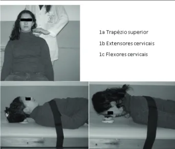
The assessment of upper trapezius muscle was performed with the volunteer in a sitting position. The dynamometer was placed on the upper scapular region, and resistance was applied over scapular depression, while asking the scapula elevation Figure a . The extensor muscles strength was assessed with the vol-unteer positioned on a stretcher in dorsal decubitus, with the dynamometer positioned below the occipital region, and the volunteer was asked to make maximum effort to perform cervical extension against the dyna-mometer Figure b . The cervical lexor muscles were assessed with the participant in ventral decubitus and the dynamometer placed under the frontal bone. At this point, the volunteer was asked to lex the cervical spine against the dynamometer resistance Figure c . )n both tests, the dynamometer was positioned so that the cervical spine remained in neutral position and, in the meantime, the participant was asked to keep the distance between the mento and stern to avoid cranio-cervical motion during the measurement process. )n order to control the utilization of the trunk muscles, a safety belt was placed on the volunteer’s shoulder, maintaining the trunk ixed during the test.
Each muscle was assessed bilaterally, repeating twice. The isometric contraction was kept for sec-onds and the volunteers were encouraged to employ maximum strength during the test by means of voice command, push, push, push . Each registration was followed by a one-minute resting interval. Data col-lection was performed in the same sequence for all the volunteers and by the same examiner.
The averages were calculated for both measures, which were expressed in kilogram-force Kgf and normalized by each volunteer’s body mass Kg each muscle strength value/body mass . The normaliza-tion of strength data is fundamental to allow the com-parison of individuals of different physical completions.
)nter and intra-examiner reliability of measure-ments of cervical muscles strength in dorsal and ventral decubitus, performed with a digital hand dynamometer, were previously assessed in a group of healthy individuals, showing excellent levels of reproducibility and reliability )CC> . . the study objectives and had a free and informed
consent signed.
Assessment Procedures
Disability related to neck pain was assessed through the Neck Disability )ndex. This is a ques-tionnaire translated and validated for the Brazilian population comprising items regarding the interference of pain while performing daily activities, except for item , which assesses headache intensity. The alternatives are numbered from to , describ-ing increasdescrib-ing degrees of neck pain interference on the activity assessed. The score consists of the sum of scores in each of the items, ranging from - points. The individual is rated as mild disability from to points; moderate disability, from to points; severe disability, from to points; and complete disability ranging from to points .
The diagnosis of TMD was made through clinical exam, according to the protocol established by the research diagnostic criteria for temporomandibular disorders RDC/TMD , performed by a previ-ously trained examiner. The algorithms of the instru-ment were employed to establish the diagnosis and TMD classi ication myofascial, disc or joint disorder .
From the results of RDC, scores were calculated for the Temporomandibular )ndex T) . T) com-prises three sub-indices: functional index F) , muscle index M) and joint index J) . Each sub-index value is calculated by adding the scores of the items assessed, divided by the total number of items. T) consists of the mean value of the three sub-indices, ranging from to , with the highest possible punctuation being , denoting higher severity of dysfunction. F) includes items regarding the amplitude of man-dibular movement, characterizing pain or limitation and deviations during mouth opening movement. M) measures the pain related to bilateral digital palpa-tion of the intra and extra-oral masticatory muscles, in a total of sites. J) assesses the pain evoked by digital palpation of the lateral pole and posterior liga-ment, and the occurrence of noise in each TMJ.
test for normality of variables. The strength results for each muscle were normalizedby each individual body mass and expressed in terms of average and standard deviation. For analyzing the correlation between data regarding cervical-scapular muscles strength and T), the Spearman correlation was used. The correlation was considered strong for correla-tion coef icient values r . ; moderate when . < r < . , and weak when r was . . With all the tests a signi icance level of α < . was considered. For statistical data analysis, the Statsoft STAT)ST)CA
. ® software was used.
Results
Fourteen women with mechanical neck pain NP and women without neck pain CG took part in this study. Among women with NP, showed mild disability, and showed moderate disability.
The results of descriptive statistics regarding the clinical-demographic characteristics – age, weight, the neck disability index ND) scoring and the temporomandibular index T) – are shown in table . The groups did not differ re-garding body weight. As for ND) and T) scores, significantly higher values in NPG than those in CG were observed.
Statistical Analysis
After data collection, a descriptive analysis of the participants’ clinical-demographic data was performed. Next, the Kolmogorov-Smirnov test was used for analyzing the normality of data regarding the primary outcome variables, and the Chi-square test to compare the frequency of TMD diagnosis be-tween the groups. A comparison bebe-tween the mea-surements of cervical-scapular muscle strength was performed by the Mann Whitney test, and the T) values were compared by t-test, according to the
Table 1- Clinical-demographic characteristics of participants in control group and neck pain group
Group Average (SD) CI 95% Value p
Age (years)
NPG 27.5±3.89 25.26 – 29.74
0.050 CG 25.4±6.04 22.05 – 28.76
Weight (Kg)
NPG 62.93±14.03 54.83 – 71.03
0.188 CG 58.13±12.37 51.28 – 64.98
NDI (points)
NPG 11.43±5.12 8.47 – 14.39
0.000*
CG 1.67±1.37 0.71 – 2.6
TI (points)
NPG 0.393±0.22 0.26 – 0.52
0.038* CG 0.245±0.14 0.17 – 0.26
Note: NDI, Neck disability index; TI, temporomandibular index; NPG, Mechanical Neck Pain Group; CG, Control Group; *signifi cantly different, α = 0.05
Table 2 - Diagnostic classification of TMD for control group (CG) and mechanical neck pain group (NPG)
Diagnoses of DTM NPG
(n= 14)
CG (n=15)
Ia (Myofascial pain) 1 2
IIa (Disc displacement with
reduction) 0 1
IIIa (Arthralgia) 1 0
Ia, IIa 0 1
Ia, IIIa 2 0
Ib, IIIa 4 0
IIa, IIIa 1 1
Without diagnosis 5 10
Multiple diagnoses (>3) 7 0
Note: NPG showed lower isometric muscle strength of cervical fl exor (p = 0.044) and extensor (p = 0.006) muscles than CG (Figure 2)
0,07
0,09 0,09
0,15
0,11 0,11
0,10 0,10
NPG CG NPG CG NPG CG NPG CG DUT NDUT Cervical Extensors
Cervical Flexors 0,16
0,14 0,12 0,1 0,08 0,06 0,04 0,02 0
0.00 0.10 0.20 0.30 0.40 0.50 0.60 0.70 0.80 0.20
0.18 0.16 0.14 0.12 0.10 0.08 0.06 0.04 0.02 0.00
Temporomandibular Index
p=0.046 r=-0.547
0.0 0.1 0.2 0.3 0.4 0.5 0.6 0.7 0.8 0.20
0.18 0.16 0.14 0.12 0.10 0.08 0.06 0.04 0.02 0.00
Temporomandibular Index
p=0.007 r=-0.695
0.0 0.1 0.2 0.3 0.4 0.5 0.6 0.7 0.8 0.20
0.18 0.16 0.14 0.12 0.10 0.08 0.06 0.04 0.02 0.00
Temporomandibular Index
p=0.023 r=-0.606 The frequency of TMD diagnosis in women with
mechanical neck pain was . %, while in the con-trol group it was . %. This percentage difference has not achieved a statistical signi icance level ac-cording to Chi-square test X = . , p = . .
The volunteers’ distribution according to the diag-nostic classi ication of TMD by RDC/TMD is described in Table .
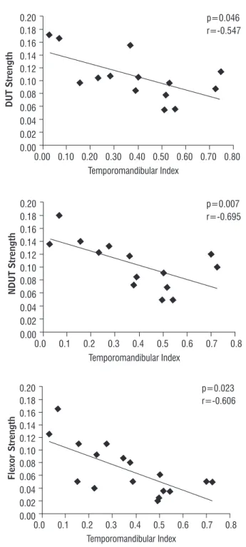
From the analysis of figure , it can be ob-served that in NPG, T) scores showed a moderate negative correlation . < r < . , with strength values assessed for every muscle, except for the cervical extensors. )n CG no significant correla-tion was found.
frequency described in the present study shows values similar to those described for the general population regarding signals and symptoms of TDM . % . According to the literature, individuals with me-chanical neck pain present with hyperalgesia in the trigeminal region, and lower levels of pain sensitivity to the pressure of masseter and temporal muscles , the reduction of which is associated with the intensity and duration of symptoms in the cervical region. Thus, the chronicity of neck pain may po-tentialize the trigeminal sensitization, favoring pain triggering and a dysfunction of the stomatognathic system, which in this study was con irmed by the greater severity of TMD found in NPG. This result denotes how important is the utilization of indices or scales that quantify TMD severity , as they are able to identify subtle changes, even in the ab-sence of a diagnosis for this dysfunction. Moreover, T) allows for the characterization and de inition of the component that contributes the most for TMD severity – muscular, articular or functional – favor-ing the selection of strategies and speci ic therapeu-tic interventions in the stomatognathic system, and monitoring the progress of therapeutic outcomes.
Women with NP showed lower values for isomet-ric strength of cervical lexor and extensor muscles, compared to CG, with greater reduction for exten-sor muscles. Changes in cranio-cervical strength and motor control in the context of neck pain have been described in the literature , , , , , , , and they are linked to an increase in disability levels caused by chronic neck pain . These modi ications, including a reduction in muscle strength, may be ex-plained by the mechanism of neural inhibition caused by pain and by the change in motor strategies related to cervical spine control and stability while executing the task .
The investigation of the presence of TMD , and the analysis of muscle strength in the context of neck pain , - , , have been performed. Additionally, this research results show the moder-ate negative correlation between the T) indicating DTM severity and the isometric strength of cervical lexor, dominant upper trapezius and non-dominant upper trapezius muscles in NPG. The same correla-tion was not observed in CG, which results may have been in luenced by the smaller number of volunteers with the diagnosis of TMD in this group.
Therefore, the reduction of cervical-scapular mus-cle strength can be explained in part by the increase in
Figure 3 - Dispersion graph and description of correlation val-ues between the severity of TMD (Temporomandibular Index) and normalized isometric strength of dominant upper trapezius (DUT), non-dominant upper trapezius (NDUT), cervical extensor, and fl exor muscles in women with neck pain
Discussion
The main indings of this study show that women with neck pain present lower strength of cervical muscles and greater TMD severity compared with women without neck pain. Additionally, in the pres-ence of neck pain, greater TMD severity was related to lower strength of cervical-scapular muscles.
Although the prevalence of TMD diagnosis in women with and without neck pain is not statisti-cally different . % versus . %, respectively , the analysis of T) revealed a difference between the groups regarding the degree of involvement caused by TMD, which was more severe in NPG. These ind-ings are consistent with the fact that % of women with neck pain present with multiple simultaneous diagnoses of TMD > , with the association between myofascial pain and arthralgia being the most fre-quent disorders , while in CG this was not veri ied.
Previously, Vicsher et al. showed that the fre-quency of signals and symptoms of TMD in individuals with cervical spine disorders are comparable to those found in the adult population without any disorder. )n contrast, Feirão et al. veri ied the presence of TMD in % of patients with neck pain receiving physical therapy treatment. )n the present study, the volunteers were selected in the community and were not receiving treatment for their dysfunction. These factors might have been decisive for the differences among the prevalence found. Regarding the CG, the 0.0 0.1 0.2 0.3 0.4 0.5 0.6 0.7 0.8 0.20
0.18 0.16 0.14 0.12 0.10 0.08 0.06 0.04 0.02 0.00
Temporomandibular Index
the damages in the generation of cervical-scapular muscles strength in women with neck pain, since these variables were not analyzed in a control group of healthy women. Thus, further research is suggested comparing individuals with and without TMD in order to better de ine the implications of this dysfunction for the decrease of muscle strength and neck pain.
)n view of this study results, professionals must be aware that signals and symptoms in the cervical and mandibular area cannot be considered alone. The determination of TMD severity in women with neck pain, as well as the assessment of cervical-scapular muscles strength may help determine the actions necessary to reduce the disability, and plan effective interventions for the treatment of both dysfunctions.
Conclusion
No differences were observed in the prevalence of TMD in women with and without neck pain. Women with neck pain, however, presented lower strength of cervical lexor and extensor muscles and greater severity of TMD compared with those without neck pain. Moreover, this study showed that the increase of TMD severity is related to a decrease in the strength of cervical lexor and upper trapezius muscles in women with neck pain. The presence of this asso-ciation reinforces the importance of the assessment of TMD severity and the strength of cervical-scapular muscles in women with neck pain. Such information can contribute for the elaboration of effective thera-peutic strategies for the treatment of these disorders.
References
. O'Leary S, Falla D, Jull G. The relationship between super icial muscle activity during the cranio-cervi-cal lexion test and clinicranio-cervi-cal features in patients with chronic neck pain. Man Ther. ; : - . . Cohen SP. Epidemiology, diagnosis, and treatment of
neck pain. Mayo Clin Proc. ; : - . . (ogg-Johnson S, Van der Velde G, Carroll LJ, (olm LW,
Cassidy JD, Guzman J, et al. The burden and determi-nants of neck pain in the general population: results of the Bone and Joint Decade - Task Force on Neck Pain and )ts Associated Disorders. J Manip Physiol Ther. ; Suppl :S - .
the severity of TMD in women with neck pain and vice versa. Speci ically, lower muscle strength of cervical lexors, associated with higher severity of TMD, can be understood as an extension of functional damage to SCM muscle, present in cases of TMD. Previous evidence , con irmed the role of muscle SCM – a primary cer-vical spine lexor – as a synergist in the stabilization of the head during the mastication function, as well as the changes in its electromyographic activity in the presence of TMD , , . )n addition, the posture control of the head and cervical spine is directly related to the stability promoted by an adequate synergism, the muscle ability to generate strength and the resistance of agonist and antagonist cervical muscles , . Considering that the mandible is a mobile bone with articulation with the skull by means of TMJ, the effective control of mandibular func-tions depends on a stable cervical base . Thus, the worst performance in the test of cervical lexion strength can also be related to the functional compensations re-quired for the maintenance of the masticatory system stability and ef iciency , .
Other factors that may be associated to the lowest recruitment and capacity of torque generation during cervical function are the electromiographic hyper-activity at rest and the hyperalgesia of SCM and upper trapezius muscles commonly observed in individuals with myogenic TMD , - . Thus, the inhibition of the muscle mechanism may be contributing to the poorer performance in the generation of cervical lexion strength in more severe case of TMD associated with neck pain.
The reduction in the maintenance time of contrac-tion and the resistance of cervical lexor and extensor muscles in individuals with TMD associated with cervi-cal dysfunction was previously demonstrated , . )n contrast, Armijo-Olivo et al. did not ind differences between maximal strength of cervical lexor muscles in individuals with TMD and asymptomatic individuals, or a correlation between mandibular impairment and muscle strength. )n these studies, TMD severity was not assessed, and the authors hypothesized that differ-ent conditions in the registration of strength and/or a sample of patients with greater severity of TMD might in luence the results. Further investigation is needed to clarify the role of this function in the modi ication of maximal strength of the cervical musculature.
. Milanesi JM, Weber P, Pasinato F, Corrêa ECR. Severi-dade da desordem temporomandibular e sua relação com medidas cefalométricas craniocervicais. Fisioter Mov. ; : - .
. Corrêa EDR, Bérzin F. Temporomandibular disorder and dysfunctional breathing. Braz J Oral Sci. ;
: - .
. Giannakopoulos NN, (ellmann D, Schmitter M, Kr“ger B, (auser Thomas, Schindler (J. Neuromuscular in-teraction of jaw and neck muscles during jaw clench-ing. J Orofac Pain. ; : - .
. Chandu A, Suvinen T), Reade PC, Borromeo GL. Elec-tromyographic activity of frontalis and sternocleido-mastoid muscles in patients with temporomandibular disorders. J Oral Rehabil. ; : - . . Strini PJ, Strini PJ, Barbosa TS, Gavião MB. Assessment
of thickness and function of masticatory and cervical muscles in adults with and without temporomandibu-lar disorders. Arch Oral Biol. ; : - . . Svensson P, Wang K, Sessle BJ, Arendt-Nielsen L.
As-sociations between pain and neuromuscular activ-ity in the human jaw and neck muscles. Pain. ;
: - .
. Kobayashi M, Yabushita T, Zeredo JL, Toda K, Soma K. Sple-nius muscle activities induced by temporomandibular joint stimulation in rats. Brain Res Bull. ; : - . . De-la-Llave-Rincon A), Alonso-Blanco C, Gil-Crujera A, Ambite-Quesada S, Svensson P, Fernandez-de-Las-Penas C. Myofascial trigger points in the masti-catory muscles in patients with and without chronic mechanical neck pain. J Manip Physiol Ther. ;
: - .
. Carrara SV, Conti PCR, Barbosa JS. Termo do ° Con-senso em Disfunção Temporomandibular e Dor Oro-facial. Dental Press J Orthod. ; : - . . Wiesinger B, Malker (, Englund E, Wanman A. Does a
dose-response relation exist between spinal pain and temporomandibular disorders? BMC Musculoskelet Disord. ; : .
. Visscher CM, Ligthart L, Schuller AA, Lobbezoo F, De Jongh A, Van (outem CM, Boomsma D). Comorbid disorders and sociodemographic variables in tem-poromandibular pain in the general Dutch popula-tion. J Oral Facial Pain (eadache. ; : - . . Fejer R, Kyvik KO, (artvigsen J. The prevalence of neck
pain in the world population: a systematic critical re-view of the literature. Eur Spine J. ; : - . . Salo PK, (akkinen A(, Kautiainen (, Ylinen JJ. Effect
of neck strength training on health-related quality of life in females with chronic neck pain: a randomized controlled -year follow-up study. (ealth Qual Life Outcomes. ; : .
. Cagnie B, Cools A, De Loose V, Cambier D, Danneels L. Differences in isometric neck muscle strength be-tween healthy controls and women with chronic neck pain: the use of a reliable measurement. Arch Phys Med Rehabil. ; : - .
. Lindstrom R, Schomacher J, Farina D, Rechter L, Falla D. Association between neck muscle coactivation, pain, and strength in women with neck pain. Man Ther. ; : - .
. Lindstroem R, Graven-Nielsen T, Falla D. Current pain and fear of pain contribute to reduced maximum volun-tary contraction of neck muscles in patients with chronic neck pain. Arch Phys Med Rehabil. ; : - . . Jørgensen R, Ris ), Falla D, Juul-Kristensen B.
Reliabil-ity, construct and discriminative validity of clinical testing in subjects with and without chronic neck pain. BMC Musculoskelet Disord. ; : .
. Ylinen J, Takala EP, Kautiainen (, Nykanen M, (ak-kinen A, Pohjolainen T, et al. Association of neck pain, disability and neck pain during maximal effort with neck muscle strength and range of movement in women with chronic non-speci ic neck pain. Eur J Pain. ; : - .
. Armijo-Olivo S, Fuentes JP, Da Costa BR, Major PW, Warren S, Thie NM, et al. Reduced endurance of the cervical lexor muscles in patients with concurrent temporomandibular disorders and neck disability. Man Ther. ; : - .
. Falla D, Farina D. Neuromuscular adaptation in experi-mental and clinical neck pain. J Electromyogr Kinesiol.
; : - .
. Ferão M)B, Traebert, J. Prevalence of temporomandibu-lar dysfunction in patients with cervical pain under phys-iotherapy treatment. Fisioter Mov. ; : - . . La Touche R, de-Las-Penas C,
Fernández-Carnero J, Díaz-Parreno S, Paris-Alemany A, Arendt-Nielsen L. Bilateral mechanical-pain sensitivity over the trigeminal region in patients with chronic me-chanical neck pain. Journal Pain. ; : - . . Boudreau SA, Falla D. Chronic neck pain alters muscle activation patterns to sudden movements. Exp Brain Res. ; : - .
. Schomacher J, Falla D. Function and structure of the deep cervical extensor muscles in patients with neck pain. Man Ther. ; : - .
. Ries LGK, Alves MC, Bérzin F. Asymmetric activation of temporalis, masseter, and sternocleidomastoid muscles in temporomandibular disorder patients. Cranio. ; : - .
. Pallegama RW, Ranasinghe AW, Weerasinghe VS, Sithe-eque MA. )n luence of masticatory muscle pain on electromyographic activities of cervical muscles in patients with myogenous temporomandibular dis-orders. J Oral Rehabil. ; : - .
. Milanesi JM, Corrêa ECR, Borin GS, Souza JA, Pasinato F. Atividade elétrica dos músculos cervicais e amplitude de movimento da coluna cervical em indivíduos com e sem DTM. Revista Fisioterapia e Pesquisa. ; : - . . Silveira A, Armijo-Olivo S, Gadotti )C, Magee D. Mas-ticatory and cervical muscle tenderness and pain sensitivity in a remote area in subjects with a tem-poromandibular disorder and neck disability. J Oral Facial Pain (eadache. ; : - .
. Armijo-Olivo SL, Fuentes JP, Major PW, Warren S, Thie NM, Magee DJ. )s maximal strength of the cervical lexor muscles reduced in patients with temporomandibular disorders? Arch Phys Med Rehabil. ; : - . . Armijo-Olivo SL, Magee DJ. Cervical
musculoskel-etal impairments and temporomandibular disor-ders. J Oral Maxillofac Res. ; : - .
Received: / / Recebido: 16/12/2013
Approved: / / Aprovado: 03/09/2015
. Armijo-Olivo S, Silvestre RA, Fuentes JP, Da Costa BR, Major PW, Warren S, et al. Patients with temporoman-dibular disorders have increased fatigability of the cer-vical extensor muscles. Clin J Pain. ; : - . . Pehling J, Schiffman E, Look J, Shaefer J, Lenton P,
Fricton J. )nterexaminer reliability and clinical valid-ity of the temporomandibular index: a new outcome measure for temporomandibular disorders. J Orofac Pain. ; : - .
. Freire AB, De Nardi AT, Bou leur J, Chiodelli L, Pasinato F, Corrêa ECR. Abordagem isioterapêutica multimodal: efeitos sobre o diagnóstico e a gravidade da disfunção temporomandibular. Fisioter Mov. ; : - . . La Touche R, de-las-Penas C, Fernández-Carnero J, Escalante K, Angulo-Diaz-Parreno S, Paris-Ale-many A, et al. The effects of manual therapy and exercise directed at the cervical spine on pain and pressure pain sensitivity in patients with myofascial temporomandibu-lar disorders. J Oral Rehabil. ; : - . . Cook C, Richardson JK, Braga L, Menezes A, Soler X,
Kume P, et al. Cross-cultural adaptation and valida-tion of the Brazilian Portuguese version of the Neck Disability )ndex and Neck Pain and Disability Scale. Spine. ; : - .
. Vernon (. The Neck Disability )ndex: state-of-the-art, - . J Manipulative Physiol Ther. ;
: - .
. Dworkin SF, Le Resche L. Research diagnostic criteria for temporomandibular disorders: review, criteria, examinations and speci ications, critique. J Cranio-mandib Disord. ; : - .
. Cools AM, Johansson FR, Cambier DC, Velde AV, Palmans T, Witvrouw EE. Descriptive pro ile of scapulothoracic position, strength and lexibility variables in adolescent elite tennis players. Br J Sports Med. ; : - . . Cassafuz T), Delavati VK. Con iabilidade da medida da força dos músculos lexores e extensores cervic-ais por meio da dinamometria manual digital [TCC]. Uruguaiana: Universidade Federal do Pampa; . . De Wijer A, Steenks M(, Bosman F, (elders PJM, Faber