RADIOLOGY PAGE
855
MRI fi ndings in Marion’s disease: the bulb and the
doughnut signs
Ernesto Lima Araujo Melo, Ligia Persici Rodrigues, Ricardo Reges Maia de Oliveira
Centro Avançado de Diagnóstico por Imagem (Boghos Dom Luis), (ELAM), Instituto Dr. José Frota (LPR) and Faculdade de Medicina Christus & Faculdade de Medicina da Universidade Federal do Ceará (RRMO), Fortaleza, CE, Brazil
_______________________________________________________________________________
A 77-year-old female presented to our facility with a fi ve-year history of progressive urinary bladder retention and voiding diffi cul-ties. She underwent a pelvic magnetic resonance imaging (MRI) examination as a part of her lower urinary tract symptoms evaluation, and for treat-ment planning.
MRI with a torso phasedarray superfi -cial coil was performed in a 1.5T magnetic fi eld equipment. We obtained multiplanar T1- and T2-weighted pre-contrast sequences, with and with-out fat suppression techniques, axial diffusion weighted images, axial apparent diffusion coef-fi cient (ADC) map, and multiplanar T1-weighted fat suppressed images after the administration of intravenous gadolinium.
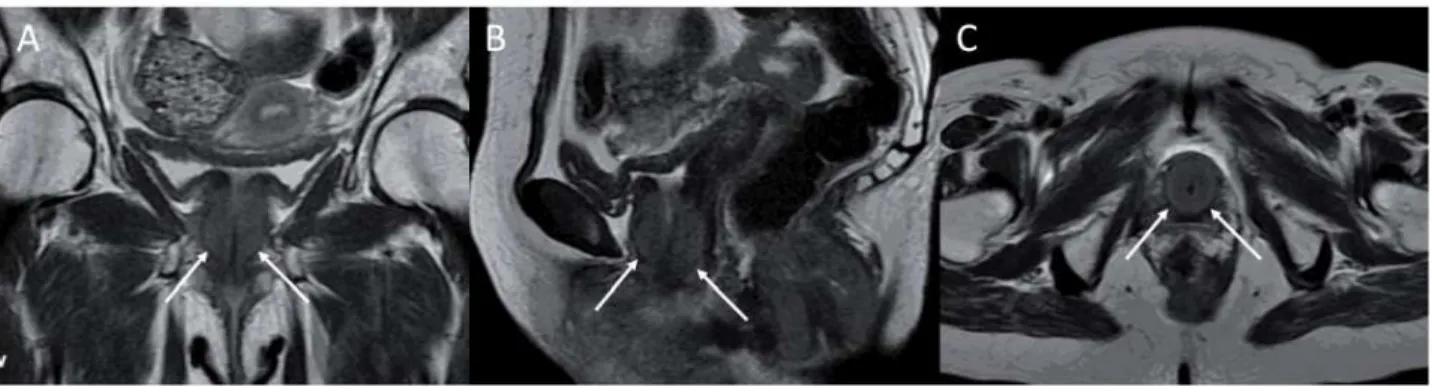
MRI revealed an exuberant concentric hypertrophy of the entire urethra. This charac-teristic imaging fi nding resembled a bulb of a sphygmomanometer on the sagittal and coronal planes, and a doughnut on the axial ones (Fig-ure-1). The affected musculature was isointense on T1- and slightly hyperintense on T2-weighted images compared with unaffected muscles. The diffusion sequence showed restriction to free wa-ter molecules movements, making the hypertro-phied muscles hyperintense on this sequence and hypointense on the ADC map (Figure-2). Addi-tionally this patient also exhibited a marked con-centric hypertrophy of the internal anal sphincter
muscle, probably related to the same bladder neck neuropathological mechanism. This latter fi nding was previously misdiagnosed in another facility as a rectal tumor. After the intravenous contrast administration, there was slight enhancement of the hypertrophied muscle. The combination of the MRI fi ndings together with the patient´s symptoms and urodynamic testing was consis-tent with the diagnosis of primary bladder neck obstruction (PBNO) due to severe muscular hy-pertrophy.
DISCUSSION
PBNO was fi rst described in men by Mari-on (1), a cMari-onditiMari-on wherein the bladder neck fails to open adequately during voiding, resulting in increased striated sphincter activity or obstruc-tion of urinary fl ow in the absence of another anatomic obstruction, such as that caused by be-nign prostatic enlargement in men or genitouri-nary prolapse in women (2,3).
856
IBJU |RADIOLOGY PAGE
Figure 1 – Coronal (a), sagittal (b), and axial (c), T2-weighted MRI images showing exuberant concentric hypertrophy of the muscular planes surrounding the urethra along its complete extension (arrows). This characteristic fi nding resembled the bulb of a sphygmomanometer in “a” and “b”, and a doughnut in “c”.
Figure 2 – (a) Axial diffusion sequence showed a ring-like hyperintensity of the peri-urethral hypertrophied muscles. (b) The axial ADC map demonstrated hypointensity in the affected muscles. In both images the doughnut sign is clearly depicted (arrows).
ABBREVIATIONS
ADC: apparent diffusion coeffi cient
MRI: magnetic resonance imaging
PBNO: primary bladder neck obstruction both. Occasionally, the initial presentation may
be urinary retentions (3). Presenting symptoms appear to be similar in men and women, with a combination of voiding and storage symptoms being common (4). Currently PBNO is diagnosed primarily by videourodynamics and cystourethroscopy, but the increasing use of MRI can introduce new imaging fi ndings (5).
REFERENCES
1. Marion G. Surgery of the neck of the bladder. Br J Urol. 1933; 5: 351-7.
2. Nitti VW: Primary bladder neck obstruction in men and wom-en. Rev Urol. 2005; 7(suppl 8): S12-7.
3. Huckabay C, Nitti VW. Diagnosis and treatment of primary blad-der neck obstruction in men. Curr Urol Rep. 2005; 6: 271-5. 4. Dewan PA, Rajimwale A, Gent RJ, Lequesne GW: Marion´s
disease presenting as prenatal hydronephrosis. Pediatr Surg Int. 1994; 9: 290-2.
5. N Tasali, Cubuk R, Sinanoglu O, Sahin K, Saydam B: MRI in stress urinary incontinence: endovaginal MRI with an intracavitary coil and dynamic pelvic MRI. Urol J. 2012; 9: 397-404.
______________________ Correspondence address: