10 artigo 465
ORIGINAL ARTICLE
1 - Assistant Physician of the Shoulder Group of the Orthopedics and Traumatology Service, HSPE – IAMSPE – São Paulo, SP, Brazil. 2 - Head of the Shoulder Group of the Orthopedics and Traumatology Service, HSPE – IAMSPE – São Paulo, SP, Brazil.
3 - Intern of the Shoulder Group of the Orthopedics and Traumatology Service, HSPE – IAMSPE – São Paulo, SP, Brazil.
Work carried out at the Orthopedics and Traumatology Service of the Hospital do Servidor Público Estadual Francisco Morato de Oliveira – IAMSPE - São Paulo. Correspondence: Rua Pedro de Toledo, 1800 - CEP 04029-000 - São Paulo, SP - E-mail: [email protected]
Received for publication: 12/07/2010, accepted for publication: 07/14/2011
RESULTS OF SURGICAL TREATMENT OF DENERATIVE
ARTHROPATHy OF THE ROTATOR CUFF USING
HEMIARTHROPLASTy- CTA
®Rômulo Brasil Filho1, Fabiano Rebouças Ribeiro1, Antonio Carlos Tenor Junior1, Cantidio Salvador Filardi Filho2, Guilherme Barbieri Leme da Costa3, Thiago Medeiros Storti3, André da Costa Garcia3, Hilton Vargas Lutfi3
The authors declare that there was no conflict of interest in conducting this work
This article is available online in Portuguese and English at the websites: www.rbo.org.br and www.scielo.br/rbort ABSTRACT
Objective: To assess results of CTA® partial shoulder arthroplasty for treatment of degenerative arthropathy of
the rotator cuff. Methods: Between December 2006 and
June 2009, 23 shoulders of 23 patients were submitted to
CTA® type partial shoulder arthroplasty for treatment of
arthropathy secondary to rotator cuff injury. Post-operative follow up time ranged from 6 to 35 months. Mean age was 74.1 years. Patients were predominantly female, rep-resenting 78.3% of cases. The right limb was affected in 18 patients. All patients had undergone at least 6 months of physiotherapy without improvement of the algetic picture, and being submitted to surgery by the same surgical team. None of the patients had history of surgery on the affected shoulder. The method elected for assessing patients
dur-ing post-operative follow up was based on UCLA scordur-ing criteria. Results: Improvement in pain was observed in all patients after arthroplasty. Mean UCLA pain score was 9.22 (ranging from 10 to 8). Mean function was 6 (10 to 2). Active frontal flexion was 2.39 (highest score 4 and lowest 0). Mean frontal flexion force was 4.09, maximum was 5 and minimum 3. Mean score on the UCLA was 26.52. 95%
were satisfied with the surgery. Conclusion: CTA® type
partial shoulder arthroplasty produced satisfactory results in the treatment of degenerative arthropathy of the rotator cuff and had a low rate of complications.
Keywords – Arthroplasty; Replacement; Joint Diseases; Rotator Cuff
INTRODUCTION
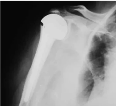
Degenerative arthropathy of the rotator cuff con-sists of collapse of the glenohumeral joint secondary to massive chronic rotator cuff injury. It causes eleva-tion of the humeral head, joint destruceleva-tion, synovial fluid changes, subchondral cysts, flattening of the greater tubercle, osteophytes, acetabularization of the
coracoac-romial arch and osteopenia(1-3) (Figure 1).It occurs
more frequently among female patients after the age of 60 years, and it manifests with pain, crepitation and diminution of the range of motion(4).
Several hypotheses for explaining the development of arthropathy due to rotator cuff lesions have been put forward. In the rheumatological literature, the
term Milwaukee shoulder was introduced to describe a condition presented by four elderly women who had massive rotator cuff injuries, destructive gleno-humeral arthritis and recurrent effusion in the
shoul-der (Geyser sign)(5). The most accepted hypotheses
suggest that accumulation of hydroxyapatite crystals inside the capsule, synovium and joint cartilage would allow these crystals to be released into the synovial fluid. The crystals would be phagocytized by syno-vial cells and would thus accumulate inside them and stimulate the release of proteolytic enzymes, includ-ing collagenase and protease. These enzymes would lead to joint, capsule and cuff destruction(5-7).
Neer et al(3) described a hypothesis for how
67
Figure 1 – Anteroposterior radiograph on a shoulder with ar-thropathy due to a rotator cuff injury.
HEMIARTHROPLASTY- CTA®
patients. Loss of the dynamic stabilizers of the gleno-humeral joint would lead to repeated trauma on the joint surface, thus causing cartilage loss. Furthermore, loss of closed joint space would lead to poor nutrient diffusion to the joint cartilage. Secondarily to shoulder disuse, the subchondral bone would then become more osteoporotic, thus resulting in erosion of the humeral head and completing the development of arthropathy.
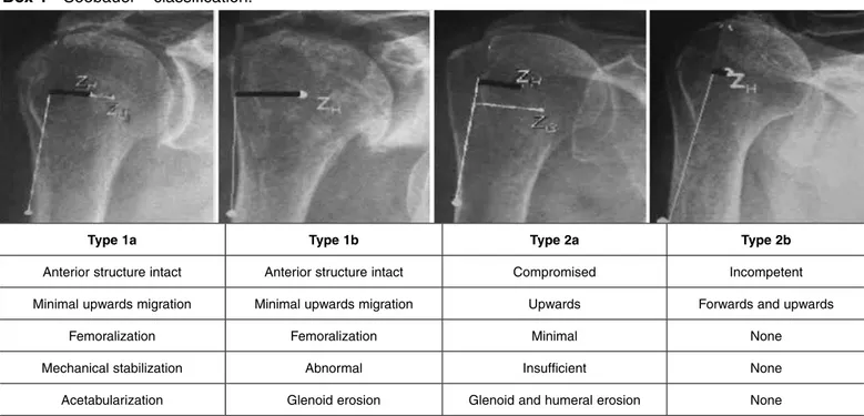
The arthropathy classification most used is the
sys-tem described by Seebauer(8) (Box 1). It assesses the
integrity of the anterior shoulder stabilizers and co-racoacromial arch, the presence of dynamic stability and the upwards migration of the humeral head. In stage IA, the head is centered on the glenoid; in IB, the head migrates medially in relation to the glenoid; in IIA, the humeral head migrates upwards, but is stabilized by the coracoacromial arch; and in IIB, the humeral head migrates forwards and upwards.
Conservative treatment is initially administered to all patients, using physiotherapy and analgesics. When there is no improvement of the pain and/or range of motion, surgical procedures are chosen(9,10).
Arthroscopic debridement is an alternative when the patient’s main complaint is pain, and consists of joint and bursa cleaning, tuberculoplasty and teno-tomy of the biceps. The result may be a transitory improvement of pain, without interfering with the range of motion, but with frequent recurrences over the first two years(10,11).
The options for replacement arthroplasty are CTA®
(cuff tear arthroplasty) and a reverse prosthesis. CTA®
prostheses (Figure 2) are used when the arthropathy has not compromised the stability of the glenohu-meral joint, the glenoid erosion is only partial and the coracoacromial arch is complete. These are partial prostheses, with a larger humeral head so that there is greater contact with the coracoacromial arch, thus enabling better lever-arm action by the deltoid muscle on arm elevation movements. A reverse prosthesis is used when there is no stability in the glenohumeral joint and the glenoid anatomy is compromised. It is characterized by modification of the center of gleno-humeral rotation medially and distally, through po-sitioning the glenoid component (glenosphere) with the aim of boosting the acting of the deltoid muscle(9).
The aim of the present study was to assess the re-sults obtained from CTA® partial shoulder arthroplasty
to treat degenerative arthroplasty of the rotator cuff.
Figure 2 – Postoperative radiography on a shoulder, showing CTA® hemiarthroplasty.
METHODS
Between December 2006 and June 2009, 23
shoul-ders of 23 patients underwent CTA® partial shoulder
Box 1 - Seebauer classification.
Figure 3 – Distribution of the 23 patients according to the See-bauer classification.
attributing points according to the pain, degree of mo-bility, shoulder function, strength and patient satisfac-tion. The maximum score is 35 points. To measure the degree of joint range of motion, the method described by the American Academy of Orthopedic Surgeons was used(13). To compare the pre and postoperative results regarding UCLA and the orthopedic physical examina-tion with range of moexamina-tion (OPE), the nonparametric Wilcoxon test was used(14). The rejection level for the nullity hypothesis was 0.05 (significance level of 95%).
Type 1a Type 1b Type 2a Type 2b
Anterior structure intact Anterior structure intact Compromised Incompetent
Minimal upwards migration Minimal upwards migration Upwards Forwards and upwards
Femoralization Femoralization Minimal None
Mechanical stabilization Abnormal Insufficient None
Acetabularization Glenoid erosion Glenoid and humeral erosion None
39,1
21,7
39,1 patients’ mean age was 74.1 years, ranging from 62
to 84 years. Females predominated, accounting for 78.3% of the cases (18 patients). The dominant limb was affected in 18 patients and the non-dominant limb in five patients (Table 1).
Out of the 23 patients evaluated before the opera-tion, five were classified as type IA, nine as type IB and the remaining nine as type IIA. There were no cases of type IIB (Figure 3).
All the patients had previously undergone physio-therapy for at least six months, without improvement of their painful condition, and they all had a clini-cal and imaging diagnosis (radiographs and nuclear magnetic resonance) of degenerative arthropathy of the rotator cuff.
The inclusion criterion was that the patients should be symptomatic, with a Seebauer classification of IA, IB or IIA, who had not improved with rehabilitation treatment administered for a minimum of six months. The following were taken to be exclusion criteria: degenerative arthropathy of the rotator cuff that im-proved with clinical treatment; previous surgery or neurological lesions in the affected limb; arthropathy classified as Seebauer IIB; and insufficiency of the deltoid muscle.
69
HEMIARTHROPLASTY- CTA®
RESULTS
It was observed that there was a significant in-crease in the mean UCLA score, from 10.39 points before the operation to 26.52 points after the opera-tion (Figure 4). The result was considered to be good in the cases of nine patients (39.1%), fair in thirteen (56.6%) and poor in one (4.3%).
With regard to joint range of motion measure-ments, there was an increase in mean active eleva-tion, from 57.61º before the operation to 77.83º after the operation (Figure 5 and Table 2).
The lateral rotation increased from 19.78º before the operation to 26.09º after the operation (Figure 6 and Table 2).
The mean medial rotation did not present any change and remained at the level of the third
lum-bar vertebra (L3) from before to after the operation (Table 1).
It was observed that all the patients’ pain im-proved after arthroplasty, in relation to their pre-operative pain. The mean score on the UCLA table relating to pain was 9.22, with maximum of 10 and minimum of 8. Regarding function, the mean was 6, the maximum was 10 and the minimum was 2. For active frontal flexion among these patients, the mean was 2.39, the maximum was 4 and the minimum was 0. Regarding the strength of frontal flexion, the mean was 4.09, the maximum was 5 and the minimum was 3 (Figure 7 and Table 3).
Twenty-two of the 23 patients (95%) were satis-fied with the surgery. The patient who presented the lowest score on the UCLA scale (15 points) and was not satisfied with the surgery presented atrophy of
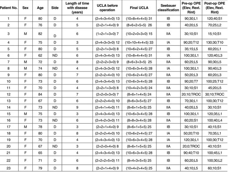
Table 1. Data on patients.
Patient No. Sex Age Side
Length of time with disease
(years)
UCLA before
operation Final UCLA
Seebauer classification
Pre-op OPE (Elev, Rext.
Rint)
Post-op OPE (Elev, Rext.
Rint)
1 F 80 D 4 (2+4+3+4+0) 13 (10+8+4+4+5) 31 IB 90;30;L1 120;40;S1
2 F 76 D 5 (2+2+1+4+0) 9 (8+6+2+5+5) 26 IB 40;20;L5 70;25;L2
3 M 82
D 6 (1+2+1+3+0) 7 (10+2+0+3+0) 15 IIA 30;10;S1 15;10;S1
4 F 75 D 3 (2+4+3+3+0) 12 (10+10+4+4+5) 33 IA 90;20;T12 130;30;T10
5 F 80 D 5 (2+2+1+3+0) 8 (10+6+2+4+5) 27 IB 35;15;L5 60;20;L1
6 F 62 ND 6 (2+4+3+4+0) 13 (10+8+4+4+5) 31 IA 100;30;L1 120;40;L3
7 M 72 D 8 (2+2+2+3+0) 9 (8+6+3+3+5) 25 IIA 60;25;L5 90;30;L5
8 M 74 ND 4 (2+4+3+3+0) 12 (10+6+3+4+5) 28 IA 100;30;L1 90;40;L3
9 F 80 D 7 (2+2+2+4+0) 10 (10+6+2+4+5) 27 IIA 50;20;L3 60;20;L3
10 F 73 D 6 (2+4+3+4+0) 13 (10+6+3+4+5) 28 IB 90;20;T7 100;25;T12
11 F 70 D 4 (2+2+1+3+0) 8 (10+4+2+3+5) 24 IIA 30;10;S1 45;20;L5
12 F 84 D 7 (2+2+0+3+0) 7 (8+6+1+4+5) 24 IIA 20;10;TROC 30;10;TROC
13 F 67 D 6 (2+2+2+4+0) 10 (8+6+3+5+5) 27 IB 70;30;L1 100;30;T12
14 F 73 ND 9 (2+4+1+4+0) 11 (8+6+1+5+5) 25 IIA 40;05;L5 30;10;S1
15 M 75 D 3 (2+4+3+4+0) 13 (10+6+3+4+5) 28 IB 100;30;L1 120;35;L1
16 F 73 ND 6 (2+4+2+3+0) 11 (8+8+3+4+5) 28 IIA 60;20;S1 100;40;L4
17 M 78 D 3 (2+2+1+4+0) 9 (8+6+1+5+5) 25 IB 30;10;S1 40;15;S1
18 F 80 D 8 (2+2+2+4+0) 10 (10+6+2+4+5) 27 IA 50;20;T10 70;35;L1
19 F 70 D 4 (2+4+3+4+0) 13 (10+6+3+4+5) 28 IA 120;30;L1 100;30;T12
20 F 67 ND 3 (2+2+0+4+0) 8 (8+6+1+5+5) 25 IIA 20;0;TROC 40;10;S1
21 F 65 D 4 (2+4+3+4+0) 13 (10+6+3+4+5) 28 IB 90;40;T10 100;45;L1
22 F 71 D 6 (2+2+2+5+0) 11 (8+4+3+5+5) 25 IB 60;20;L5 100;30;L2
23 F 79 D 8 (2+2+1+4+0) 9 (10+4+2+4+5) 25 IIA 40;10;L5 60;10;S1
Source:SAME IAMSPE / medical iles
Figure 4 – Mean and standard deviation of the UCLA scores before and after the operation.
Figure 5 – Mean and standard deviation of the elevation before and after the operation.
Figure 6 - Mean and standard deviation of the lateral rotation before and after the operation.
Figure 7. Mean and standard deviation of the UCLA scores be-fore and after the operation.
Table 2 – Elevation and external rotation values at the times evaluated.
Variable Time n Mean SD Median Minimum Maximum p*
Elevation Before 23 57.61 30.74 50 10 120 < 0.001
After 23 77.83 33.60 90 15 130
External Before 23 19.78 9.94 20 0 40 < 0.001
rotation After 23 26.09 11.48 30 10 45
(*) descriptive probability level from the nonparametric Wilcoxon test
the deltoid muscle, although his electroneuromyog-raphy result was normal. There was no case of pros-thesis dislocation, infection or neurovascular deficit in the operated limb.
DISCUSSION
CTA® arthroplasty is used when the arthropathy
has not compromised the anterosuperior stability of the glenohumeral joint, with partial erosion of the
glenoid and a complete coracoacromial arch. This is a partial prosthesis with a humeral head that extends as far as the greater tuberosity, in order to provide con-tact with the coracoacromial arch, thereby enabling lever-arm improvement of the deltoid muscle in arm elevation movements.
Visotsky et al(2) found that 89% of their results were satisfactory, through using conventional partial arthro-plasty for treating patients classified as Seebauer IA, Before
Time
Time
Time
Pain Function Active flex-ion
Flexion strength
Satisfaction
Domain
External r
otation
El
e
v
a
ti
o
n
Escore
Before
Before
After After
After
71
HEMIARTHROPLASTY- CTA®
IB and IIA. They achieved improvements in pain, elevation and lateral rotation of the shoulder. Like in our study, they did not include patients classi-fied as Seebauer IIB. In our sample, we observed that only one case (patient no. 3; classified as type IIA) presented an unsatisfactory result. This patient achieved an improvement in pain, but with wors-ening of elevation movement, and was dissatisfied with the limitation on joint range of motion that was obtained. This patient also presented atrophy of the deltoid muscle, although the electroneuromyography result was normal.
Zuckerman et al(15) used the postoperative
luation method based on the UCLA criteria to eva-luate conventional partial arthroplasty, and obtained a mean of 22 points, with a mean range of motion of 86º of active elevation and 29º of active lateral rotation. They reported that 87% of the patients were satisfied in relation to improvement of pain.
In our sample, the results obtained from CTA®
par-tial shoulder arthroplasty presented a mean of 26.52 points and a satisfaction rate of 92.2% in relation to pain improvement (Table 1).
In our study, we used rigid exclusion criteria, which we believe to be very important for favorable
results from CTA® arthroplasty. CTA®
hemiarthro-plasty requires anterior stabilizers to impede anterior subluxation of the prosthesis: this role is played by the coracoacromial arch and the subscapular muscle.
In addition, a functioning motor is required, such as the deltoid muscle, along with a complete
axil-lary nerve. Field et al(16) evaluated 16 patients who
underwent hemiarthroplasty to treat arthropathy due to rotator cuff injury, and found that four of the six patients with unsatisfactory results had undergone previous acromioplasty, with release of the cora-coacromial ligament. They concluded that coracoac-romial arch competence and deltoid muscle function are important components for the stability of hemi-arthroplasty, when performed to treat arthropathy due to cuff injuries.
The studies by Zuckerman et al(15) and
Sanchez-Sotelo et al(17) showed that hemiarthroplasty
pro-vides adequate pain improvements but only moder-ate gains in functional range of motion, in patients with arthropathy due to cuff injuries. On the other
hand, in relation to the Delta® reverse prosthesis,
studies(18-21) have shown better gains in active range of movement, which leads to functional improve-ment in these patients. However, the complication rates remain relatively high (> 17%), and studies with longer follow-up and greater experience with these implants are still necessary.
CONCLUSION
CTA® partial shoulder arthroplasty has satisfactory results when used to treat degenerative rotator cuff arthropathy, with a low complication rate.
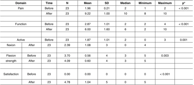
Table 3. Domain values for UCLA scores at the times evaluated.
Domain Time N Mean SD Median Minimum Maximum p*
Pain Before 23 1.96 0.21 2 1 2 < 0.001
After 23 9.22 1.00 10 8 10
Function Before 23 2.87 1.01 2 2 4 < 0.001
After 23 6.00 1.60 6 2 10
Active Before 23 1.87 1.01 2 0 3 0.001
flexion After 23 2.39 1.08 3 0 4
Flexion Before 23 3.70 0.56 4 3 5 0.003
strength After 23 4.09 0.60 4 3 5
Satisfaction Before 23 0.00 0.00 0 0 0 < 0.001
After 23 4.78 1.04 5 0 5
REFERENCES
1. Hamada K, Fukuda H, Mikasa M, Kobayashi Y. Roentgenographic findings in massive rotator cuff tears. A long-term observation. Clin Orthop Relat Res. 1990;(254):92-6.
2. Visotsky JL, Basamania C, Seebauer L, Rockwood CA, Jensen KL. Cuff tear arthropathy: pathogenesis, classification, and algorithm for treatment. J Bone Joint Surg Am. 2004;86(Suppl 2):35-40.
3. Neer CS, Craig EV, Fukuda H. Cuff-tear arthropathy. J Bone Joint Surg Am. 1983;65(9):1232-44.
4. Pollock RG, Deliz ED, McIlveen SJ, Flatow EL, Bigliani LU. Prosthetic replace-ment in rotator cuff-deficient shoulders. J Shoulder Elbow Surg. 1992;1(4):173-86. 5. Garancis JC, Cheung HS, Halverson PB, McCarty DJ. “Milwaukee shoulder”
– association of microspheroids containing hydroxyapatite crystals, active col-lagenase, ad neutral protease with rotator cuff defects. III. Morphologic and biochemical studies of an excised synovium showing chondromatosis. Arthritis Rheum. 1981;24(3):484-91.
6. Halverson PB, Cheung HS, McCarty DJ, Garancis J, Mandel N. “Milwaukee shoulder” – association of microspheroids containing hydroxyapatite crystals, active collagenase, ad neutral protease with rotator cuff defects. II. Synovial fluid studies. Arthritis Rheum. 1981;24(3):474-83.
7. McCarty DJ, Halverson PB, Carrera GF, Brewer BJ, Kozin F. “Milwaukee shoulder” – association of microspheroids containing hydroxyapatite crystals, active collagenase, and neutral protease with rotator cuff defects. I. Clinical aspects. Arthritis Rheum. 1981;24(3):464-73.
8. Seebauer L. Biomecanical classification of cuff tear arthropaty [abstract]. In: Global Shoulder Society Meeting; 2003 July 17-19 Salt Lake City, UT-USA. 9. Arntz CT Jackins S, Matsen FA. Prosthetic replacement of the shoulder for the
treatment of defects in the rotator cuff and the surface of the glenohumeral joint. Bone Joint Surg Am. 1993;75:485-91.
10. Boileau P, Sinnerton RJ, Chuinard C, Walsh G. Arthroplasty of the shoulder. J Bone Joint Surg Br. 2006;88(5):562-75.
11. Walch G, Madonia G, Pozzi I, Riand N, Levigne C. Arthroscopic tenotomy of the tendon of the long head of the biceps in rotator cuff ruptures. Amsterdam: Elsevier; 1997. p.350-5.
12. Ellman H, Kay SP. Arthroscopic subacromial decompression for chronic impinge-ment. Two- to five-year results. J Bone Joint Surg Br. 1991;73(3):395-8 13. Hawkins RJ, Bokor DJ. Clinical evaluation of shoulder problems. In: Rockwood
CA, Matsen FA, editors. The shoulder. 2nd ed. Saint Louis: W.B. Saunders; 1998. p.164-98.
14. Rosner B. Fundamentals of Biostatistics. 2nd ed. Boston: PWS Publishers; 1986. 584p.
15. Zuckerman JD, Scott AJ, Gallagher MA. Hemiarthroplasty for cuff tear arthropa-thy. J Shoulder Elbow Surg. 2000;9(3):169-72.
16. Field LD, Dines DM, Zabinski SJ, Warren RF. Hemiarthroplasty of the shoulder for rotator cuff arthropathy. J Shoulder Elbow Surg. 1997;6(1):18-23.
17. Sanchez-Sotelo J, Cofield RH, Rowland CM. Shoulder hemiarthroplasty for gle-nohumeral arthritis associated with severe rotator cuff deficiency. J Bone Joint Surg Am. 2001;83(12):1814-22.
18. Boileau P, Watkinson DJ, Hatzidakis AM, Balg F. Grammont reverse prosthesis: design, rationale, and biomechanics. J Shoulder Elbow Surg. 2005;14(1 Suppl S):147S-61S.
19. De Buttet M, Bouchon Y, Capon D, Delfosse J. Grammont shoulder arthroplasty for osteoarthritis with massive rotator tears-report of 71 cases [abstract]. J Shoul-der Elbow Surg. 1997; 6:197.
20. Rittmeister M, Kerschbaumer F. Grammont reverse total shoulder arthroplasty in patients with rheumatoid arthritis and nonreconstructible rotator cuff lesions. J Shoulder Elbow Surg. 2001;10(1):7-22.