I
„
Searching for innovative
anti-tumoral drugs in marine microalgae”
Farah Kamberović
Master Thesis
European Master of Quality Control in Analytical
Laboratories (EMQAL)
Supervision:
Prof. Luisa Afonso Barreira
Dr. Carla Viegas
II
DECLARATION ABOUT THE AUTORSHIP
“Searching for innovative antitumoral drugs in marine
microalgae”
Declaration of Authorship
I declare that I am the author of this work, which is original. The work cites other authors and works, which are adequately referred in the text and are listed in the bibliography.
___________________________________________________________________________
(Farah Kamberović)
The University of Algarve has the right to keep and publicize this work through printed copies in paper of digital form, or any other means of reproduction, to disseminate it in scientific repositories and to allow its copy and distribution with educational and/or research objectives, as long as they are non-commercial and give credit to the author and editor.
III
AKNOWLEDGEMENTS
In the first place, I would like to thank Erasmus Mundus and EMQAL for the given opportunity and scholarship to be part of this program and pursue this Master thesis. Special thanks to my supervisors, Prof. Luisa Barreira and Dr. Carla Viegas for their patience, time, transmitted knowledge and unconditional support during all this time. Thanks to all MarBiotech group and Prof. João Varela for their coperation and help.
To Dr. Josep Manyé, Dra. Violeta Lorén and Anna Garcia (IGTP, Barcelona), for providing me with support and lab material (DMEM, gloves, etc) when we were in shortage. Thank you for all your advice, help, ideas, words of encouragement and for being “present” during my first and second Master thesis.
Forever grateful to my mother, who introduced me to the world of science and who is my life-time mentor.
IV ABSTRACT
Cancer is one of the leading causes of death globally. Current available chemotherapeutics are aggressive and not specific to cancer cells, causing damage and death of healthy cells as well. As a consequence, the number of side-effects in patients arise. Another important therapeutic issue is the development of resistance and/or development of secondary malignancies. In some types of cancer, such as hepatocellular carcinoma (HCC) and acute monocytic leukaemia (AML), chemotherapy is associated with high mortality rate. This points out to the need to search and identify new sources of anti-cancer drugs with high selectivity and toxicity only for malignant cells, while conserving healthy cells.
Marine microalgae are a rich source of different bioactive metabolites (e.g. poly-unsaturated fatty acids, carotenoids, polysaccharides, phenols, sterols, vitamins) with anti-inflammatory, anti-bacterial, anti-diabetic and anti-hypertensive properties, among others. During the past few years, marine microalgae have been featured in cancer research. In this research, we studied the cytotoxic effect of six selected microalgae species against adherent (HepG2) and suspended (THP-1) human cancer cell lines.
The ethanolic extract of Phaeodactylum tricornutum was the most bioactive with an IC50 of
19.4±2.2 µg/mL for HepG2 cells. In addition, this extract was highly selective for HepG2 cells (SI=4.40) in comparison with a non-tumoural derived cell line (S17). The active extract was further subjected to bio-guided fractionation process to obtain four fractions: hexane, dichloromethane, ethyl-acetate and water with ethanol. Among these fractions, the dichloromethane fraction displayed high cytotoxicity towards both HepG2 and THP-1 cell lines with IC50 of 27.5±1.6 and 22.3±1.8 µg/mL, and selectivity of SI>4.54 and SI>5.60,
respectively.
In order to tentatively identify compounds responsible for the observable cytotoxic effect, the dichloromethane fraction was analysed by gas chromatography – mass spectrometry (GC/MS). Thirteen molecules with potential anti-cancer properties were identified, belonging to six different classes of metabolites: saturated fatty acids (SFA), polyunsaturated fatty acids (PUFAs), sterols, vitamins (dl-α-Tocopherol), phenols and terpenoid alcohols. The most abundant compounds detected were hexadecanoic acid, 9-hexadecenoic acid and 5,8,11,14,17-Eicosapentaenoic acid (EPA).
V ABBREVIATIONS
GS Growth signals
G0 Quiescent (resting) phase in the cell cycle
G1 First growth period of the cell cycle
pRB Retinoblastoma protein
Apaf-1 Apoptotic peptidase activating factor 1
VEGF Vascular endothelial growth factor
TSP-1 Thrombospondin 1
RSV Rous sarcoma virus
v-SRC Rous sarcoma virus oncogene
HPV Human papillomavirus
CIN Cervical intraepithelial neoplasia
SSBs Single-strand breaks in the DNA
DSBs Double-strand breaks in the DNA
HCC Hepatocellular carcinoma
AML Acute monocytic leukaemia
HepG2 Hepatic carcinoma cell line
THP-1 Human monocytic leukaemia cell line
S-17 Murine bone marrow stromal cell line
SV-40 Simian virus 40
ATCC American type culture collection
CYP Liver cytochrome P450 enzymes
PMA Phorbol 12-myristate 13-acetate
Fas Surface death receptors involved in apoptosis
FADD Death receptors ligand molecules involved in apoptosis
Topo I Topoisomerase enzyme I
Topo II Topoisomerase enzyme II
DEPBG 4′‐demethylepipodophyllotoxin benzylideneglucoside
NP Natural products
FDA Food and drug administration
EPA Eicosapentaenoic acid
VI PUFAs Polyunsaturated fatty acids
EA Ethyl acetate
ETH Ethanol
DCM Dichloromethane
Hex Hexane
DMSO Dimethyl sulfoxide
DMEM Dulbecco’s modified eagle medium RPMI Roswell park memorial institute medium
MTT 3-(4,5-Dimethylthiazol-2-yl)-2,5-Diphenyltetrazolium bromide
RT Room temperature
FBS Foetal bovine serum
PBS Phosphate buffer saline
IC50 Half maximum inhibitory concentration
SI Selectivity index
LLE Liquid-liquid extraction
GC/MS Gas chromatography mass spectrometry
PHA Phaeodactylum tricornutum
ISO Isochrysis sp.
POC Porphyridium sp.
SKLT Skeletonema costatum
T CTP4 Tetraselmis sp. CTP4
VII INDEX
1. INTRODUCTION……… 1
1.1. Cancer biology……….. 1
1.1.1. Hallmarks of cancer cells………. 1
1.1.2. Causes of cancer……….. 4
1.1.3. Types of cancer……… 5
1.1.4. Hepatocellular arcinoma………. 6
1.1.5. Monocytic leukaemia……….. 7
1.2. Cancer cell lines ………. 8
1.2.1. HepG2 cells………. 10
1.2.2. THP-1 cells………. 12
1.3. Cancer therapies……….. 14
1.3.1. Chemotherapeutic agents……… 14
1.3.2. Etoposide………. 15
1.4. Marine environment as promising source of new drug leads…….. 17
1.4.1. Marine microalgae as a novel source of anticancer……… 19
1.4.2. Marine microalgae used in this study... 23
1.5. Objectives... 24
2. MATERIALS AND METHODS……… 25
2.1. Chemicals and reagents ……….. 25
2.1.1. Algal biomass……….. 25
2.2. Preparation of extracts……… 25
2.3. Selection of extracts for testing………... 26
2.4. Bioassays……….. 27
2.4.1. Cell cultures………. 27
2.4.2. In vitro cytotoxicity………. 28
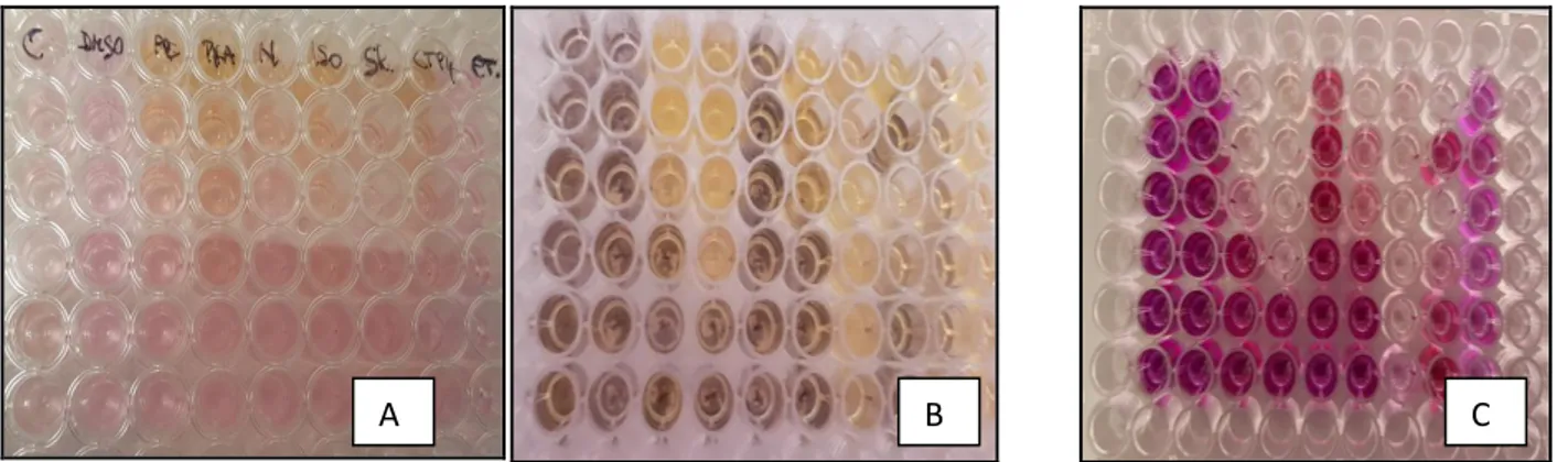
2.4.2.1. MTT assay……… 28
2.4.2.2. Calculations and data interpretation………... 30
2.5. Liquid-liquid extraction (LLE)……….. 32
VIII
2.6. Characterization with gas chromatography – mass spectrometry (GC/MS)……… 34 2.6.1. Derivatization of samples……… 34 2.6.2. GC/MS analysis………... 35 2.6.2.1. Expression of results……….. 35 2.7. Statistical analysis………... 35
3. RESULTS AND DISCUSSION………... 36
3.1. Initial screening for cytotoxicity against HepG2 and S17 cell lines – ethyl acetate (EA) extracts……….. 36 3.2 Screening for cytotoxicity of P. tricornutum (PHA) ethanol extract against HepG2 and S17 cell lines... 40 3.3. Screening for cytotoxicity of P. tricornutum (PHA) ethanol extract against THP-1 cell line... 43 3.4. Evalution of cytotoxic activity of fractions obtained from P. tricornutum ethanol extract against HepG2, THP-1 and S17 cell lines... 46 3.5. Microscopic examination of morphological changes using inverted microscope... 49 3.6. Identification of compounds present in the extract using gas chromatography-mass spectrometry (GC/MS)... 51 4. CONCLUSIONS………... 57
5. ANNEX………... 59
IX INDEX TABLES AND FIGURES
Table 1: Summary of most commonly used cancer cell lines with origin in different cell types……….
10
Table 2: Main characteristics of HepG2 cell line (ATCC® HB-8065™)… 11 Table 3: Main characteristics of THP-1 cell line (ATCC® TB-202™)…… 13 Table 4: Characterization of chemotherapeutic agents with some examples. 15 Table 5: Bioactive compounds obtained from microalgae and their effect on
human health………
20
Table 6: In vitro cytotoxicity of microalgae on cancer cell lines……… 21 Table 7: Properties of solvents used in liquid-liquid extraction (LLE) and
operating conditions for rotary evaporator………
33
Table 8: In vitro cytotoxic activity (IC50; µg/mL) of the ethyl acetate (EA)
extracts of the species P. tricornutum, Isochrysis sp.,
Porphyridium sp., Skeletonema costatum, Nannochloropsis sp., Tetraselmis sp. CTP4 and etoposide, on HepG2 and S17 cell
lines...
40
Table 9: In vitro cytotoxic activity (IC50; µg/mL) of ethanol (ETH) extracts
of P. tricornutum, Isochrysis sp. and etoposide on HepG2 and S17 cell lines...
42
Table 10: Cytotoxicity (IC50; µg/mL) of P. tricornutum ethanol extract and
etoposide against HepG2, THP1 and S17 cell lines...
44
Table 11: Cytotoxicity (IC50; µg/mL) of fractions (hexane, dichloromethane,
ethyl acetate, water+ethanol) from P. tricornutum ethanol extract and etoposide against HepG2, THP1 and S17 cell lines...
49
Table 12: Characterisation of compounds present in dichloromethane
fraction by (GC/MS)...
X
Figure 1: HepG2 cells in culture………... 12
Figure 2: THP-1 cells in culture……… 13
Figure 3: Extract preparation from dried algal biomass... 26 Figure 4: Microalgae species used to test cytotoxicity on HepG2 and THP-1
cell lines ………...
27
Figure 5: MTT assay performed on 96-well plate (HepG2 cell line)…………
30
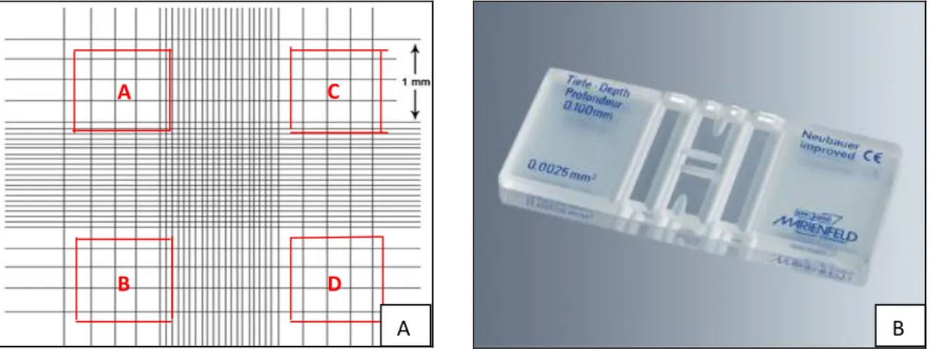
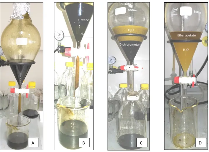
Figure 6: Neubauer chamber: counting cells ... 31 Figure 7: Liquid-liquid extraction (LLE) for Phaeodactylumtricornutum
ethanol extract………...
34
Figure 8: Cell viability (%) after 48 h exposure to different concentrations (3.9 – 125 µg/mL) of the ethyl acetate (EA) extracts of the species
P. tricornutum, Isochrysis sp., Porphyridium sp., Skeletonema costatum, Nannochloropsis sp., and Tetraselmis sp. CTP4) and
etoposide, on HepG2 and S17 cell lines...
39
Figure 9: Cell viability (%) after 48 h exposure to different concentrations (3.9 – 125 µg/mL) of the ethanol (ETH) extracts of the species P.
tricornutum and Isochrysis sp., on HepG2 and S17 cells...
42
Figure 10: Cell viability (%) after 48 h exposure to different concentrations (0.24– 125 µg/mL) of the ethanol (ETH) extract of P. tricornutum and etoposide on HepG2 and S17 cells...
44
Figure 11: Cell viability (%) after 48 h exposure to different concentrations (3.9– 125 µg/mL) of the fractions (hexane, dichloromethane, ethyl-acetate, water+ethanol) from P. tricornutum ethanol (ETH) crude extract and etoposide, on HepG2, THP-1 and S17 cells...
50
Figure 12: Examination of morphological changes on cells with light inverted microscopy...
1
1. INTRODUCTION
1.1. Cancer biology
Cancer is a global health issue and the second leading cause of death in high-income countries and the third leading cause of death in low- and middle-income countries worldwide1. It is estimated that cancer was responsible for 9.6 million deaths in 20182. Lung, prostate, colorectal, stomach and liver cancer are the most common types of cancer in men, while breast, colorectal, lung, cervix and thyroid cancer are the most common among women2. By 2030, it is estimated that the incidence of cancer in the world will grow to over 21.7 million new cases of disease and lead to 13 million deaths1.
According to current evidence, between 30% and 50% of cancer deaths could be prevented simply by avoiding tobacco products, reducing alcohol consumption, maintaining a healthy body weight, exercising regularly, and addressing infection-related risk factors2.
The term cancer derives from the Greek word "karkinos", which means crab. Cancer encompasses over 100 distinct diseases having in common the abnormal growth of cells that invade adjoining parts of the body and/or spread to other organs. Other common terms used for cancer are malignant tumours and neoplasms. Cancer can affect almost any organ in the human body and has many anatomic and molecular subtypes, that require specific management strategies. The oldest written description of cancer known to exist dates back to about 1600 BC, but it is believed to be based on a much earlier document, from ca. 3000 BC. Hippocrates first used the words carcinos and carcinoma to describe cancer, referring to the thick blood vessels that surround and feed the tumours and that resemble the claws of a crab3,4.
1.1.1. Hallmarks of cancer cells
Cancer cells differ from normal cells in many ways due to the fact these cells do not respond properly to the signals that regulate normal cell behaviour, and consequently cancer cells are able to proliferate continuously avoiding cell death1,3,4.
Hanahan and Weinberg (2000) established six hallmarks that are common for all human tumours and are responsible for their malignant properties: self-sufficiency in growth signals (GS), insensitivity to antigrowth signals, ability to avoid apoptosis, limitless replication potential, angiogenesis and capacity to produce metastasis.
2
Self-sufficiency in growth signals. Normal cells need mitogenic growth signals (GS) in order to proliferate. Most soluble GSs are produced by one cell type in order to stimulate the proliferation of another cell type in a process called heterotypic signalling. Cancer cells, on the other hand, have acquired the ability to synthesize their own GSs to which they respond by proliferating and creating a positive feedback signalling loop called autocrine stimulation5. Insensitivity to antigrowth signals. In order to maintain normal tissue homeostasis, many antigrowth signals are synthesized and recognized by transmembrane cell surface receptors to prevent excessive proliferation. These antigrowth signals can block proliferation either by forcing cells from proliferative cycle (G1) back to quiescent state (G0) or by inducing
permanent loss of proliferative potential of the cell5,6. One of the most studied mechanisms of negative cell cycle control is the retinoblastoma protein (pRb) pathway. It has been shown that pRb is responsible for a major G1 checkpoint, blocking proliferation and progression from G1 to S-phase of cell cycle. Cancer cells can disrupt the pRb pathway by releasing signalling molecules like TGFβ that prevents phosphorylation, which in turn inactivates pRb. In this way tumour cells turn themselves insensitive to antigrowth factors and are able to keep proliferating5,7.
Avoiding apoptosis. There are various strategies through which cancer cells avoid apoptosis. One of the most common mechanisms is through a mutation of the p53 tumour suppressor gene5,6. The p53 is a transcription factor that binds to damaged DNA and promotes tumour suppression by two distinct mechanisms. One mechanism includes inducing cell cycle arrest in G1 phase and preventing S-phase entry; the other one is promoting apoptosis dependent on the Apoptotic Peptidase Activating Factor 1 (Apaf-1)/caspase-9 pathway and involves
mitochondrial cytochrome c release. However, it remains unclear why certain cells undergo apoptosis in response to p53 activation while other cells undergo p53-dependent cell cycle arrest. More than half of all types of human cancers have a mutation or missing gene for p53 resulting in a damaged or missing p53 protein5,6,8. As an alternative, cancer cells can
compromise the activity of p53 by increasing its inhibitors or by silencing the activators of p53. One typical example of this is E6 protein produced by human papilloma virus, which inactivates p53 and enables cancer cells to evade apoptosis and spread8.
Limitless replication potential. The three acquired capabilities of cancer cells described above (self-sufficiency in growth signals, insensitivity to antitumor growth factors and avoiding apoptosis) are major responsible factors for tumour malignancies, but still not enough to cause macroscopic tumours. Research performed with cultured cell lines over the years revealed that after a certain number of cell divisions (60-70) cells stop growing and enter a
3
phase called senescence9. After this they slowly start to die. The reason behind this limited number of replications is, as it was discovered later, progressive telomeres shortening which leads to an inability of DNA polymerases to completely replicate the 3' ends of chromosomal DNA. Cancer cells have acquired a mechanism to avoid senescence by maintaining telomere length. This is achieved by increased expression of the telomerase enzyme, which adds hexanucleotide repeats on the ends of telomeric DNA, keeping the length of telomeres always above a certain threshold and assuring constant cell replication.5,6,9,10.
Angiogenesis. The process through which new blood and lymphatic vessels are formed from pre-existing vessels is called angiogenesis. This process is essential during foetal
development, the female reproductive cycle, and for tissue repair. One of the most specific and critical regulators of angiogenesis is the vascular endothelial growth factor (VEGF), which regulates endothelial proliferation, permeability, and survival; and thrombospondin-1 (TSP-1), which is an endothelial growth inhibitor. During normal physiological processes these two factors are always at equilibrium assuring tissue homeostasis. When tumours are present, as they grow, their need for oxygen, nutrients and waste disposal is increasing. Because of this, an “angiogenic switch“ is activated in order to sustain the expanding neoplastic growth. This is achieved by changing the balance between VEGF and TSP-1, mainly through altered gene transcription6,11.
Metastasis. Almost all types of solid tumours during their development enter a process called metastasis. Metastasis involves the spread of cancer cells from the primary tumour to the surrounding tissues and to distant organs, and it is the primary cause of cancer morbidity and mortality. The complete metastatic process consists of the following steps: first, cancer cells detach from the primary tumour, and intravasate into the circulatory and lymphatic system where they evade immune responses, then extravasation of cells at distant capillary beds occurs while invading and proliferating in distant organs. Several hypotheses ,which explain the origin of metastasis, exist: epithelial mesenchymal transition, an accumulation of
mutations in stem cells, a macrophage facilitation process, and a macrophage origin involving either transformation or fusion hybridization with neoplastic cells6,12.
4 1.1.2. Causes of cancer
Cancer is a heterogeneous group of diseases with diverse aetiology and pathogenesis. Each type of cancer has different mechanism of arising and progressing. Because of that, one general cause of cancer cannot be established as a rule.
There are different hypothesis and explanations for cancer evolution4:
Genetic changes
Epigenetic changes
Viruses infection
DNA lesions
Genetic changes. For the past 30 years cancer has been thought to arise from a single cell which goes through a series of different genetic alterations causing tumour proliferation, invasion, metastasis and drug resistance. This theory is called clonal genetic model. On the other hand, the multiple-mutation model, which was proposed later, was based on a belief that cancer arises as a result of many different mutations accumulating in different cells over time. According to this model, it should take decades for a tumour to arise, and then it should also take decades for tumour progression, which is not the case. The genetic instability model has been suggested as an alternative model. This model proposes that cancer arises due to an accumulation of genetic changes over time, but in specific genes that were found to be altered in many cancers – these genes are called oncogenes and tumour-suppressor genes4. Proto-oncogenes stimulate cell growth, division, and survival. When a proto-oncogene mutates, it becomes permanently activated, which enables cells to grow without control and become oncogenic. Oncogenes are activated by chromosome rearrangements or gene duplication. Tumour suppressor genes normally help to prevent unrestrained cellular growth and promote DNA repair and cell cycle checkpoint activation. When they lose their function, cells tend to acquire abnormal behaviour13.
Epigenetic changes . Epigenetics is the study of non-sequence-based alterations that are inherited through cell division and affect gene expression. Epigenetic alterations do not involve changes in the underlying DNA sequence but only phenotype change. Epigenetic change is a regular and natural occurrence, but it can also be influenced by several factors including age, the environment/lifestyle, and disease state. The most studied epigenetic modifications include: DNA hypomethylation, hypermethylation and hypomethylation of specific genes, chromatin alterations and loss of imprinting. All these lead to aberrant
5
Viral infections. There are some viruses that possess oncogenes and are able to cause cancer. The first discovered oncovirus was Rous sarcoma virus (RSV), which causes avian sarcoma and was named by its discoverer, Peyton Rous. Thanks to this discovery, many years of research led to another important discovery – the first oncogene, v-SRC as well as its cellular precursor, c-SRC3. Another example of an oncogene virus is the human papillomavirus (HPV) that causes a series of transformations on squamous epithelial cells in woman's cervix. These transformations, which are called cervical intraepithelial neoplasia (CIN), are
reversible in the beginning, and the affected part of tissue can be successfully removed. However, if CIN is not detected on time, it can progress to irreversible cervical cancer14. DNA lesions. One of the most frequent causes amongst all types of cancers are DNA lesions which cannot be controlled and arise from human genome instability. It is estimated that there are tens of thousands of DNA lesions per cell per day that can corrupt human genetic information3.
Factors responsible for DNA lesions occurrence are:
chemical reactions (hydrolysis of nucleotide bases and non-enzymatic methylations);
occasional mismatches introduced by DNA polymerases during replication and by DNA strand breaks generated as a consequence of abortive activities of
topoisomerases I and II;
endogenous agents (reactive oxygen species – ROS, generated through aerobic cellular respiration);
exogenous agents (UV light, tobacco-derived chemicals and ionizing radiation). DNA lesions include adducts, oxidized bases, abasic sites, DNA crosslinks, single-strand breaks (SSBs) and, less frequently, double-strand breaks (DSBs)3.
1.1.3. Types of cancer
Cancers are named after the area in which they begin and the type of cells they are made of, even if they spread to other parts of the body.
Carcinoma refers to a cancer that starts in the skin or the tissues that line internal organs (liver, lungs, kidneys, intestines, reproductive organs, etc.).
Sarcoma refers to a cancer of connective tissues such as bones, muscles, cartilage, and blood vessels.
Leukaemia is a cancer of bone marrow, affecting the production of white blood cells (monocytes, platelets, granulocytes).
6
Lymphoma is a cancer of the immune system cells (T lymphocytes and B lymphocytes)15. For the purpose of this Master thesis, the focus was on two different types of cancer:
hepatocellular carcinoma (HCC), which is fast-growing solid tumour primary in liver, and acute monocytic leukaemia (AML), a haematological malignant disorder or “suspended” tumour. The major difference between HCC and AML is obviously in their phenotype and area where they begin (liver and bone marrow, respectively). Apart from that, it is interesting to mention that the prevalence of haematological malignancies is much lower comparing to solid tumours, and accounts for only 6,2% of all deaths caused by cancer. However, the number of publications in the field of haematological malignancies is much higher than those on solid tumours as specimens of malignant cells can be obtained easily from peripheral blood of patients16.
1.1.4. Hepatocellular carcinoma
Hepatocellular carcinoma (HCC) is the most common type of primary liver carcinoma. It is usually found in people with chronic liver disease and it is a leading cause of cancer-related deaths in the world. Despite the fact that huge progress has been made in terms of
prevention, screening and treatment of HCC, incidence and mortality of this disease continue to rise. This carcinoma occurs more often in males than in females (2.4:1), with a higher incidence in Eastern and Southern Asia, Middle and Western Africa, Melanesia, and Micronesia/Polynesia17.
The main important risk factor for developing HCC is cirrhosis. This is a condition where liver is severely damaged and scarring tissue is more abundant than healthy one,
compromising liver function. Cirrhosis develops over time due to liver damage caused by infection with hepatitis B and C viruses and alcohol consumption. Approximately 80% of patients infected with hepatitis C develop chronic hepatitis, and ~20% end up developing cirrhosis. The development of HCC occurs almost exclusively in livers with already established cirrhosis17.
The HCC diagnosis is confirmed without pathologic confirmation. Screening includes radiologic tests (ultrasound, computerized tomography, and magnetic resonance imaging), and the presence of a serological marker (fetoprotein at 6-month intervals)17.
In spite of all available screening techniques, the majority of patients are not properly screened and HCC is many times diagnosed at an already advanced stage when patients are symptomatic and liver is severely damaged. A problem at this late stage is that there is no effective treatment to improve the survival rate of patients. The treatment option with higher
7
successful rate is surgical resection or liver transplant, which is not always possible. Nonsurgical treatments in the form of chemotherapy are usually not successful for HCC patients. Moreover, higher mortality rates in HCC are associated with chemotherapy. Currently, scientists worldwide are working on finding a new potential therapeutic agent which could improve life of HCC patients17.
1.1.5. Acute monocytic leukaemia
Acute myeloblastic leukaemia (AML) is a group of bone marrow neoplasms of myeloid precursors of white blood cells. One of the most common types of AML is acute monocytic leukaemia (AML-M5) that usually occurs in young children (< 2 years). However, the
condition is rare and represents approximately 2.5% of all leukaemias during childhood and it has an incidence of 0.8 – 1.1 per million per year18.
The AML-M5 is defined by the presence of more than 20% (WHO classification) or more than 30% (French-American-British, FAB classification) of myeloblasts in the bone marrow aspirate while normal bone marrow myeloblast count is less than 5%. Malignancy can develop as a result of congenital (Down syndrome, Fanconi's anaemia, congenital
neutropenia) and / or acquired (prenatal exposure to tobacco, alcohol, radiation) factors18. The symptoms are not specific and include asthenia, pallor, fever, dizziness and respiratory failure. More specific symptoms are the appearance of unexplained bruises and/or (excessive) bleeding, coagulation disorders (DIC), neurological disorders and gingival hyperplasia. Diagnostic methods include blood analysis, bone marrow aspirate for cytochemical, immunological and cytogenetical analysis, and cerebrospinal fluid (CSF) investigations18. Treatment options for AML consist of aggressive multidrug chemotherapy regimens, which are associated with high mortality and morbidity. The main drugs used for the treatment of AML are: arecytarabine, anthracyclins (daunorubicin, idarubicin and mitoxantrone) and etoposide. Therapy protocols include 3-7 courses of drug administration with intervals of 3-4 weeks. Chemotherapy can have serious consequences as severe bone marrow suppression, leading to leukocytopenia, neutropenia and thrombocytopenia. The overall 5-year survival rate for AML patients is 30-60% and long-term event free survival rate is only 20-50%. Therefore, new therapeutics are needed to increase the probability of cure in this disorder19.
8
1.2. Cancer cell lines as in vitro models for cancer research
Different experimental model systems, both in vivo and in vitro, exist for studying
pathobiology of cancer. In vivo models include primary tumours, patient-derived xenografts and mice. The most used in vitro models are: paraffin-embedded samples, cancer cell lines and tumour primary cell cultures. Each of these models is used for different studies due to limitations associated with possible genetic manipulation20.
For example, primary tumours (samples obtained by surgery from patients) represent the state of the tumour in vivo with its heterogeneity, but only at a specific evolutionary moment of the tumour. This sample is difficult to obtain, its amount is limited and genetic manipulation is almost impossible21,22.
Patient-derived xenografts in nude mice models are often used for drug testing, providing the
in vivo microenvironment for human tumour original cells but, on the other hand, they have
an important limitation – these mice are immune-compromised which contributes to an overall inflammation process in cancer.
Animal models with spontaneous or induced tumours are used in the pathobiology research of cancer and for testing new therapeutics in vivo. However, using animal models in research means following strict ethical rules and the number of animals is always limited. Apart from that, it is difficult to extrapolate obtained results to humans20.
As for in vitro systems, primary cell cultures are good models since they are derived directly from the tumours of the patient, which means they maintain some of the heterogeneity of the original tumour. However, the tissue environment is lost while culturing and samples are scarce and difficult to obtain20.
This brings us to the conclusion that every experimental model for cancer research has certain advantages and disadvantages and none of them is completely representative of the
phenotype of the tumour.
Nevertheless, cancer cell lines are the most adequate in vitro model for cancer research for many reasons. Cell lines are permanently established cell cultures that differ from cell strains in that they are immortalize. E6/E7 gene of human papillomavirus16 (HPV-16), or small and large T-antigen of the simian virus 40 (SV-40) are some examples of strategies used for cell transformation and immortalization . This means that cell lines will proliferate indefinitely given appropriate fresh medium and space, which makes them easy to handle and manipulate. Due to the progress in immortalization and cell culture techniques, almost every tissue can be
9
cultured nowadays and there is a large number and variety of cell lines immediately available for studying different types of cancer21.
The most important characteristic of cancer cell lines is their high genomic similarity with the original tumour. Cancer cell lines maintain the tumour-specific chromosome abnormalities and oncogene mutations during the first passages and show the same morphologic and molecular characteristics of the primary tumour. In general, cell lines conserve well all “hallmarks of cancer” (described in Section 1.1.1), with an exception of angiogenesis due to the fact it requires the presence of stromal tissues20,21.
Cancer cell lines also contain cancer stem cells, which makes them an excellent model for development and testing of anticancer drugs, which was proven by many scientists over two decades ago. Finlay and Bagulay (1984) demonstrated that cancer cell lines display a similar response to anticancer drugs when compared to the original tumour.
However, cancer lines do have some limitations. As the number of cell passages tends to increase, cells start to lose their phenotypic properties and become genomically instable. This leads to the rise of molecular changes including modifications in cell signalling pathways. Because of that, cell culturing for long time periods should be avoided21,23.
To summarize, chosing the appropriate in vitro model in cancer research is the most important step for the screening of new potential cancer therapies. The results from the research done on cancer cell lines can be extrapolated to in vivo human tumours and this has a great importance for drug testing and translational studies. This characteristic has been recognized by many biomedical and pharmaceutical companies20.Nevertheless, proper molecular characterization (genetic, epigenetic) of cell lines is fundamental before their use in order to determine the polygenetic aetiology of the type of cancer studied and the
molecular mechanisms involved. This enables assessment of different types and subtypes of cancer as well as suitability of cell line established for studying that particular cancer type. Characterization is especially important for the development of new anticancer drugs as it can aid in revealing new targets and it also helps to understand the mechanisms involved in the resistance patterns of cells to certain chemotherapeutics22.
As mentioned earlier in this section, there are a large number of cancer cell lines available in different culture collections. However, some of them are poorly characterized and may not bare the same molecular characteristics as the tumours they represent. In Table 1, the most commonly used cancer cell lines are summarized20.
10
To conduct this Master Thesis, two cell lines from this list (Table 1) were studied in detail – HepG2 and THP-1 cells, representing hepatocellular carcinoma and acute monocytic leukaemia, respectively.
Table 1. Summary of most commonly used cancer cell lines with origin in different cell types20
Cancer cell line Species of origin Type of cancer Morphology
HeLa Homo sapiens Cervix adenocarcinoma Epithelial
MCF-7 Homo sapiens Brest adenocarcinoma Epithelial
U87MG Homo sapiens Glioblastoma-astrocytoma Epithelial
HT-29 Homo sapiens Colon adenocarcinoma Epithelial
A549 Homo sapiens Lung carcinoma Epithelial
HepG2 Homo sapiens Hepatocellular carcinoma Epithelial K-562 Homo sapiens Chronic myeloid leukaemia Lymphoblast
THP-1 Homo sapiens Acute monocytic leukaemia Lymphoblast
PC3 Homo sapiens Prostate adenocarcinoma Epithelial
A375 Homo sapiens Malignant melanoma Epithelial
1.2.1. HepG2 cell line
HepG2 is an immortalized cell line consisting of human liver carcinoma cells. It is the most widely used human hepatoma cell line and it was derived from a liver hepatocellular
carcinoma of a 15 year old Caucasian male23–25.
Hepatocellular carcinoma (HCC), the major type of liver cancer, has emerged as the second most common cause of cancer-related death. The only approved drug for treating advanced HCC, developed so far, is Sorafenib. As previously described in Section 1.1.4, chemotherapy used for treating HCC is not successful and is associated with high mortality in patients; moreover, liver transplant is the only effective treatment option. Therefore, it is highly desirable to develop new drugs for this disease24.
The HepG2 cell line used in this study was obtained from the American Type Culture Collection (ATCC® HB-8056™). The morphology of HepG2 cells is epithelial and contains 55 chromosome pairs. Cells are adherent and grow as monolayers and in small aggregates (Figure 1). HepG2 cells can be grown successfully at large scale, and secrete many plasma proteins, such as transferrin, fibrinogen, plasminogen and albumin26. A detailed summary of HepG2 characteristics provided by ATTC is given in Table 2.
11
Table 2. Main characteristics of HepG2 cell line (ATCC® HB-8065™)26 Characteristics Description
Karyotype modal number = 55 (range = 50 to 60); has a rearranged chromosome 1
Receptor expression insulin; insulin-like growth factor II (IGF II)
Genes expressed alpha-fetoprotein (alpha fetoprotein); albumin; alpha2 macroglobulin
(alpha-2-macroglobulin); alpha1 antitrypsin (alpha-1-antitrypsin); transferrin; alpha1 antichymotrypsin; (alpha-1-antichymotrypsin); haptoglobin; ceruloplasmin;
plasminogen;,complement (C4); C3 activator; fibrinogen; alpha1 acid glycoprotein (alpha-1 acid glycoprotein); alpha2 HS glycoprotein
(alpha-2-HS-glycoprotein); beta lipoprotein (beta-lipoprotein); retinol binding protein (retinol-binding protein
Cellular products alpha-fetoprotein (alpha fetoprotein); albumin; alpha2 macroglobulin (alpha-2-macroglobulin); alpha1 antitrypsin (alpha-1-antitrypsin); transferrin; alpha1 antichymotrypsin; (alpha-1-antichymotrypsin); haptoglobin; ceruloplasmin;
plasminogen;
complement (C4); C3 activator; fibrinogen; alpha1 acid glycoprotein (alpha-1 acid glycoprotein); alpha2 HS glycoprotein (alpha-2-HS-glycoprotein); beta lipoprotein (beta-lipoprotein); retinol binding protein (retinol-binding protein)
The HepG2 cell line has several advantages comparing to primary cultured human
hepatocytes: it grows continuously, has almost an unlimited life span and a stable phenotype. It is easily available; culture conditions are simpler than primary hepatocytes, and are easily standardized among laboratories. These features make the HepG2 cell line appropriate for drug screening purposes24,25.
Despite the fact they express many liver-specific functions, the major drawback of this cell line is the lack of functional expression of almost all relevant human liver cytochrome P450 enzymes (CYP) that are involved in phase I of the oxidative metabolism of xenobiotics (drugs) system. There are about 50 CYP genes in the human genome encoding for catalytically active monooxygenase haemic proteins; however, only a few of them are capable to metabolize drugs and xenobiotics, which takes place in the liver. As a
consequence, HepG2 cell lines show very limited drug metabolism activities and they are resistant to most anticancer drugs23–25.
Primary human hepatocytes are still considered the best in vitro model for studying new therapeutic agents for hepatocellular carcinoma because they can provide information on how
12
the potential drug will be metabolized in vivo. Despite this, HepG2 cell lines are usually the first option and they are the most commonly used for screening and selection of new drugs as they provide the fastest way to identify drug leads with favourable pharmacokinetic and metabolic properties; sometimes, they are also the only option25.
Figure 1.HepG2 (ATCC® HB-8056™)in culture. Cells grow as monolayer forming aggregates: A) Low cell density; B) High cell density. Source: www.atcc.org.
1.2.2. THP-1 Cell line
THP-1 is a human monocytic leukaemia cell line that was firstly cultured from the peripheral blood of a 1-year-old male with acute monocytic leukaemia27,28. Acute monocytic leukaemia (AML) is a malignant disorder that affects hematopoietic cells (bone marrow). Usually occurs in young children (< 2 years) and it is associated with high mortality due to aggressive chemotherapeutic treatment. New therapies are needed to improve the probability of cure for this disease18,19.
The THP-1 cell line is a non-adherent, suspension cell line (Figure 2)29. The cells used in this study were obtained from the American Type Culture Collection (ATCC® TIB-202™). A summary of the main characteristics of THP-1 cell lines is given in Table 3.
The THP-1 cell line is one of the few leukaemia cell lines available and it is a good model for studying acute monocytic leukaemia since it retains monocytic properties (phagocytosis, activation of T-lymphocytes, lysozyme production, presence of alpha naphthyl butirate esterase) for prolonged period of time in culture conditions (>14 months)28.
13
Table 3.Main characteristics of THP-1 cell line (ATCC® TB-202™)29
In addition, upon stimulation with phorbol 12-myristate 13-acetate (PMA), monocyte (THP-1) cells start to differentiate into macrophage-like cells,becoming adherent. They resemble native monocyte-derived macrophages with respect to numerous criteria which makes THP-1 a valuable model for studying the role of macrophages in human immune response27,28. Moreover, both monocytes and macrophages play an important part in cancer progression. In tumour settings, monocytes are recruited from peripheral blood to the tumour tissue, where they differentiate into tumour-associated macrophages (TAM). This subclass of macrophages is a heterogeneous cell population regarding their phenotype and pro-tumour function. They have abilities to support tumour initiation, local progression and distant metastasis. Therefore, targeting monocytes and macrophages in cancer is a promising therapeutic approach30.
Figure 2. THP-1 (ATCC® TB-202™) in culture. Cells grow in suspension: A) Low cell density; B) High cell density. Source: www.atcc.org.
Characteristics Description Karyotype diploid (46, XY); Receptor expression complement (C3); Fc
Genes expressed lysozyme,HLA A2, A9, B5, DRw1, DRw2
Cellular products lysozyme
Antigen expression HLA A2, A9, B5, DRw1, DRw2
14 1.3. Cancer therapy
There are many types of cancer treatment. The types of treatment that patients receive will depend on the type of cancer and its stage of progression31.
According to the National Cancer Institute31 available cancer therapies are:
Surgery (procedure in which the malignant tissue is removed from the body);
Radiation therapy (using high-energy particles or waves, such as X-rays, γ-rays, electron beams or protons, in order to destroy or shrink cancer cells);
Chemotherapy (special drugs are administered orally or intravenously to stop the proliferation of malignant cancer cells);
Hormone therapy (slows or stops the growth of cancer cells; used only for prostate and breast cancer which need hormones to grow);
Immunotherapy (aims to boost human immune system in order to increase production of T cytotoxic cells, natural killer (NK) cells, or specific antibodies which target cancer cells);
Stem cell transplant (bone marrow transplant to induce production of new hematopoietic cells, which can increase ability to fight off cancer cells). Some patients with cancer will have only one treatment while some will receive a
combination of treatments, such as surgery with chemotherapy and/or radiation therapy31. Among all currently used cancer treatments, chemotherapy continues to play an extremely important role. However, its effectiveness is limited in some cases by the existence of drug resistance, making it necessary to define optimal combinations for therapeutic strategies in order to ensure an efficient elimination of the tumour1.
1.3.1. Chemotherapeutic agents
In general, chemotherapeutic agents kill malignant cells causing apoptosis. This was demonstrated in vitro with tumour cell lines, as well as in vivo, in patients receiving
chemotherapy. Apoptosis is a normal cell response to irreparable DNA damage, which leads to cell death. There are two major cell-intrinsic pathways for inducing apoptosis. One involves inducing the expression of cell surface death receptors (Fas) that bind to ligands (FasL), which in turn activate adaptor molecules (FADD) and finally procaspase-8. Another pathway involves the release of cytochrome c from mitochondria, binding Apaf-1, and activatingprocaspase-9. Once activated, caspase-8 or caspase-9 can in turn activate caspases-3, 6, and 732. Caspases are a unique family of cysteine-dependent proteases that are
15
responsible for proteolytic cleavages during apoptosis. These cleavages include internucleosomal DNA degradation and overall cytoskeleton break up leading to the
formation of apoptotic bodies. Caspases are present in the cell as inactive zymogens and go through a cascade of catalytic activations at the onset of apoptosis33.
Chemotherapeutics are generally classified in five groups: alkylating agents, antimetabolites, mitotic inhibitors, topoisomerase inhibitors and tumour antibiotics. Each group has a different mechanism of action on cancer cells (Table 4).
The most widely used antineoplastic drug is etoposide, from the topoisomerase inhibitors group34.
Table 4.Classification of chemotherapeutic agents with some examples. Source: www.chemocare.com35 Drug class Drug Mechanism
Alkylating agents Cyclophosphamide, Cisplatin Alkylation of DNA/RNA leading to cross-link between DNA strands
Mitotic inhibitors Vincristine, Docetaxel Destruction of tubulin causing mitotic arrest in metaphase
Topoisomerase inhibitors
Topotecan, Etoposide Inhibition of topoisomerase I and II causing DNA degradation
Antimetabolites Methotrexate,Hydroxyurea Inhibit ion of DNA replication and prevention of cell growth
Cancer antibiotics Mitomycin, Actinomycin D Inhibition of RNA synthesis which prevents cell mitosis
1.3.2. Etoposide as a topoisomerase II inhibitor
DNA topoisomerases are essential enzymes that regulate the topological state of genetic material by introducing transient breaks in the DNA molecule. They are involved in fundamental biological processes such as DNA replication, transcription, DNA repair and chromatin remodelling. Two major topoisomerase forms are present in all cells: type I (Topo I) which makes single-strand cuts in DNA, and type II (Topo II) which modulates DNA topology by passing an intact helix through a transient double-stranded break created in the DNA backbone. As a result, Topo II is able to regulate DNA catenation/decatenation, relaxation/supercoiling and knotting/unknotting. The covalent topoisomerase-cleaved DNA complex is a short-live intermediate and the breaks created are quickly repaired after passing the helix. Etoposide does not directly inhibit the action of Topo II but rather stabilizes this covalent cleavage complex introducing more breaks in DNA, which become permanent and irreparable. When these permanent DNA breaks are present at sufficient concentration, they trigger a series of events that culminate in cell death by apoptosis32,36,37.
16
Etoposide is an analogue of 4-demethylepipodophyllin benzylideneglucoside (DEPBG), which is a plant toxin obtained from Podophyllum (American Mayapple or Mandrake). Podophyllotoxins have been used as medication by various cultures for over 1,000 years and were found to be topically effective for skin cancers, although, when ingested, they displayed high toxicity37.
Etoposide was first synthesized by Sandoz Pharmaceuticals in 1966 and approved for cancer therapy in 1983 by the U.S. Food and Drug Administration. During clinical trials, etoposide demonstrated antineoplastic activity in various types of leukaemias, lymphomas and solid tumours. Namely, acute monocytic leukaemia (AML), Hodgkin's disease, non-Hodgkin's lymphoma, lung cancer (both small cell and non-small cell), gastric cancer, breast cancer and ovarian cancer, sarcoma and neuroblastoma. It could be administered both orally and
intravenously to patients36,37.
However, etoposide, as all chemotherapy drugs, has certain drawbacks. The first issue addresses drug specificity. All mammals have two Topo II isoenzymes, Topo II α and β that are differently regulated during cell growth. Topo II α is a proliferation marker, greatly elevated in tumour cells, whereas the TopoII β is presented in proliferating, as well as post-mitotic cells. Both isoenzymes are the target of etoposide which leads to off-target toxicity destroying not only malignant cells but also healthy cells including white blood cells needed for immunity36,37. As a consequence, the number of side-effects arise in patients: hair loss, low white blood cell count, vomiting, nausea, mouth sores, diarrhoea, fatigue, low blood pressure, etc.38. Another important therapeutic issue is the development of resistance and/or development of secondary malignancies, such as AML37.
These side effects point out to the need to search, find and identify new sources of anticancer drugs with high selectivity and toxicity only for malignant cells, while conserving healthy immune cells.
Moreover, in the last decades, with the continuous growth of cancer cases and concerns over resistance to certain drugs, development of secondary cancers and the unwanted side effects observed, scientists are turning to alternative sources, trying to find potential drug leads in natural products (NP)1.
17
1.4. The marine environment as promising source of new drug leads
Natural products (NPs) have been used as a source of therapeutic agents for the treatment of a wide spectrum of illnesses for thousands of years. Plants, in particular, have formed the basis of traditional medicine with the earliest records from Mesopotamia, around 2600 BCE, where usage of approximately 1000 plant-derived substances for curing different disorders was documented.39,40Even nowadays, many drugs present on the market are plant-derived NPs or their derivatives.
However, since natural product-based drug discovery is sometimes difficult due to their complexity, pharmaceutical industry has shifted its focus toward synthetic compound libraries for discovery of new drug leads. Unfortunately, obtained results did not meet expectations, especially for autoimmune diseases or cancer. This resulted in a declining number of new drugs on the market and, at the same time, revitalizing interests for NP. Bioactive compounds obtained from NPs are interesting due to their complex chemical structures. This complexity is a result of a phenomenon called biodiversity, where the interactions between organisms and their environment happen all the time, leading to the production of chemical compounds within the organisms, that enhance their survival and competitiveness in the environment39,41,42.
Apart from terrestrial organisms, marine organisms have emerged as potential new sources of different NPs over the past two decades.
The systematic investigation of marine environments in order to identify novel biologically active compounds started only in the mid-1970s with the development of new technologies in scuba diving techniques and engineering of manned submersibles and remotely operated vehicles (ROVs) which facilitated exploring and sampling of deep waters1,40,43.
During the period 1977–1987, about 2,500 new metabolites were reported from a variety of marine organisms. In 2015, a review performed by Blunt et al. revealed that the number of compounds of marine origin reached 30,000 with more than 300 patents waiting for approval. Moreover, in their last review from 2018, they reported additional 1277 new compounds isolated and described from marine microorganisms and phytoplankton, green, brown and red algae, sponges, cnidarians, bryozoans, molluscs, tunicates, echinoderms, mangroves and other intertidal plants and microorganisms, only for the year 2016. These numbers suggest that the marine environment indeed is a rich source of NPs with uncommon and unique chemical features that are not found in terrestrial species. The reason for this is a large
18
are represented in aquatic environments, with 15 exclusively marine. 17 are found in marine and non-marine environments (with 5 of these having more than 95% of their species only in marine environments), and only one is exclusively non-marine (Onychophora). It has been predicted that approximately 8.9 million of eukaryotic species exist, of which approximately 2.2 million are marine organisms. This means that around 86% of the species on the earth, and 91% in the ocean, have not yet been described1,41.
The coexistence of so many species in marine habitats and constant competition between them, as well as demanding environmental conditions (UV light, salinity, temperature, high pressure), resulted in the development of different mechanisms by marine organisms to defend themselves against predation or overgrowth of competing species. These mechanisms are actually chemical adaptions, defined as secondary metabolites that involve different classes of chemical compounds. They are generally specific for a particular taxonomic family, genus, species or even organism, and constitute a very small fraction of the total biomass of the organism. Predominantly, production of these NPs with excellent
pharmacological potential occurs in sessile or slow-moving organisms (e.g. algae, sponges, cnidarians, tunicates and bryozoans) that, without effective escape mechanisms or structural protection, ensure their protection through chemical defence. Moreover, high
pharmacological activity of NPs obtained from marine organisms comes from the fact that these compounds are released to the water and are rapidly diluted, therefore, they have to be highly potent to retain their efficacy1,40,41,43.
To date, different types of secondary metabolites were isolated from marine organisms: terpenoids, alkaloids, polyketides, peptides, shikimic acid derivatives, sugars, steroids, and a large mixture of biogenesis metabolites. These compounds were found to exhibit many biological activities (antimicrobial, anti-tumoural, anti-diabetic, anticoagulant, antioxidant, anti-inflammatory, antiviral, antimalarial, anti-tubercular, anti-aging antifouling, and antiprotozoal) with huge industrial and therapeutic potentials1,41,43.
The current pipeline for new compounds, from the initial demonstration that a molecule may have therapeutic potential to the production and approval, a drug needs to pass pre-clinical testing, complex clinical trials in humans, and post-trial approval by the regulatory organisms - Food and Drug Administration (FDA) (in US) and European Medicines Agency (EMA) (EU). This process can take 10 to 15 years with less than 12% of the potential drugs receiving final approval44.
To date, seven approved marine-derived pharmaceuticals are in clinical use, four of which are anticancer drugs. The first marine derived anticancer agent developed for clinical use,
19
cytarabine or Ara-C, is a synthetic analogue of a C-nucleoside from the Caribbean sponge,
Cryptothethya crypta, approved in 1969 and still in use for the treatment of acute myelocytic
leukaemia and non-Hodgkin’s lymphoma. Another example is Aplidine (dehydrodidemnin B) developed by PharmaMar©. This depsipeptide hydrodidemnin was isolated from the
Mediterranean tunicate Aplidiumalbicans, and used in the treatment of multiple myeloma (phase III of clinical trials), and for solid and haematological malignant neoplasias, like T-cell lymphoma (phase II of clinical trials)45.
1.4.1. Marine microalgae as a novel source of anti-tumoural drugs
Among marine organisms, algae and microalgae are one of the most important resources of the ocean, economically and ecologically. Microalgae are microscopic, photosynthetic organisms found in both marine and freshwater environments and represent the major component of phytoplankton. They are primary producers, responsible for up to 50% of global carbon fixation. Their photosynthetic mechanism is similar to that of land-based plants, converting solar energy into biomass, mainly because of their simple cellular structure and being submerged in an aqueous environment with access to water, CO2, and other
nutrients46,47.
Microalgae are a polyphyletic and highly diverse group of prokaryotic and eukaryotic organisms48. The classification into divisions is based on various properties: pigmentation, chemical nature of photosynthetic storage products, the organization of photosynthetic membranes, and other morphological features. The most abundant microalgal classes are:
Cyanophyceae (blue-green and red algae), Chlorophyceae (green algae), Bacillariophyceae
(diatoms), Chrysophyceae (including golden algae) and Phaeophyceae (brown algae)46,49. Microalgae are considered as one of the most promising sources for new products and applications due to their chemical composition. These organisms are rich in important biomolecules such as polyunsaturated fatty acids (PUFAs), pigments (chlorophylls and carotenoids), polyphenolic compounds, phycobilins, vitamins, sterols and polysaccharides. It has been found that these biomolecules have not only nutritional value but also display bioactivities and have a potential use as therapeutic agents in the biomedical area. Many of these algae-derived compounds have been associated with numerous health promoting effects including anti-obesity, anti-diabetic, antihypertensive, anti-hyperlipidaemia, antioxidant, anticoagulant, anti-inflammatory, immune-modulatory, anti-estrogenic, thyroid-stimulating, neuroprotective, osteoarthritic, osteoporosis, antiviral, antimicrobial and anti-tumoural (Table 5)44,50,51.
20
Table 5.Bioactive compounds obtained from microalgae ant their effect on human health49–51 Bioactive compound isolated Health-promoting effect Microalgae Carotenoids
β-carotene antioxidant activity Dunaliellasalina; Haematococcus pluvialis; Tetraselmisspp. astaxanthin antioxidant, immunomodulation,
and cancer prevention H.pluvialis; Chlorella vulgaris fucoxanthin antioxidant, immunomodulation,
and cancer prevention
Isochrysisgalbana; Phaeodactylum tricornutum
lutein antioxidant activity Chlorella pyrenoidosa; Tetraselmis spp. violaxanthin antioxidant activity
Chlorella ellipsoidea; Nannochloropsisoculata; Tetraselmis
spp.
PUFAs
eicosapentaenoic acid (EPA)
reduce risk of certain heart diseases, anticoagulation,anti-inflammatory, neuroprotective
P.tricornutum;Porphyridiumcruentum; I. galbana
oleic acid antioxidant activity C. vulgaris; H.pluvialis; Spirulinaplatensis linoleic acid antimicrobial activity D.salina; S.platensis
palmitic acid antimicrobial activity D.salina
palmitoleic acid Antihypertensive S.platensis
Docosahexaenoic acid (DHA)
reduce risk of certain heart diseases, anticoagulation,anti-inflammatory, neuroprotective S.platensis Proteins Phycobiliproteins immunomodulation activity, anticancer activity, hepatoprotective, anti-inflammatory, and antioxidant properties S.platensis; Porphyridium spp. Polysaccharides sulfated polysaccharide antiviral, antitumor, antihyperlipidemia, and anticoagulant C. pyrenoidosa; Porphyridium spp. Insoluble fiber
reduce total and LDL cholesterol C. vulgaris
Vitamins
tocopherols (vitamin E) antioxidant activity S.platensis; Porphyridium spp., Tetraselmisspp
Phenolic compounds
benzoic acid derivatives, hydroxybenzaldehydes, and cinnamic acid derivatives
antioxidant activity S.platensis
Along the last five decades it is estimated that more than 3,000 NPs have been discovered from algae and among all of the mentioned biological activities, anti-tumoural activity seems to be one of the most promising44. Many studies demonstrated the in vitro cytotoxicity of different extracts (water, ethanol, ethyl acetate, hexane, acetone) obtained from microalgal biomass on tumoural cell lines (Table 6).
21
Table 6. In vitro cytotoxicity of microalgae on cancer cell lines
Microalgae Extract tested Cell line IC50 (μM or
μg/mL)
Compound responsible (if
applicable)
Observed effects on cells (if applicable
Spirulina platensis52
water Colon carcinoma(HCT116), hepatocellular carcinoma (HepG2)
18.8 μg/mL for HCT116; 22.3 μg/mL for HepG2 Porphyridium
purpureum53
isolated and purified carotenoid
Human melanoma (A2058) 40 µM zeaxanthin Chromatin condensation, nuclear blebbing, DNA nucleosomal fragmentation, activation of
caspase-3 (apoptosis) Nitzschia sp54 isolated and purified
carotenoid
Human glioma (U251) fucoxanthin Induction of apoptosis with PARP cleavage and caspase activation Isocrysis
galbana55
acetone HepG2 81.3 μg/mL Showed high selectivity (3.1)
comparing to non-tumoural, murine stromal S17 cell line Chaetoceros
calcitrans56
hexane, dichloromethane, ethyl
acetate and methanol
Breast adenocarcinoma (MDA-MB-231) , mouse breast carcinoma (4T1), HepG2,
cervix epithelial carcinoma (HeLa), human prostate carcinoma (PC-3), human lung adenocarcinoma (A549), human colon adenocarcinoma (HT-29)
60 µg/mL, ethyl acetate extract on the MDA-MB-231
cancer cell line
Extract did not show cytotoxicity on non-tumorigenic cells (mouse
embryo fibroblast (3T3))
Porphyridium cruentum64
sulfoglyco-lipidic fraction (SF)
Colon adenocarcinoma (DLD-1), human breast adenocarcinoma (MCF-7), human prostate adenocarcinoma (PC-3), and
human malignant melanoma (M4)
20-46 µg/mL (range applies to all tested cell cultures)
sulfolipids Inhibition of DNA α polymerase
Skeletonema costatum67
organic extract human non-small-cell bronchoplumanory carcinoma line
(NSCLC-N6)
G1 cell cycle arrest
Phaeodactylum tricornutum
ethanol HepG2 250 µg/mL Sulphated
polysaccharide Nannochloropsis
oculata
methanol human chronic myeloid leukemia cell line (K562)
50 µg/mL Induction of apoptosis with PARP cleavage and caspase activation
22
Despite the number of compounds isolated from microalgae and biological activities attributed to these, they still have not entered clinical trials or been marketed.,51,57.
There are several reasons for this, including the time and cost it takes to reach the market, difficulties in harvesting the organism, low titres of natural product in producing organisms, difficulties in isolation and purification procedures, problems in obtaining a sustainable supply of the compound, high toxicity of the active compound, ecological impact on natural populations, and insufficient investment by pharmaceutical companies45. In spite of the high number of microalgae species identified, only a few of them have been successfully
commercialized for biotechnological applications.
Examples are Chlorella spp. (production of lutein)57, Spirulina platensis (production of
phycobiliproteins)51, Dunaliella salina (natural source of β-carotene)46,57, Haematococcus pluvialis (production of astaxanthin)46,57, Porphyridium cruentum ( production of sulphated
polysaccharides, phycocyanin, and phycoerithrin)49,51, Phaeodactylum tricornutum (production of eicosapentaenoic acid)58, etc.
A reason behind this small number of commercialized microalgae species is a complex process of growth optimization of these organisms and in obtaining the compound of interest in large quantities. The chemical composition of microalgae is not an intrinsically constant factor, it varies among strains and batch cultures, seasons, and it is affected by environmental and culture conditions (temperature, pH, mineral content of water, light exposure, and
agitation). Therefore, it is very important to select adequate culturing methods (e.g. open or closed photobioreactors, indoors or outdoors) with carefully controlled conditions, so that the metabolism of the microalgae favours a high production of the particular compound of commercial interest (e.g. PUFAs antioxidants, polysaccharides)46,50,51. Once the biomass is enriched in the target compound, the next step is to optimize the conditions to extract the valuable component with high yields while maintaining its activity. An efficient separation process of the desired compound should be able to process a large volume of biomass, yield a product with a high dry weight percentage, and require modest investment, low consumption of energy, and low maintenance cost46,50,51.
Another difficulty in the production of microalgae is their recovery and preservation. The low productivity of biomass means that recovery systems need to efficiently manage very large volumes of medium with a very low concentration of biomass. This alone constitutes a great challenge to engineers who need to combine various recovery operations such as
(freeze-23
drying or spray-drying) the microalgae cells produced, so as to maintain the quality of the biomass and the activity of compounds of interest49–51.
1.4.2. Microalgae used in this study to assess cytotoxicty
In this study, ethyl acetate and ethanol extracts of six microalgal species were selected to be screened for cytotoxicity in cancer cell lines: Nannochloropsis sp., Isochrysis sp.,
Skeletonema costatum, Porphyridium sp., Tetraselmis sp. CTP4, and Phaeodactylum tricornutum. In literature, up to date, there are no reported cytotoxicity assays that involve
testing ethyl acetate extracts of these species on both HepG2 and THP-1 cells. For example, previous studies from MarBiotech research group reported toxicity of acetone, hexane, water and diethyl ether extracts of Isochrysis galbana on HepG2 cells, but research did not involve ethyl acetate extract and THP-1 cells55. In another study, a sulfoglycolipidic fraction (SF) isolated from Porphyridium cruentum was screened for anti-proliferative activity on human breast adenocarcinoma (MCF-7), human prostate adenocarcinoma (PC-3), and human malignant melanoma (M4) Beu cell-lines59. No studies are available reporting anti-proliferative activity of this microalgae on HepG2 and THP1. Sulphated polysaccharides extracted in ethanol from Phaeodactylum tricornutum, showed strong anticancer activity on HepG2 cells, but not on THP-1 cells60. Also, Phaeodactylum tricornutum methanol extract was toxic to human promyelocytic leukemia cell line (HL60), while Nannochloropsis oculata methanol extracts were toxic to human chronic myeloid leukemia cell line (K562)61. As for
Skeletonema costatum, organic extract of this diatom was studied in vitro for its effect on
asynchronous cells of a human non-small-cell bronchopulmonary carcinoma line (NSCLC-N6) and showed inhibition of growth in G1 stage of cell cycle62(Table 6). To the extent of our knowledge, Tetraselmis sp. CTP4 was not assessed for cytotoxic activity so far. Based on all stated above, we decided to screen for cytotoxic effects of ethyl acetate and ethanol extracts of already explained microalgal species on HepG2 and THP-1 cancer cells.
24 1.5. Objectives
The objective of this thesis is to search for innovative drug leads for hepatocellular carcinoma and acute monocytic leukaemia treatment. For this purpose, different extracts (ethanol, ethyl acetate) from already commercialized, available microalgal biomass (Porphyridium sp., Skeletonema costatum, Nannochloropsis sp., Phaeodactylum
tricornutum, Tetraselmis sp. CTP4 and Isochrysis sp.) were screened for cytotoxicity on
HepG2 and THP-1 tumoural cell lines. In addition, a cell line (S-17) derived from a murine bone marrow (non-tumoural) was used to assess selectivity. Results were compared with etoposide, a widely used agent in chemotherapy.
In order to study the anti-tumoural effect of microalgae extracts on HepG2 and THP-1 cells and propose a new anticancer drug lead, we have established the following specific objectives:
1. To test different concentrations of ethanol and ethyl acetate extracts from microalgae (Porphyridium sp., Skeletonema costatum, Nannochloropsis sp.,
Phaeodactylum tricornutum, Tetraselmis sp. CTP4 and Isochrysis sp.) on HepG2
and THP-1 cell lines in order to determine the effects on cell viability.
2. To test different concentrations of ethanol and ethyl acetate extracts on S-17 non-tumoural cell line to assess selectivity.
3. To fractionate the most active extracts and produce active fractions with a more limited number of compounds.
4. To chemically characterize the most active fraction and tentatively identify a potential drug lead in microalgae extracts/fractions.