UNIVERSIDADE DA BEIRA INTERIOR
Ciências da Saúde
Mecanismos moleculares da progressão do cancro
da bexiga e modulação metabólica induzida pelo
extrato de chá branco
Vanessa Raquel Conde
Dissertação para obtenção do Grau de Mestre em
Ciências Biomédicas
(2º ciclo de estudos)
Orientador: Prof.ª Doutora Branca M. Silva
Co-orientadores: Prof. Doutor Pedro F. Oliveira e Prof. Doutor Marco G. Alves
O conteúdo do presente trabalho é da exclusiva responsabilidade da autora:
____________________________________________________________
v
Dedicatória
vii
Acknowledgements
I would like to thank my supervisor Professor Branca Silva for all the support, patience and
knowledge that helped me throughout this year. Professor, you will always have my upmost
gratitude for giving me the opportunity of working and learning in such a great environment.
To my co-supervisor Professor Pedro Oliveira, I would like to thank for all the knowledge,
opinions, suggestions, patience and willingness to help in all the work that I developed.
Finally, I would like to express my gratitude to my co-supervisor Professor Marco Alves, for the
critic review of text, immense patience, knowledge, help, and for always reminding me that
hard work and great success go hand-in-hand.
I gratefully acknowledge Professor José Alberto Pereira and Professor Elsa Ramalhosa from
Escola Superior Agrária at Instituto Politécnico de Bragança, for generously providing both cell
lines needed to complete this work.
Also, a great thank you is in order to my lab colleagues Ana Martins, Tito Jesus, Raquel
Bernardino, Luís Rato and Tânia Dias, for all the help and support along the way.
A very special thank you to Gonçalo Tomás for the infinite support, patience and help. I could
never have done it without you.
To Cátia Rocha and Raquel Nunes, thank you for the friendship, support, talks, laughs and
tears. All these years in Covilhã will never be forgotten.
To Pedro Rocha, thank you for always reminding me that brothers don’t necessarily have to
share the same blood, and never letting me give up.
Last, but not least, I would like to address my parents, godparents, brother and sister. Thank
you for all the sacrifice, understanding and unconditional love, that many times encouraged
me to go on.
ix
Resumo
O cancro da bexiga constitui uma das formas mais comuns de cancro. A maioria dos casos corresponde a tumores superficiais papilares, mas existe a possibilidade de estes evoluírem para um fenótipo muito mais agressivo e potencialmente fatal. É sabido que o metabolismo cancerígeno está intrinsecamente relacionado com elevado fluxo glicolítico, um fenómeno conhecido como efeito Warburg. Mesmo na presença de quantidades de oxigénio suficientes para realizar o processo de fosforilação oxidativa mitocondrial, estas células utilizam a glucose como principal fonte de energia, e exportam grandes quantidades de lactato. Deste modo, o estudo do metabolismo das células cancerígenas da bexiga e a forma como se associa com a progressão para estadios mais agressivos é fundamental para o desenvolvimento de novos métodos de diagnóstico e estratégias terapêuticas. Por outro lado, sabe-se que esta doença é influenciada por factores dietéticos, de entre quais o consumo de chá tem sido destacado em vários estudos. O chá, uma bebida obtida através da infusão de folhas de
Camellia sinensis, é amplamente conhecido pelas suas propriedades anticancerígenas. De
facto, vários estudos reportaram que o extrato de chá verde e alguns dos seus componentes podem causar apoptose, interrupções no ciclo celular e modular vias de sinalização específicas em células cancerígenas da bexiga. Foi também demonstrado que a ação destes componentes pode resultar na inibição da metastização e dos processos angiogénicos tumorais. Pensa-se que o chá branco, apesar de não estar tão estudado, possa possuir propriedades anticancerígenas mais intensas que os outros tipos de chá.
O primeiro objetivo deste trabalho foi analisar o metabolismo glicolítico de duas linhas celulares humanas de cancro da bexiga, representativas de diferentes estadios de progressão do cancro: RT4, representativas de um estadio primitivo, e TCCSUP, representativas de um estadio altamente invasivo. Com este propósito, foi estabelecido o perfil glicolítico das duas linhas celulares. Para tal, o meio extracelular foi analisado através de Ressonância Magnética Nuclear e os níveis de glucose, piruvato, alanina e lactato produzidos foram quantificados. Procedeu-se ainda à análise das expressões dos transportadores de glucose 1 e 3 (GLUT1 e GLUT3), do transportador de monocarboxilato 4 (MCT4) e das enzimas fosfofrutocinase 1 (PFK), glutamato-piruvato transaminase (GPT) e lactato desidrogenase (LDH) através da técnica de Western Blot. Com este estudo pretende-se contribuir para a identificação dos alvos moleculares terapêuticos para evitar ou contrariar a progressão do cancro da bexiga. Os nossos resultados demonstraram que, apesar de os níveis de consumo de glucose terem sido semelhantes em ambas as linhas celulares, os níveis de GLUT1, PFK e GPT estavam severamente reduzidos nas células TCCSUP, que representam um estadio altamente invasivo de cancro da bexiga. Além disso, estas células consumiam grandes quantidades de piruvato, levando à produção de grandes quantidades de lactato e alanina. O segundo objetivo deste trabalho consistiu no estudo preliminar dos efeitos de diferentes concentrações de extrato aquoso de chá branco na sobrevivência e no perfil glicolítico de ambas as linhas celulares, de
x
forma a obter novas perspetivas acerca dos mecanismos através dos quais o chá branco exibe os seus efeitos anticancerígenos. Pretende-se, por fim, possibilitar o desenvolvimento de novos suplementos alimentares ou farmacêuticos para combater o cancro da bexiga. Deste modo, as células foram tratadas com diferentes concentrações de extrato aquoso de chá branco durante 48 horas. Os efeitos citotóxicos do extrato de chá branco foram avaliados através de um ensaio com sulforrodamina B. Depois de identificadas as concentrações de mais apropriadas para o estudo, as células foram tratadas com essas concentrações durante 24 horas. As expressões de GLUT1, MCT4, PFK e LDH foram determinadas através de Western Blot. Os estudos acerca da citotoxicidade revelaram que as concentrações de 0.25 mg/ml e 1 mg/ml de extrato induziram significativa morte celular no estadio mais primitivo de cancro da bexiga, representado pelas células RT4, mas a indução de morte celular significativa nas células TCCSUP foi atingida apenas com a concentração de 1 mg/ml. De notar, os níveis de expressão do GLUT1, PFK, LDH e MCT4 não foram significativamente alterados com o tratamento em nenhuma das linhas celulares.
Os nossos resultados demonstram que a progressão do cancro da bexiga está associada a diversas alterações no metabolismo das células, particularmente no consumo de piruvato. Para além disso, verificámos que o consumo de glucose não é alterado na progressão de um estadio primitivo para um estadio altamente invasivo no cancro da bexiga, mas são produzidos níveis significativamente elevados de lactato e alanina, indicativos de um metabolismo mais acelerado. Estes factores podem indicar de que forma as células cancerígenas da bexiga respondem a ambientes agressivos, como estados de hipoxia. Além disso, os estudos de citotoxicidade revelaram que, apesar de o extrato de chá branco ser capaz de induzir morte celular em ambos os estadios de cancro da bexiga, é necessária uma concentração mais elevada para induzir a morte celular no estadio mais agressivo; isto sugere que as células de diferentes estadios de cancro da bexiga podem apresentar diferenças em termos de mecanismos de sobrevivência e/ou proliferação. Os nossos resultados preliminares indicam que esta indução de morte celular pelo extrato de chá branco não parece estar associada a alterações nos níveis de expressão de transportadores ou enzimas relacionadas com o processo glicolítico, mas são necessários mais estudos para comprovar estes resultados.
Este trabalho demonstra que existem alterações metabólicas significativas na via glicolítica das células cancerígenas da bexiga, à medida que o cancro progride, e que o metabolismo do piruvato tem um papel preponderante. Estes resultados fornecem evidências importantes de que o metabolismo, particularmente um shift do consumo de glucose para piruvato, está envolvido na progressão de um estadio primitivo para altamente invasivo no cancro de bexiga. Foi ainda demonstrado que o chá branco é capaz de induzir a morte celular tanto em estadios primitivos da doença como em estadios mais avançados, embora neste último as concentrações de chá branco necessárias sejam mais elevadas. Estes são factos importantes para o futuro desenvolvimento de novas estratégias terapêuticas para o cancro da bexiga.
xi
Palavras-chave
xiii
Resumo Alargado
O cancro da bexiga constitui uma das formas mais comuns de cancro e pode surgir sob diversas formas, sendo o carcinoma de transição celular a mais comum. Cerca de 80% dos casos de cancro da bexiga são tumores superficiais papilares; os restantes 20% constituem tumores altamente invasivos e agressivos. Cerca de 10 a 15% dos tumores superficiais evoluem para um fenótipo muito mais agressivo e potencialmente fatal. As alterações sofridas pelas células saudáveis que originam células cancerígenas uroteliais são principalmente atribuídas a mutações genéticas e factores ambientais; no entanto, os mecanismos metabólicos responsáveis pela progressão do cancro da bexiga permanecem, na sua maioria, desconhecidos. É conhecido que o metabolismo cancerígeno está intrinsecamente relacionado com o elevado fluxo glicolítico, um fenómeno conhecido como o efeito Warburg. Mesmo na presença de quantidades de oxigénio suficientes para realizar o processo de fosforilação oxidativa mitocondrial, estas células preferem utilizar a glucose como fonte de energia e exportam quantidades excessivas de lactato. Este fenómeno já foi verificado em tumores urinários, refletido nos níveis elevados de intervenientes metabólicos da glicólise e que, nalguns casos, foram correlacionados com o aumento da malignidade dos tumores. O estudo do metabolismo das células cancerígenas da bexiga e a forma como se associa com a progressão para estados mais agressivos é fundamental para o desenvolvimento de novos métodos de diagnóstico, estratégias terapêuticas e até mesmo para o desenvolvimento de estratégias que possam ajudar a prever a sobrevivência dos doentes. Por outro lado, estudos demonstraram que o desenvolvimento desta doença é influenciado por factores dietéticos, dos quais o consumo de chá tem sido destacado. O chá, uma bebida obtida através da infusão de folhas de Camellia sinensis, é amplamente conhecido pelas suas propriedades promotoras da saúde. Dentre estas propriedades, os efeitos anticancerígenos do chá estão bem documentados. A planta C. sinensis pode originar quatro tipos diferentes de chá: chá branco, chá verde, chá oolong e chá preto. Todos os tipos de chá possuem atividade anticancerígena mas pensa-se que o chá branco, devido à sua composição fitoquímica rica em antioxidantes, possui propriedades anticancerígenas superiores. De facto, a atividade anticancerígena dos chás verde, oolong e preto está documentada; contudo, os estudos acerca dos efeitos anticancerígenos do chá branco, mesmo que previsíveis, são escassos. Particularmente na área do cancro da bexiga, vários estudos reportaram que o extrato de chá verde e alguns dos seus componentes podem causar apoptose, interrupções no ciclo celular e modular vias de sinalização específicas em células cancerígenas da bexiga. Além disso, a ação destes componentes também resultou na inibição da metastização e dos processos angiogénicos tumorais.
Deste modo, neste trabalho é feito o estudo do metabolismo glicolítico de duas linhas celulares humanas de cancro da bexiga, representativas de diferentes estadios de progressão do cancro: RT4, representativas de um estadio primitivo, e TCCSUP, de um estadio altamente
xiv
invasivo. Este estudo poderá contribuir para a identificação de alvos moleculares terapêuticos para evitar ou contrariar a progressão do cancro da bexiga. Também foram realizados estudos preliminares acerca dos efeitos de diferentes concentrações de extrato de chá branco na sobrevivência e no perfil glicolítico de ambas as linhas celulares, de forma a obter novas perspetivas acerca dos mecanismos através dos quais o chá branco exibe os seus efeitos anticancerígenos. Estes estudos poderão possibilitar o desenvolvimento de novos suplementos alimentares ou farmacêuticos para combater o cancro da bexiga.
Com estes propósitos, estabelecemos o perfil glicolítico das duas linhas celulares humanas de cancro da bexiga, RT4 e TCCSUP. Os níveis de glucose, piruvato, alanina e lactato produzidos foram analisados no meio extracelular através de Ressonância Magnética Nuclear. A expressão dos transportadores de glucose 1 e 3 (GLUT1 e GLUT3), do transportador de monocarboxilato 4 (MCT4) e das enzimas fosfofrutocinase 1 (PFK), glutamato-piruvato transaminase (GPT) e lactato desidrogenase (LDH) foi avaliada por Western Blot. Depois, as células foram tratadas com concentrações de extrato de chá branco de 0.025 mg/ml, 0.1 mg/ml, 0.25 mg/ml e 1 mg/ml durante 48 horas. A citotoxicidade foi avaliada através de um ensaio com sulforrodamina B. Depois de identificar as concentrações de chá branco responsáveis por uma morte celular significativa nas duas linhas celulares (1 mg/ml) e por não provocar morte significativa em nenhuma delas (0.1 mg/ml), as células foram tratadas com estas concentrações de extrato de chá branco durante 24 horas. A expressão do transportador GLUT1, do transportador MCT4 e das enzimas PFK e LDH foram também determinadas através de Western Blot. Os nossos resultados demonstraram que, apesar de os níveis de consumo de glucose terem sido semelhantes em ambas as linhas celulares, os níveis do GLUT1, da PFK e da GPT estavam severamente reduzidos na linha celular TCCSUP, representativa de um estadio altamente invasivo de cancro da bexiga. Além disso, estas células consumiam grandes quantidades de piruvato, levando à produção de grandes quantidades de lactato e alanina. O rácio lactato/alanina também foi significativamente superior nestas células, ilustrando um estado de maior stress oxidativo. Os estudos acerca da citotoxicidade induzida por extrato de chá branco revelaram que as concentrações de 0.25 mg/ml e 1 mg/ml de extrato induziram morte celular no estadio mais primitivo de cancro da bexiga, representado pelas células RT4, enquanto que nas células TCCSUP a morte celular induzida pelo extrato de chá branco apenas foi atingida na concentração de 1 mg/ml. Além disso, os níveis de expressão do GLUT1, da PFK, da LDH e do MCT4 não foram significativamente alterados com o tratamento com extrato de chá branco em nenhuma das linhas celulares.
Este trabalho demonstra portanto que a progressão do cancro da bexiga inclui muitas alterações no metabolismo das células, das quais o consumo de piruvato deve ser destacado. Também são apresentadas evidências de que o consumo de glucose não é alterado, mas são produzidos níveis significativamente elevados de lactato e alanina, indicativos de um metabolismo mais acelerado. Estes factores podem indicar de que forma as células
xv
cancerígenas da bexiga respondem a ambientes agressivos, como estados de hipoxia. Além disso, os estudos de citotoxicidade revelaram que, apesar de o extrato de chá branco ser capaz de induzir morte celular em ambos os estadios de cancro da bexiga, é necessária uma concentração mais elevada para induzir a morte celular no estadio mais agressivo. Estes resultados sugerem que as células de diferentes estadios de cancro da bexiga podem apresentar diferenças em termos de mecanismos de sobrevivência e/ou proliferação, deste modo respondendo às ações do chá branco de formas diferentes. Os nossos resultados preliminares sugerem que esta indução de morte celular pelo extrato de chá branco não parece estar associada a alterações nos níveis de expressão de transportadores de glucose/lactato ou enzimas na glicólise e conversão de lactato, mas são necessários mais estudos para comprovar estes resultados.
Em conclusão, este trabalho vem demonstrar que existem alterações metabólicas significativas na via glicolítica das células cancerígenas da bexiga, à medida que o cancro progride, e que o metabolismo do piruvato tem um papel preponderante. Foi ainda demonstrado que o chá branco é capaz de induzir a morte celular, tanto em estadios primitivos da doença como em estadios mais avançados, embora neste último as concentrações de chá branco necessárias tenham sido mais elevadas. Estes resultados fornecem evidências importantes de que o metabolismo, particularmente um shift do consumo de glucose para piruvato, está envolvido na progressão de um estadio primitivo para altamente invasivo no cancro de bexiga. Por outro lado, fica claro que o chá branco possui propriedades anticancerígenas. Estas são descobertas relevantes para o futuro desenvolvimento de novas estratégias terapêuticas para o tratamento do cancro da bexiga.
xvii
Abstract
Bladder cancer is among the most common types of cancer and it can appear under different forms, being the transitional-cell carcinoma the most usual. The majority of the bladder cancer cases are superficial, low-grade tumors, but they may evolve to more aggressive and potentially fatal tumors. Cancer metabolism is intrinsically related to high glycolytic flux, a phenomenon known as the Warburg effect. Even in the presence of enough oxygen to sustain oxidative phosphorylation, these cells prefer to use glucose as main energy source, resulting in the export of very high levels of lactate. Knowledge of bladder cancer cells metabolism and how it is associated with progression to different and more aggressive states is still lacking and may help to develop new therapeutic approaches. Bladder cancer development is thought to be influenced by dietary factors, from which tea consumption has been highlighted. Tea is a beverage obtained from the infusion of the leaves or buds of the
Camellia sinensis and is widely known for its anticancer properties. Particularly in bladder
cancer, several studies reported that green tea extract and some of its components may cause cell apoptosis, cell cycle arrest, modulate cell specific pathways and inhibit metastization processes. White tea (WT), although not as well studied as the other types of tea, is thought to possess the highest anticancer properties among all types.
Herein we propose to study the glycolytic metabolism of two human urinary bladder cancer cell lines, representative of different cancer progression stages: RT4 (primitive stage) and TCCSUP (highly invasive stage). With these purpose, we established the glycolytic profile of the two human bladder cancer cell lines. Therefore, levels of glucose, pyruvate, alanine and lactate in extracellular media were measured by Proton Nuclear Magnetic Resonance. The expression of glucose transporters 1 and 3 (GLUT1 and GLUT3), monocarboxylate transporter 4 (MCT4), phosphofructokinase 1 (PFK), glutamic-pyruvate transaminase (GPT) and lactate dehydrogenase (LDH) was determined by Western blot. This may help to identify a molecular pharmacological/therapeutic target to counteract or avoid the progression of bladder cancer. Our results demonstrate that although glucose consumption levels were similar in both cell lines, the levels of GLUT1, PFK and GPT were severely reduced in the TCCSUP cell line, representative of the highly invasive cancer stage. Moreover, these cells consumed high quantities of pyruvate, yielding elevated amounts of lactate and alanine. We also propose to conduct preliminary studies on the effects of different WT extract concentrations on the survival and glycolytic profile of both cancer cell lines. This may yield new insights on the mechanisms through which WT exhibits its anticancer effects, raising hypothesis about its use, or of any of its components, in anticancer food supplements or drugs. For citotoxity studies, the cells were treated with different concentrations of WT extract during 48 hours. WT induced cytotoxicity was evaluated through a sulforhodamine B assay. After selecting two suitable WT extract concentrations, the cells were treated for 24 hours. The expression of GLUT1, MCT4, PFK and LDH was determined. Studies on WT cytotoxicity revealed that 0.25
xviii
mg/ml and 1 mg/ml of WT extract successfully induced cell death in the primitive cancer stage, represented by the RT4 cell line, but cell death induction on TCCSUP cell line was only achieved by 1 mg/ml WT extract. Moreover, expression levels of GLUT1, PFK, LDH and MCT4 were not significantly altered by the treatment with WT extract on RT4 or TCCSUP cell lines.
Our work demonstrates that bladder cancer progression includes several alterations in the cells’ metabolism, from which pyruvate consumption seems to be a major factor. Also, compelling evidence is provided that glucose uptake is not altered and higher levels of alanine and lactate are produced, which are indicators of a more accelerated metabolism, and may indicate how bladder cancer cells respond to aggressive environments such as hypoxia. Moreover, our cytotoxicity studies revealed that, although WT extract is capable of inducing cell death in both stages of bladder cancer, a higher concentration of WT extract is necessary to achieve cell death in the highly invasive stage than in the primitive stage. These results illustrate that cells in different cancer stages may present differences in their survival and/or proliferative mechanisms. Our preliminary results suggest that this cell death induction by WT extract does not seem to be accompanied by alterations in the protein expression levels of glucose transporters or enzymes related to the glycolytic process, but more studies are needed to clarify these results. Nonetheless, our work clearly demonstrates the metabolic alterations that occur in the glycolytic machinery of bladder cancer cells, as the cancer progresses. Moreover, WT successfully induces cell death in primitive and more advanced bladder cancer stages. This provides important new insights on cancer metabolism and evidence regarding WT anticancer properties, both extremely important for the future development of new therapeutic strategies for bladder cancer.
Keywords
xx
Contents
List of Figures ... xxiii
List of Tables ... xxv
Abbreviations ... xxvii
I. Introduction ... 1
1. Bladder Cancer ... 2
1.1. General aspects ... 2
1.2. Bladder cancer and the Warburg effect ... 4
1.3. Risk and preventive factors ... 7
2. Tea: types, composition and health benefits ... 8
2.1. Types of tea ... 8
2.2. Chemical composition ... 10
3. Tea and bladder cancer ... 14
3.1. Epidemiological studies ... 14
3.2. In vitro and in vivo studies ... 16
II. Aims of the study ... 24
III. Materials and Methods ... 25
1. Chemicals……….26
2. WT extract ... 26
3. Cell lines and experimental design ... 26
4. 1H NMR spectroscopy and spectral analysis ... 27
5. Total protein extraction and quantification ... 27
6. Western Blot ... 28
7. Sulforhodamine B (SRB) citotoxity assay ... 28
8. Statistical analysis ... 29
IV. Results ... 30
1. Bladder cancer progression from a primitive to a highly invasive stage is associated with decreased expression in GLUT1 and PFK……….……….31
2. Bladder cancer progression from a primitive to a highly invasive stage is associated with severe alterations in pyruvate metabolism………...………..33
3. Production of lactate is stimulated in bladder cancer progression from a primitive to a highly invasive stage though LDH expression is decreased ... 35
4. WT extract significantly reduces bladder cancer cell growth after 48 hours. ... 37
5. Exposure of cells from primitive and highly invasive stages of bladder cancer to WT extract does not influence GLUT1 and PFK levels ... 38
6. Expression levels of LDH and MCT4 are not altered by WT extract in primitive and highly invasive stages of bladder cancer………...……….40
xxi
V. Discussion ... 42
VI. Conclusions ... 51
VII. References ... 54
xxiii
List of Figures
Figure 1. Main pathways of human bladder carcinogenesis. ... 3 Figure 2. Schematic overview of the regulatory points and most important products formed in
glycolysis. ... 5
Figure 3. Schematic representation of the intrinsic relations between glycolysis and other
metabolic pathways known to occur in cancer cells ... 6
Figure 4. Processing methods that yield the different types of tea. ... 9 Figure 5. Chemical structures of the main tea catechins ... 11 Figure 6. Chemical structures of the main theaflavins ... 12 Figure 7. Chemical structure of caffeine ... 13 Figure 8. Schematic illustration of the main effects of tea components in a bladder cancer
cell ... 21
Figure 9. Glucose metabolism and uptake in bladder cancer cells representative of a
primitive (RT4) and a highly invasive stage (TCCSUP)...32
Figure 10. Pyruvate metabolism in bladder cancer cells representative of a primitive (RT4)
and a highly invasive stage (TCCSUP). ... 34
Figure 11. Lactate metabolism and transport in bladder cancer cells representative of a
primitive (RT4) and a highly invasive stage (TCCSUP). ... 36
Figure 12. Effects of WT extract in the cell growth of bladder cancer cells from a primitive
(RT4) and a highly proliferative (TCCSUP) stage, after 48h...37
Figure 13. Glucose metabolism and uptake in bladder cancer cells representative of a
primitive (RT4) and a highly invasive stage (TCCSUP) treated with different concentrations of WT extract. ... 39
Figure 14. Expression of lactate dehydrogenase (LDH) and monocarboxylate 4 (MCT4) in
bladder cancer cells representative of a primitive (RT4) and a highly invasive stage (TCCSUP) treated with different concentrations of WT extract. ... 41
Figure 15. Summary of bladder cancer cells metabolism and the alterations induced by the
xxv
List of Tables
Table 1. Epidemiological studies regarding regular tea consumption and human bladder cancer. The types of tea, studies and results obtained are presented. ... 15 Table 2. Summary of the main effects observed in several in vivo and in vitro studies focused on the effects of tea and its phytochemicals in bladder cancer. ... 18
xxvii
Abbreviations
1H NMR Akt ATP Bad Bax Bcl-XL BIM BT CIS DMEM EC ECG EGC EGCG EGF EGFR FBS FKHR GLUT GPT GT H2O2 HK LDH MCT4 NADH NADPH OT PFK PK PO PP-60 RIPA ROS SRB TCC WTProton Nuclear Magnetic Resonance Protein kinase B
Adenosine Triphosphate
Bcl-2-Associated Death promoter protein Bcl-2-Associated X protein
B-cell lymphoma-extra large protein Bcl-2–interacting mediator of cell death Black Tea
Carcinoma in situ
Dulbecco’s Modified Eagle Medium (-)-Epicatechin
(-)-Epicatechin-3-gallate (-)-Epigallocatechin
(-)-Epigallocatechin-3-gallate Epidermal Growth Factor
Epidermal Growth Factor Receptor Fetal Bovine Serum
Forkhead Transcription Factor Glucose Transporter Glutamic-Pyruvate Transaminase Green Tea Hydrogen peroxide Hexokinase Lactate Dehydrogenase Monocarboxylate Transporter 4
Nicotinamide Adenine Dinucleotide (reduced form)
Nicotinamide Adenine Dinucleotide Phosphate (reduced form) Oolong Tea
Phosphofructokinase 1 Pyruvate Kinase Polyphenol Oxidase Polyphenon-60
Radio-Immunoprecipitation Assay buffer Reactive Oxygen Species
Sulforhodamine B
Transitional Cell Carcinoma White Tea
1
I. Introduction
2
1. Bladder Cancer
1.1. General aspects
Bladder cancer is among the most common types of cancer and it can appear under different forms, being the transitional-cell carcinoma (TCC) the most usual (Pelucchi et al., 2006; Crawford, 2008). The worldwide incidence and rates of urinary bladder cancer vary in the different world regions. In 2008, it was estimated 386,300 new cases and 150,200 deaths worldwide due to this type of cancer that mostly affects men (Jemal et al., 2011).
Bladder tumors may be either superficial (classified as TIS, Ta or T1) or infiltrative (classified as T2, T3 or T4), according to histopathological characteristics (Oosterlinck et al., 2002). More than 90% of all tumors in this site are TCCs that arise from epithelial bladder cells (Vaidya et al., 2013; Shin et al., 2014). In the context of TCC, there are two distinct pathways from which bladder carcinomas can arise. About 80% of the cases are originated by the papillary pathway, which originates superficial, low-grade papillary tumors; the other 20% are high-grade, invasive tumors formed by the non-papillary pathway. About 10 to 15% of the superficial tumors may evolve to a more aggressive and potentially fatal non-papillary phenotype, which invades the muscle wall of the bladder (figure 1) (Dinney et al., 2004). In superficial, nonfatal tumors, the probability of recurrence is high, and in solid, high-grade tumors, death is a risk (Jung and Messing, 2000; McConkey et al., 2010).
3
Figure 1. Main pathways of human bladder carcinogenesis. The bladder urothelium (A) begins to change
when there is a clonal expansion of a preneoplasic cell (or more). The phenotype remains very similar to that of the normal tissue, only with an abnormally superior cell number (hyperplasia, B). Low grade superficial papillary tumors arise with the continuous growth of that clone (C and D). Genetic instability of the clones (with loss of tumor suppressor genes), whether in the papillary phase or soon in the hyperplasia state, leads to an intraurotherial dysplasia state/carcinoma in situ (CIS) in the tissue (E and G). At this point, the cells will ultimately begin to provoke alterations in their surrounding environment, leading to the development of invasive, high grade bladder carcinomas. Adapted from Dinney et al., 2004.
The alterations suffered by normal, healthy urothelial cells that originate cancerous cells are mainly attributed to genetic mutations and environmental factors, such as exposure to cigarette smoke (Dinney, et al., 2004; McConkey, et al., 2010). However, the mechanisms responsible for bladder cancer progression remain largely unknown. Studying the specific molecular and metabolic pathways related to the initiation and progression of this disease is
4
crucial to develop new therapeutic strategies, as well as to identify possible biomarkers or triggers for tumor progression.1.2. Bladder cancer and the Warburg effect
Carcinogenesis is the result of several genetic and metabolic alterations (Ramanathan et al., 2005; Lopez-Lazaro, 2010), and it is widely known that the functioning of cancer cells is different from that of the normal ones. Cancer metabolism is intrinsically related to high glycolytic flux, a phenomenon known as the Warburg effect. After several years of studies, Warburg verified that cancer cells do not share the same metabolic preferences as normal cells (Warburg et al., 1927; Warburg, 1956). In normal situations, cells obtain the majority of their energy requirements from oxidative phosphorylation, which occurs in the mitochondria; only a small part of the energy is obtained from the glycolytic pathway. Glycolysis is energetically less efficient than oxidative phosphorylation, only yielding two adenosine triphosphate (ATP) molecules per molecule of glucose metabolized, while oxidative phosphorylation yields 36 ATP molecules. So, usually this pathway is mainly utilized to convert pyruvate into acetyl-CoA that enhances Krebs cycle. This cycle generates the reduced form of the intermediary nicotinamide adenine dinucleotide (NADH), which will in turn be used to fuel the mitochondrial oxidative phosphorylation, maximizing ATP production (Oliveira et al., 2014).
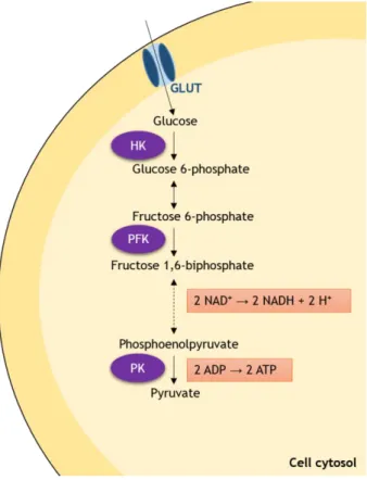
Glucose enters the cells by the action of specific glucose transporters (GLUTs), from which the high-affinity GLUTs 1 and 3 may be highlighted (Macheda et al., 2005). In the cytosol, glucose molecules suffer enzymatic conversion to pyruvate, through a series of ten chain reactions that constitute the glycolytic pathway. From these reactions, it is important to highlight that there are three main points for the regulation of the glycolytic process. These are the irreversible conversions of glucose to glucose 6-phopshate by hexokinase (HK), of fructose 6-phosphate to fructose 1,6-biphosphate by phosphofructokinase 1 (PFK) and the last step, in which pyruvate kinase (PK) catalyzes the conversion of phosphoenolpyruvate into pyruvate (Xiong et al., 2011; Oliveira, et al., 2014). This series of reactions includes the final
liberation of two ATP molecules per glucose molecule, as well as the reduction of two NAD+
molecules to two NADH molecules (figure 2). The pyruvate formed in this pathway, aside from being converted to acetyl-CoA, may also be enzymatically converted to alanine or lactate. These processes are severely important in cancer cells and will be discussed in detail below.
5
Figure 2. Schematic overview of the regulatory points and most important products formed in
glycolysis. Glucose enters the cells by the action of membrane glucose transporters (GLUTs). In the cytosol, it is irreversibly converted to glucose 6-phosphate by hexokinase (HK). In the next step, conversion to fructose phosphate occurs, which in turn will be irreversibly transformed in fructose 6-phosphate by phosphofructokinase 1 (PFK). Then, fructose 6-6-phosphate follows through a series of enzymatic reactions, which will result in the formation of two reduced nicotinamide adenine
dinucleotide molecules (NADH) from two NAD+ molecules. In the last step of this pathway, which is also
the last regulatory point, phosphoenolpyruvate is irreversibly converted to pyruvate by pyruvate kinase (PK) and two adenosine triphosphate (ATP) molecules are formed.
However, cancer cells do not seem to share this usual and expected metabolic behavior. Instead, Warburg reported that, even in the presence of enough oxygen to sustain oxidative phosphorylation, these cells use glucose as main energy source and export high levels of lactate (Warburg, et al., 1927; Warburg, 1956). Warburg and others also postulated that in cancer cells, mitochondrial respiration is either impaired, or less used. Additionally, some cancer cells present the ability to switch from glycolysis to oxidative phosphorylation, according to environmental factors (Rossignol et al., 2004; Kaldma et al., 2014; Oliveira, et al., 2014). Several alterations in the intermediates of this pathway have been reported in cancer cells, such as deregulation of GLUTs and enzyme modulation, to sustain a high glycolytic flux (Osthus et al., 2000; Atsumi et al., 2002; Langbein et al., 2006; Reis et al., 2011; Ros and Schulze, 2013; Jin et al., 2014). Of note, cancer cells are immortal,
ever-6
proliferating, highly replicable systems. In order to be able to duplicate their cellular contents, these cells have a high demand for biosynthetic intermediates. Glycolysis, besides converting glucose to pyruvate, is also a source of precursor biomolecules involved in several other metabolic pathways, which present very intrinsic relations with each other (figure 3) (Feron, 2009; Oliveira, et al., 2014).In the first step of the glycolytic pathway, glucose 6-phosphate may either follow the glycolytic way (see figure 1), or be converted to reduced nicotinamide adenine dinucleotide phosphate (NADPH) and ribose 5-phosphate by the pentose phosphate pathway. Similarly, the yielded pyruvate may either be converted to lactate by lactate dehydrogenase (LDH); to acetyl-CoA by the pyruvate dehydrogenase complex; or to alanine by glutamic-pyruvate transaminase (GPT). Alanine can be used to incorporate proteins, or be exported (Feron, 2009). Lactate is exported from the cells, through the monocarboxylate transporters 4 (MCT4) (Feron, 2009).
Figure 3. Schematic representation of the intrinsic relations between glycolysis and other metabolic
pathways known to occur in cancer cells. Glucose, as well as glutamine, are the main substrates for these cells. The high-affinity glucose transporters 1 and 3 (GLUT1 and GLUT3) transport glucose to the cells, which is then converted to pyruvate. The first glycolysis step, catalyzed by the enzyme HK, converts glucose to glucose 6-phosphate, which can then enter the pentose phosphate pathway, where its carbons are transformed in ribose 5-phosphate and utilized as sources for nucleotide synthesis. The pyruvate derived from glycolysis may either be converted to lactate, which is exported from the cells by MCT4, to alanine or to acetyl-CoA, which participates in the Krebs cycle. Adapted from Oliveira et al. 2014.
7
As in many other cancer types, the Warburg Effect has also been verified in urinary bladder tumors, reflected in altered levels of glycolysis intervenients, such as pyruvate, enzymes and GLUTs (Langbein, et al., 2006; Reis, et al., 2011; Jin, et al., 2014). In some cases, sub or overexpression of some of these intermediates of glycolysis has been correlated with an increase in tumor malignancy (Liao et al., 2011; Reis, et al., 2011; Jin, et al., 2014). Moreover, it was reported that it is possible to distinguish between healthy subjects, patients with muscle-invasive bladder cancer and patients with non-muscle-invasive bladder cancer, based on their urines’ metabolic profiles. These displayed several indicatives of excessive glycolytic and betaoxidative activities, such as high pyruvate and acetyl-CoA levels (Jin, et al., 2014). However, there are not many studies available focused on the exact alterations in the metabolism of bladder cancer during cancer progression. The study of bladder cancer cells metabolism and how it is associated with progression to different and more aggressive states is essential to the development of new diagnosis methods, therapeutic strategies and even tools to help predicting the survival of the patients (Jin, et al., 2014). Thus, more studies are needed to unveil the metabolic characteristics that ensure survival and proliferation of the bladder cancer cells, as well as their progression to more aggressive states. Herein we propose to study the glycolytic profile of two human urinary bladder cancer cells, representative of different cancer progression stages: RT4 (primitive stage) and TCCSUP (highly invasive stage). This may ultimately help to identify a molecular pharmacological/therapeutic target to counteract or avoid the progression of bladder cancer.1.3. Risk and preventive factors
The study of possible therapeutic targets to bladder cancer is extremely important and may arise from unveiling the molecular mechanisms related or promoted by the progression of this disease. Many risk factors for the development of bladder cancer in humans have been identified (Pelucchi, et al., 2006). These include smoking (Crawford, 2008; Kurahashi et al., 2009; Kobeissi et al., 2013), exposure to diesel and combustion fumes (Kobeissi, et al., 2013), genetic components (Crawford, 2008; Kobeissi, et al., 2013; Wang et al., 2013) and liquid ingestion (Hemelt et al., 2010; Wang, et al., 2013). But not all factors are consensual. For instance, there are authors that discuss liquid ingestion as a risk factor while others who defend that it may be a preventive factor instead. This occurs because some authors consider that the ingestion of great volumes of liquids may increase the amount of carcinogenic components (present in those liquids) in contact with the bladder cells (Villanueva et al., 2006) while others suggest that ingesting high quantities of liquids may help to dilute and expel potentially harmful metabolites present in the bladder (Michaud et al., 1999). Noteworthy, the increased risk also depends on the ingested liquids, especially soft drinks (Wang, et al., 2013). Similarly, some studies show that the regular intake of certain
8
beverages, such as milk (Hemelt, et al., 2010) and tea (Wang, et al., 2013), is responsible for the decrease of this risk.
There are several studies that establish a connection between tea consumption and decreased cancer risk (Kada et al., 1985; Wang et al., 1992; Suganuma et al., 1999; Steele et
al., 2000; Cooper et al., 2005; Pastore and Fratellone, 2006; Rieger-Christ et al., 2007; Khan
and Mukhtar, 2010; Mao et al., 2010; Cross et al., 2011; Dias et al., 2013). Although some authors report beneficial effects of tea in cancer, studies on bladder tumors are very scarce and conclusions are still lacking. Therefore, preliminary works regarding the effects of treating bladder cancer cells with tea will also be presented in this work.
2. Tea: types, composition and health benefits
Camellia sinensis (L.), commonly known as the tea plant, has been used for many centuries in
traditional medicine. The infusion prepared by using its leaves or buds is also known as tea. The main types of tea yielded from this plant are green tea (GT), white tea (WT), black tea (BT) and oolong tea (OT). This classification is based on differences in the manufacture and preparation processes, which result in distinctive chemical compositions. Tea’s ability to stimulate the immune system and mitigate several diseases has raised great interest. Emphasis is given to cancer research area, where tea’s beneficial properties have been verified, and several of its components have also been investigated separately (Taniguchi et
al., 1992; Wang, et al., 1992; Huang et al., 1997; Hong et al., 2001; Higdon and Frei, 2003;
Zaveri, 2006; Boehm et al., 2009; Carvalho et al., 2010; Mao, et al., 2010; Darvesh and Bishayee, 2013).
2.1. Types of tea
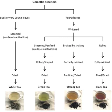
C. sinensis can originate four types of teas, depending on the tea leaves’ harvesting and processing (Pastore and Fratellone, 2006; Moderno et al., 2009; Dias, et al., 2013) (figure 4). Upon harvesting, the leaves suffer an enzymatic oxidation process, also called “fermentation” (Moderno, et al., 2009; Dias, et al., 2013). The enzyme involved in this process, polyphenol oxidase (PO), is the main responsible for the differences in the phenolic profiles of the several types of tea. Its action can be inactivated by quickly heating the leaves or buds, a post-harvesting technique commonly used in the production of GT and WT (Moderno, et al., 2009; Dias, et al., 2013).
GT, BT and OT are all obtained from C. sinensis mature dried leaves, but they possess different chemical compositions and consequently some very obvious organoleptic
9
differences, namely in taste, color and flavor. In the production of GT, the mature leaves are harvested and then steamed and rolled before drying, in order to inactivate PO and prevent oxidation. In this way, the chemical composition of GT remains similar to that of the C. sinensis’ mature leaves. On the other hand, the production of BT (also known as “fermented” tea) includes rolling and crushing the leaves, which are then allowed to “ferment” for two hours, and heated afterwards. Production of OT (also known as “semi-fermented” tea) is similar to the latter; however, the leaves are only allowed to “ferment” for one hour before being heated (Moderno, et al., 2009; Dias, et al., 2013). Finally, there is the rarest and most expensive tea, the WT. This type of tea is produced from the tips or leaf buds not fully opened, which are quickly heated to prevent withering and oxidation (Moderno, et al., 2009; Dias, et al., 2013). Therefore, WT’s chemical composition is similar to that of C. sinensis’ buds and young leaves.Figure 4. Processing methods that yield the different types of tea. White tea (WT) production requires
steaming and drying immediately after harvesting, to prevent the action of polyphenol oxidase (PO). For the production of green tea (GT), the mature leaves are harvested and then quickly heated, to inactivate PO and prevent oxidation, after which they are rolled, dried and sorted. Production of black tea (BT) and oolong tea (OT) includes crushing and rolling the leaves after harvesting and withering, a process that disrupts cellular compartmentation and brings phenolic compounds into contact with PO. Then, the leaves are allowed to “ferment” for two hours, for BT, or one hour, for OT, before being heated. Adapted from Dias et al. (Dias, et al., 2013).
10
2.2. Chemical composition
Tea’s chemical composition is very complex, containing polyphenols, proteins, polysaccharides, free amino acids, minerals, trace elements, methylxanthines and organic acids, among many others (Cabrera et al., 2006; Moderno, et al., 2009; Dias, et al., 2013). As referred above, the four types of tea present different chemical compositions, which are affected by several factors such as geographical origin, climate, growing conditions, harvesting practices, maturity stage of the plant and manufacturing processes (Lin et al., 2003; Moderno, et al., 2009; Dias, et al., 2013).
Polyphenols
Polyphenols are the most abundant and active group of compounds present in tea, and are thought to be the most important source of the health benefits attributed to this beverage (Higdon and Frei, 2003). Flavonoids are amongst the major classes of phenolic compounds, from which is important to highlight the catechins, members of the flavan-3-ol family. Catechins present very high antioxidant capacity (Cooper, et al., 2005; Pastore and Fratellone, 2006; Dias, et al., 2013), as well as anti-inflammatory, antimicrobial, antimutagenic, antimetastatic and anticarcinogenic properties (Kada, et al., 1985; Wang, et al., 1992; Suganuma, et al., 1999; Steele, et al., 2000; Cooper, et al., 2005; Pastore and Fratellone, 2006; Rieger-Christ, et al., 2007; Khan and Mukhtar, 2010; Kumar et al., 2010; Mao, et al., 2010; Afaq and Katiyar, 2011; Cross, et al., 2011; Hessien et al., 2012; Li et al., 2012; Dias, et al., 2013). There are various catechins in tea, such as epicatechin (EC), (-)-epigallocatechin (EGC), (-)-epicatechin-3-gallate (ECG) and (-)-(-)-epigallocatechin-3-gallate (EGCG) (Pastore and Fratellone, 2006; Moderno, et al., 2009), and their chemical structure is responsible for the health benefits attributed to them (especially the antioxidant power) (Yang et al., 2007; Costa et al., 2009; Aboul-Enein et al., 2013; Bubols et al., 2013). Moreover, EGCG is considered to be the most abundant and active catechin of tea, being also one of the most studied (Fernandez et al., 2000; Pastore and Fratellone, 2006; Zaveri, 2006; Moderno, et al., 2009; El-Shahawi et al., 2012; Dias, et al., 2013).
The main catechins are essentially comprised of three rings (the aromatic rings, A and B, linked to a dihydropyran heterocyclic ring, C) and are characterized by multiple hydroxyl groups on the A and B rings (Braicu et al., 2013) (figure 5). Their chemical differences are due to the presence of different groups attached to those rings (Moderno, et al., 2009; Braicu, et al., 2013; Dias, et al., 2013). In EC, we can find an ortho-di-hydroxyl group in the B ring and a hydroxyl group in the C ring; ECG contains a gallate moiety esterified in the C ring. EGC possesses a trihydroxyl group on the B ring, and EGCG possesses an esterified gallate on the C
11
ring (Braicu, et al., 2013; Dias, et al., 2013). GT and WT present higher catechin content, while OT and BT present in high quantities other phenolic compounds (Lin, et al., 2003; Unachukwu et al., 2010; Dias, et al., 2013; Li et al., 2013), which are formed by the action of PO. This enzyme is released during the crushing of the leaves for production of BT and OT and catalyzes the oxidation and polymerization of the catechins, producing theaflavins and thearubigins (Lin, et al., 2003; Li, et al., 2013).Figure 5. Chemical structures of the main tea catechins. These compounds are essentially constituted
by two aromatic rings (A, B) and a dihydropyran heterocyclic ring (C). The flavan-3-ol epicatechin is constituted by an ortho-di-hydroxyl group in the B ring (at carbons 3’ and 4’) and a hydroxyl group in the C ring (at carbon 3), and its ester derivative epicatechin-3-gallate possesses an additional gallate moiety esterified in the C ring, at carbon 3. Epigallocatechin contains a trihydroxyl group on the B ring (at carbons 3’, 4’ and 5’) and its ester derivative epigallocatechin-3-gallate additionally possesses an esterified gallate at the carbon 3 of the C ring.
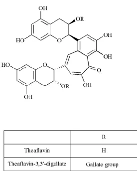
Theaflavins (figure 6) possess a basic skeleton comprised of the bicyclic benzotropolone ring, and result from catechins’ dimerization. In turn, thearubigins are still poorly chemically characterized. They are benzotropolone derivatives, and are thought to be the result of the
12
hydroxylation of theaflavins. Of note, the formation and characterization of their chemical structure needs further clarification (Li, et al., 2013).Figure 6. Chemical structures of the main theaflavins. Theaflavins result from the dimerization of the
main catechins and are constituted by a skeleton comprised of the bicyclic benzotropolone ring. The majority of theaflavins are formed from an epicatechin and an epigallocatechin. Theaflavin-3,3’-digallate is produced by dimerization of epicatechin-3-gallate and epigallocatechin-3-gallate.
Catechins’ chemical composition is very important, because it highly influences their beneficial properties. For example, their antioxidant properties, such as superoxide anion scavenging ability and quenching of hydroxyl radicals are highly correlated with the content of pyrogallol/hydroxyl groups and the presence of galloyl moieties, respectively (Nanjo et al., 1999; Moderno, et al., 2009). The number and position of the hydroxyl groups on the molecules are also thought to influence this antioxidant capacity (Braicu, et al., 2013).
13
MethylxanthinesThe methylxanthines contained in tea are caffeine, theophylline and theobromine. Caffeine (figure 7) is the main methylxanthine present in tea; its levels range between 1.0 and 3.5% in tea preparations (Fernandez, et al., 2000; Lin, et al., 2003).
Figure 7. Chemical structure of caffeine. It is a naturally occurring tea purine derivative with three
methyl groups at positions 1, 3 and 7.
Contrary to catechins and due to its chemical stability, caffeine levels are not affected by the “fermentation” process (Lin, et al., 2003), although some studies indicate that different types of tea present different caffeine levels (Lin, et al., 2003; Unachukwu, et al., 2010; Dias, et al., 2013). These discrepancies may be due to different extraction conditions, distinct analytical methods and the variability of the plants. Nonetheless, the anticarcinogenic properties of caffeine have been documented (Lu et al., 2002; Hashimoto et al., 2004). Particularly in studies of tea consumption by humans, the importance of caffeine in tea preparations was highlighted, since decaffeinated teas presented very low cancer inhibitory properties (Huang, et al., 1997). However, data on the role of caffeine on tea-associated health benefits are scarce and much work needs to be done.
In the field of cancer research, the most studied types of tea are GT and BT. Nevertheless, since WT has the highest catechin concentration among all types, it is expected to possess great anticancer properties (Dias, et al., 2013). However, to date, there are no studies focused on WT consumption and bladder cancer.
14
3. Tea and bladder cancer
3.1. Epidemiological studies
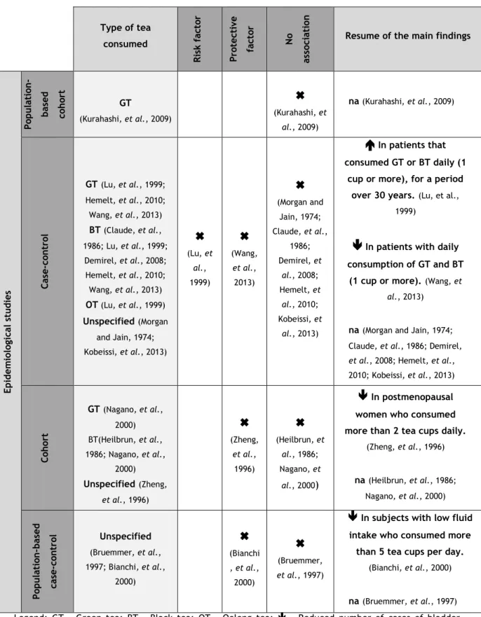
After ingestion by mice, EGCG widely distributes through the body, including the urinary bladder (Suganuma, et al., 1999). Moreover, several studies reported the beneficial effects of GT or its components on bladder cancer cells. However, particularly in the human bladder cancer research area, the anticarcinogenic properties of tea, although predictable, still lack strong supporting evidence. There are several epidemiological studies performed in this area, based on the statistical analysis of questionnaires filled by patients or former patients, regarding their dietary and lifestyle habits in the years anteceding the cancer onset. In this context, some authors reported regular tea consumption to be either a risk factor for bladder cancer (Lu et al., 1999) or a preventive factor (Zheng et al., 1996; Bianchi et al., 2000; Wang, et al., 2013). However, the majority of these studies defend that tea consumption has no association with the disease triggering, development or outcome (Morgan and Jain, 1974; Claude et al., 1986; Heilbrun et al., 1986; Bruemmer et al., 1997; Nagano et al., 2000; Demirel et al., 2008; Kurahashi, et al., 2009; Hemelt, et al., 2010; Kobeissi, et al., 2013).
15
Table 1. Epidemiological studies regarding regular tea consumption and human bladder cancer. The
types of tea, studies and results obtained are presented.
Type of tea consumed Ris k fac to r Pro tec ti ve fac to r No as so ci at io n
Resume of the main findings
Ep id em io lo gi cal st u d ies Po p u latio n -b as ed co h ort GT (Kurahashi, et al., 2009)
(Kurahashi, et al., 2009) na (Kurahashi, et al., 2009) Case -c on tro l GT(Lu, et al., 1999; Hemelt, et al., 2010; Wang, et al., 2013) BT (Claude, et al., 1986; Lu, et al., 1999; Demirel, et al., 2008; Hemelt, et al., 2010; Wang, et al., 2013) OT (Lu, et al., 1999) Unspecified(Morgan and Jain, 1974; Kobeissi, et al., 2013)
(Lu, et al., 1999)
(Wang, et al., 2013)
(Morgan and Jain, 1974; Claude, et al., 1986; Demirel, et al., 2008; Hemelt, et al., 2010; Kobeissi, et al., 2013)
In patients that consumed GT or BT daily (1cup or more), for a period over 30 years.(Lu, et al.,
1999)
In patients with daily consumption of GT and BT(1 cup or more).(Wang, et
al., 2013)
na(Morgan and Jain, 1974; Claude, et al., 1986; Demirel,
et al., 2008; Hemelt, et al.,
2010; Kobeissi, et al., 2013) Co h ort GT(Nagano, et al., 2000) BT(Heilbrun, et al., 1986; Nagano, et al., 2000) Unspecified(Zheng, et al., 1996)
(Zheng, et al., 1996)
(Heilbrun, et al., 1986; Nagano, et al., 2000)
In postmenopausal women who consumed more than 2 tea cups daily.(Zheng, et al., 1996) na(Heilbrun, et al., 1986; Nagano, et al., 2000) Po p u latio n -b as ed cas e -c on tr ol Unspecified (Bruemmer, et al., 1997; Bianchi, et al., 2000)
(Bianchi , et al., 2000)
(Bruemmer, et al., 1997)
In subjects with low fluid intake who consumed more than 5 tea cups per day.(Bianchi, et al., 2000)
na(Bruemmer, et al., 1997)
Legend: GT – Green tea; BT – Black tea; OT – Oolong tea; - Reduced number of cases of bladder cancer; - Increased number of cases of bladder cancer; na – No statistically significant association.
16
However, and although there is relevant information provided by these studies, there are also some drawbacks to be considered. The use of questionnaires makes the studies highly dependent on the subjects’ interpretation or past memory raising doubt about the veracity, due to the subjects’ forgetting or deliberately tampering the facts. Besides, most of these studies also include a complicated analysis of data, ranging from type and duration of the beverages consumed, fruit and vegetable consumption, smoking status, among others. Moreover, some of the studies do not refer the type of tea investigated (Morgan and Jain, 1974; Zheng, et al., 1996; Bruemmer, et al., 1997; Bianchi, et al., 2000; Kobeissi, et al., 2013), which hinders any association between consumption of one tea type and bladder cancer development. Finally, most of the studies were performed in very different populations, which greatly vary in terms of age, countries and habits, making very difficult the extrapolation of results and conclusions.
All these drawbacks show how important it is to develop further studies regarding human bladder cancer. Since human studies are always very difficult to conduct, and epidemiological studies present the cons considered above, good strategies to study bladder cancer, as all well as many other diseases, lay in the use of animal models and in vitro approaches. Although there has to be some care in the extrapolation of the results obtained in these studies to humans (mainly due to different metabolization of tea’s components), they may help unveil disease mechanistic and the exact effects of tea and its components in bladder cancer prevention and/or treatment.
3.2. In vitro and in vivo studies
The numerous and complex signaling pathways that exist in a cell are extremely important to maintain its homeodynamics and normal functioning. The disclosure of these pathways has become very important in the study of several diseases. Cancer cells normally display several differences in metabolism, gene expression and survival mechanisms, among others. Thus, as expected, tea and its components can inhibit carcinogenesis through a wide variety of mechanisms (Hou et al., 2005; Yang and Wang, 2011; Yang et al., 2011). Mainly acting through its polyphenol components, particularly catechins, it was demonstrated the GT’s ability to prevent the initiation and growth of bladder tumors in rats (Sato, 1999; Sato and Matsushima, 2003; Chen et al., 2011), inhibit bladder tumor development and invasion in
vitro (in some cases showing positive synergistic effects with other substances) (Roomi et al.,
2006; Chen, et al., 2011) and protect normal bladder cells, while killing the malignant ones (Coyle et al., 2008; Chen, et al., 2011). Although catechins alone are a powerful tool to oppose cancer development, GT extract and dried leaves can also be very helpful and a
17
practical way to treat cancer. They possess numerous phytocomponents with anticancer properties, being less expensive, widely available and safe (Sato, 1999).
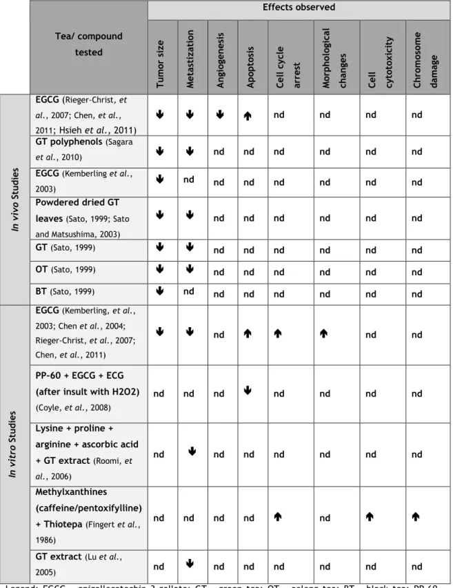
Although many studies report the anticancer properties of tea or its components in several carcinogenic models, the exact mechanisms through which they exert these effects remain to be unveiled. This is due to the fact that, aside from the lack of many studies regarding this matter, the anticancer effects suggest interference in many different pathways and processes, ranging from apoptosis, metastization and cell cycle arrest, among many others. Table 2 presents a summary of studies in this field, highlighting the main findings.
18
Table 2. Summary of the main effects observed in several in vivo and in vitro studies focused on the
effects of tea and its phytochemicals in bladder cancer.
Tea/ compound tested Effects observed Tu m or si ze Met as ti zat io n A n gi og en es is A p op to si s Cell c yc le arres t Mo rp h ol og ic al ch anges Cell cyt ot ox ic it y Ch ro m os o m e d am ag e In vi vo S tu d ies EGCG (Rieger-Christ, et
al., 2007; Chen, et al.,
2011; Hsieh et al., 2011)
nd nd nd nd
GT polyphenols (Sagara
et al., 2010) nd nd nd nd nd nd
EGCG (Kemberling et al.,
2003) nd nd nd nd nd nd nd
Powdered dried GT leaves (Sato, 1999; Sato and Matsushima, 2003) nd nd nd nd nd nd GT (Sato, 1999) nd nd nd nd nd nd OT (Sato, 1999) nd nd nd nd nd nd BT (Sato, 1999) nd nd nd nd nd nd nd In vi tr o St u d ies
EGCG (Kemberling, et al., 2003; Chen et al., 2004; Rieger-Christ, et al., 2007; Chen, et al., 2011)
nd nd nd
PP-60 + EGCG + ECG (after insult with H2O2)
(Coyle, et al., 2008)
nd nd nd nd nd nd nd
Lysine + proline + arginine + ascorbic acid + GT extract (Roomi, et
al., 2006)
nd nd nd nd nd nd nd
Methylxanthines
(caffeine/pentoxifylline) + Thiotepa (Fingert et al., 1986)
nd nd nd nd nd
GT extract (Lu et al.,
2005) nd nd nd nd nd nd nd
Legend: EGCG - epigallocatechin 3-gallate; GT - green tea; OT - oolong tea; BT - black tea; PP-60 - polyphenon-60; - Reduced/inhibited; - Increased; nd - not determined.