U
NIVERSIDADE DO
A
LGARVE
Faculdade de Ciências e Tecnologia
F
UNCTIONAL STUDY OF
AVE
SECRETED GENES
DURING MOUSE EMBRYOGENESIS
:
INSIGHTS ON THE ROLE OF
mADTK1
Lisa Gonçalves Dias da Silva
Tese orientada por Professor Doutor José António Belo
Doutoramento em Biologia
Biologia Molecular
II
Dissertação de Candidatura ao Grau de Doutor em Biologia, Área
de Biologia Molecular, pela Universidade do Algarve
PhD Thesis proposal in Biology, Molecular Biology, by the
Universidade do Algarve (Portugal)
As opiniões expressas nesta publicação são da exclusiva responsabilidade do seu Autor.
The contents of this dissertation are of the exclusive responsibility of its Author.
In theory, there is no difference
between theory and practice.
In practice, however, there is.
V
VI
scientific experience at his lab. Thank you for all the support and guidance, for all the educating scientific discussions and for believing I could carry out this project, even at the most difficult times.
To Faculdade de Ciências e Tecnologia of the Universidade do Algarve, thank you for the vote of confidence by accepting me as PhD student. To Centro de Biologia Molecular e Estrutural at Universidade do Algarve, thank you for provide such good facilities.
Fundação para a Ciência e Tecnologia, thank for the financial support, without which I couldn‟t have carried out this work.
I also want to acknowledge Intituto Gulbenkian de Ciências for its fantastic facilities, and support provided by all staff members and the scientific support all groups from Zheng Ho wing gave me when my lab was in transition to Faro. Particularly to Moises Mallo, who colaborated in the study of mADTK1, and Miguel Godinho who helped me with the Southern Blots.
Ana Cristina Borges, thank you for all the support you gave me when I first arrived at IGC. You made mouse basic embryology techniques seem much easier than they really are, thank you all you taught me. Thank you for all the patience.
Zé Inácio and Sara Marques, thank you for thoroughly reviewing this manuscript, for all comments, explanations and corrections. Without you it would have proved very difficult to deliver this manuscript. I also want to acknowledge Zé Inácio for performing all the transfection assays, and Sara Marques for helping me with the newborn analysis.
To all the members in the lab (the ones who are currently in lab and the ones who already left it), thank you for all the suggestions, advices and friendship; when experiments didn‟t work go as good as they should, you always gave the support to go on and try again, and again.
VII
unique moments of great joy and frustration when an experiment finally works, or doesn‟t.
To my mother and brother, thank you for all the patience, advices, unconditional support, comprehension and love. Thank you for sharing the moments of joy and worry, and for sharing all the setbacks and all the achievements I accomplished during this PhD. Thank you for reviewing this manuscript, for all the corrections and suggestions. Without you I couldn‟t have made it. Thank you.
IX
Sendo os organismos vertebrados caracterizados por serem organizados assimetricamente sobre três eixos distintos, anterior-posterior (A-P), dorsal-ventral (D-V) e proximal-distal (P-D), é bastante importante estudar o momento em que estes eixos se formam. Durante o desenvolvimento embrionário existem várias vias de sinalização envolvidas neste acontecimento crucial, entre elas encontra-se a superfamília “Transforming Growth Factor-β” que engloba moléculas de elevada importância, tais como Nodal e proteínas morfogenéticas do osso (BMPs).
Em ratinho, são consideradas duas estruturas organizadoras para o tronco e cabeça. O centro organizador para o tronco é o nó, uma estrutura transiente que aparece ao sétimo dia do desenvolvimento embrionário, durante o processo de gastrulação, enquanto a linha primitiva se extende.
A Endoderme Visceral Anterior (AVE) é considerada o centro organizador para a cabeça e as estruturas anteriores, sendo portanto uma estrutura de imensa importância durante o desenvolvimento embrionário. É com a formação da AVE que começam a ser aparentes as primeiras evidências de assimetria, e que se inicia a formação do primeiro eixo, o eixo A-P. É nesta estrutura embrionária que vários genes de grande importância para a correcta organização dos eixos e desenvolvimento do embrião são expressos. Entre eles encontra-se Hex, goosecoid, otx2, Hesx1/Rpx, lim1, dkk1, lefty1 e
cerberus-like1 (cerl1).Este último é um antagonista de Nodal e BMPs, e é um membro da
superfamília TGF-β.
Mutantes homozigóticos para o cerl1 não aparentam ter qualquer tipo de fenótipo, são viáveis e férteis. Contudo, é preciso salientar o facto de a ausência de fenótipo não ser sinónimo de pouca importância. Durante o desenvolvimento embrionário, vários genes compensam o papel uns dos outros, actuando sinergisticamente.
cerl1 é um gene que desempenha um papel central na regulação da via de
sinalização Nodal. Estudos prévios demonstraram que o cerl1 age em cooperação com genes lefty, também conhecidos pelo seu carácter antagonista em relação a Nodal. A via de sinalização Nodal é necessária para que o processo de gastrulação ocorra, e é essencial para a correcta formação dos eixos embrionários.
Com o intuito de aprofundar o estudo da via de sinalização Nodal, e uma vez que esta é extremamente importante durante o desenvolvimento embrionário, numa primeira
X
destas possíveis interacções sugere uma via de sinalização Nodal independente do co-receptor Cripto. O estudo de duplos mutantes para cerl1 e cripto, levou à conclusão que neste caso, a via de sinalização Nodal é parcialmente recuperada. O que indica uma potencial via de sinalização Nodal não canónica, provavelmente através do receptor serina/treonina tipo I, ALK7.
Na tentativa de caracterizar e entender melhor o papel da AVE durante o desenvolvimento embrionário de ratinho, no que diz respeito aos genes que nela são expressos, foi efectuado no laboratório um screening diferencial, onde foram seleccionados os genes que se encontravam sobre expressos na região da AVE.
Entre os vários novos genes descobertos, encontram-se o shisa e ADTK (Anterior Distal Tyrosine Kinase).
Os ortólogos, shisa-1 e shisa-2 em Xenopus, demonstraram estar envolvidos na formação de estruturas anteriores e sómitos, respectivamente. Ainda mais, revelaram ter um papel inibitório das vias de sinalização Wnt e Fibroblast Growth Factors (FGF). Foi publicado um estudo onde demonstraram que Shisa1 se liga aos receptores de FGF e Wnt no retículo endoplasmático impedindo deste modo a maturação dos receptores e, consequentemente, a sua translocação para a superfície celular, onde estes receptores desempenham as suas funções. No que diz respeito ao Shisa-2 estudos efectuados no laboratório levaram à conclusão que a expressão nos sómitos e zona pré-somítica estava directamente correlacionada com o seu papel. Experiências revelaram que baixos níveis deste gene dá origem a sómitos mais estreitos e ao deslocamento rostral de marcadores específicos da mesoderme pré-somítica posterior.
De modo a conhecer os potenciais papéis desempenhados por este gene em ratinho, foi efectuada uma análise comparativa exaustiva do padrão de expressão durante o desenvolvimento embrionário de ratinho e galinha. Verificou-se que o shisa de ratinho e de galinha têm um padrão de expressão muito idêntico, o que indica que este gene é mantido inter-espécies. Ainda mais, verificou-se que o shisa está presente em tecidos como o olho, sómitos e membros, que, tipicamente, requerem a modulação das vias de sinalização Wnt e FGF. Contudo, recentemente foi publicado um estudo em que eliminaram todas as isoformas existentes do shisa em ratinho, e nenhuns defeitos a nível
XI
A grande parte deste trabalho concentrou-se no estudo do novo gene ADTK1. Este gene codifica uma proteína com domínio catalítico tirosina cinase serina/treonina. Uma vez que os processos de fosforilação são etapas determinantes para o correcto desenvolvimento embrionário, que os domínios serina/treonina desempenham papéis fulcrais na transdução de sinal, e sendo este gene expresso na AVE, (região que contém tantos genes de particular interesse para o vários processos essenciais à embriogénese), seria do maior interesse fazer uma análise pormenorizada do padrão de expressão deste gene durante as várias fases do desenvolvimento embrionário, bem como uma análise funcional. Deste modo, verificou-se que o gene ADTK1 está expresso desde muito cedo no desenvolvimento embrionário, e permanece expresso até mais de metade do processo. O padrão de expressão do ADTK1 intrigou-nos desde logo, uma vez que está presente em várias estruturas de particular interesse para o desenvolvimento. Verificou-se que este gene está preVerificou-sente na AVE, o que é um controlo positivo para o screening efectuado, aparece pontualmente na linha primitiva quando esta ainda não se encontra na sua extensão completa, depois do qual fica restrito à parte anterior do embrião, marcando zonas como dobras neurais, primórdios do olho, sómitos, arcos faríngeos, pericárdio, membros superiores e inferiores. Tais resultados sugeriram um potencial papel na regulação de estruturas como a cabeça, tubo neural, coração e desenvolvimento dos membros. Dados preliminares em Xenopus e galinha corroboraram esta hipótese, uma vez que os ortólogos de Xenopus quando subexpressos, apresentam dificuldades em fechar o tubo neural, enquanto os ortólogos de galinha, quando subexpressos, apresentam defeitos a nível do coração. Procedeu-se então à inactivação do ADTK1 em ratinho através de recombinação homóloga. Os animais heterozigóticos não apresentam quaisquer tipos de defeitos, são viáveis e férteis. O cruzamento destes animais gerou homozigóticos que foram sacrificados nas primeiras 48 horas. Pelo menos durante este período de tempo, os animais homozigóticos são viáveis e não apresentam comportamentos diferentes dos restantes animais da mesma ninhada. A análise dos homozigóticos para o ADTK1 demonstra que a maioria destes animais não exibe qualquer defeito exterior óbvio. A cabeça apresenta-se bem fechada, os membros parecem estar bem formados e o tubo neural está correctamente fechado. No entanto, em alguns mutantes verificaram-se alterações no olho e orelha. Uma análise preliminar
XII
estes se posicionavam correctamente, e não foram identificadas diferenças significativas a nível do coração, pulmões, fígado, intestinos e estômago. Ao comparar os rins dos animais homozigóticos com os correspondentes wild-type, observou-se que todos os animais analisados apresentavam rins maiores e com uma cor mais vermelha. A análise histológica preliminar dos rins, sugere deficiências ao nível da zona medular do rim, e diminuição de estruturas glomerulares. Comparando com a literatura, verificou-se que o fenótipo obtido nos rins de homozigóticos ADTK1, é bastante semelhante ao descrito para o gene wnt4. Animais mutantes para o wnt4 apresentam defeitos nas células do estroma medular do rim. Este gene está envolvido no processo de desenvolvimento dos rins, nomeadamente no desenvolvimento e proliferação de células do músculo liso. Tal sugere um potencial papel na regulação do desenvolvimento dos rins. Este é um processo bastante complexo e que ainda se encontra muito pouco estudado, sabendo-se que as vias de sinalização Wnt, FGF e BMP estão envolvidas neste processo.
Tal como acontece com tantos outros genes, o gene ADTK1 não reproduz os defeitos que os correspondentes ortólogos em Xenopus apresentam. É provável que tal se deva ao facto de, nos mamíferos, existirem genes que compensam as funções uns dos outros, de modo a minimizar as anomalias, e ultrapassar possíveis defeitos que dessem origem à interrupção do desenvolvimento embrionário.
Palavras-chave: AVE, ratinho, desenvolvimento embrionário, gastrulação,
XIV
three different axes, it is important to study the onset of these axes. Several pathways are involved in this crucial event, among them is Transforming Growth Factor-β (TGF-β) superfamily which comprises several key molecules as Nodal and Bone Morphogenetic Proteins (BMP).
The Anterior Visceral Endoderm (AVE) is a very important structure during mouse embryonic development, where first asymmetries start to show and axes start to be established. Several genes such as mouse cerberus-like, a member of the TGF-β superfamily, and a Nodal and BMP antagonist, are expressed in this tissue and are of major importance for correct development of the embryo. cerl-1 is a critical player in the Nodal signaling pathway, which is of major importance during gastrulation and the onset of axes establishment. Interestingly, the analysis of potential interactions between Cer-l and Nodal co-receptor Cripto, suggested the possibility of a Cripto independent Nodal signaling pathway, in establishing the anterior-posterior (A-P) and dorso-ventral (D-V) axes during embryonic development.
In an attempt to further study the AVE a pool of novel genes upregulated in this tissue was discovered by a differential screening. Among these were found shisa and
ADTK1.
Previous work in Xenopus has shown that Shisa-1 inhibits both FGF and Wnt signaling pathways, by regulating the maturation of their receptors. Downregulation of 1 leads to defects in anterior structures. In the lab, it was demonstrated that Shisa-2 is involved in the correct development of somites. During this work, it was performed an exhaustive comparative analysis of shisa expression patterns in mouse and chick. Cross-species comparison showed that the expression pattern is conserved in mouse and chick, indicating that the Shisa family genes are typically expressed in tissues known to require the modulation of Wnt and FGF signaling, such as somites, eye and limbs.
The largest part of this work consisted of the analysis of ADTK1 role in mouse. A detailed analysis of its expression pattern was performed, indicating several processes in which this gene might be involved in, such as the formation of anterior structures, limb and heart development and neurulation. This hypothesis was consistent with data from
Xenopus and chick orthologs, where downregulation of ADTK1 leads to defects in
XV
phenotype, some presented defects in the eye and ear. Skeletal analysis of ADTK1 null mutants demonstrated defects in bone length; these mutants present shorter limbs than their wild-type littermates. Regarding the internal organs, defects in kidney development were detected in all analyzed mutants. ADTK1 mutant kidneys were larger and had a reddish color, comparing to the wild-type littermates. Furthermore, preliminary histological analysis suggests that the kidney medullar region is affected, and that glomerular structures are diminished, when comparing to the wild type. The process of kidney development is a complex one, not yet fully understood. Several signaling pathways, such as Wnts, Shh, BMPs and FGFs, are involved in the process of nephrogenesis.
Once again, as it happens with so many genes, ADTK1 null mutants don‟t mimic the defects presented by downregulation in Xenopus and chick orthologs. It is possible that the lack of striking phenotype can be accounted for the fact that, in mammals, there are several genes with redundant activity, preventing, this way, potential anomalies, and overcoming defects that could lead to embryonic development arrest.
Keywords: AVE, mouse, embryonic development, gastrulation, ADTK1, shisa,
XVII
Abstract... XIII Table of Contents ... XVI Table of Figures ... XXI Abbreviations ... XXIV
1. Introduction ... 1
1.1 Mouse Embryogenesis ... 4
1.2 Early mouse development ... 5
1.2.1 Gastrulation ... 8
1.2.2 The origin of body axis and the organizer... 11
1.2.3 The Node ... 13
1.2.3 Anterior visceral endoderm ... 15
1.3 The importance of signaling pathways ... 18
1.3.1 Nodal signaling ... 21
1.3.2 BMP signaling ... 26
1.3.3 Wnt signaling ... 28
1.4 Later embryonic development ... 31
1.4.1 Neurulation ... 31 1.4.2 Somitogenesis... 34 1.4.3 Limb Formation... 35 1.4.4 Heart Development ... 36 1.4.5 Kidney Development... 38 1.5 Objectives ... 39
1.5.1 Nodal signaling pathway ... 39
1.5.2 The importance of AVE secreted genes ... 39
XVIII
2.3 Whole Mount in situ hybridization ... 43
2.4 Histology ... 45
2.4.1 Gelatin embedding ... 45
2.4.2 Paraffin embedding ... 45
2.4.3 Kidney histology ... 46
2.5 Targeting Construct ... 46
2.6 Mouse Embryonic Fibroblasts Preparation ... 47
2.7 ES cell targeting ... 48
2.8 DNA extraction from ES cells ... 49
2.9 Southern Blot ... 49
2.9.1 Radioactive Hybridization... 50
2.9.2 Non-Radioactive Hybridization - DIG High Prime DNA Labeling . 51 2.10 Genotyping ... 52
2.10.1 DNA extraction from the tail ... 52
2.10.2 DNA extraction from mouse embryos ... 52
2.10.3 PCR mix ... 53
2.11 RNA Extraction ... 54
2.12 Reverse Transcription PCR ... 55
2.13 Skeletal analysis ... 56
3. Results I - Cripto-Independent Nodal Signaling Promotes Positioning of the A-P Axis in the Early Mouse Embryo ... 57
3.1 Abstract ... 59
3.2 Introduction ... 59
3.3 Results and Discussion ... 63
XIX
3.3.3 cerl1;cripto mutants rescue Nodal signaling ... 68
3.3.4 cryptic and alk7 expression profiles argue against a compensatory role of these factors in cerl1;cripto embryos... 72
3.3.5 Cripto-independent Nodal signaling guides positioning of the A-P axis………….. ... 74
3.3.6 Cripto-independent Nodal signaling is inhibited by Cerl1... 75
4. Results II - Comparative Expression of Mouse and Chicken Shisa Homologues During Early Development ... 77
4.1 Abstract ... 79
4.2 Introduction ... 79
4.3 Results and Discussion ... 82
4.3.1 Cloning and Sequence Analysis of Mouse and Chicken shisa ... 82
4.3.2 Expression of mshisa During Mouse Development ... 85
4.3.3 Expression of cshisa During Chick Development ... 87
5. Results III - Functional analysis of the role of the novel gene mADTK1 during mouse development ... 91
5.1 Abstract ... 93
5.2 Introduction ... 94
5.3 Results and Discussion ... 98
5.3.1 ADTK1 Sequence Analysis ... 98
5.3.2 ADTK1 expression pattern... 103
5.3.3 ES Cell targeting ... 114
5.3.4 Offspring analysis from ADTK1 +/- ES cell injection ... 119
6. General Discussion ... 128
7. Future Perspectives ... 136
XX In situ Probes ... 164
XXII
Figure 1.1 - The dwarf embryo as imagined by Leonardo da Vinci in the 15th century ... 2
Figure 1.2 - The sperm as miniature human being by Hartsoeker from the 17th century ... 3
Figure 1.3 - Development of the pre-implantation blastocyst in mice ... 5 Figure 1.4 - Transcriptional circuitry of cell fate decisions ... 6 Figure 1.5 – Implanting blastocyst to pre-DVE stage ... 7 Figure 1.6 - Early post-implantation development in the mouse: Post DVE to early streak stage ... 9
Figure 1.7- Cell Movements and Molecular Signals Controlling Axis Formation . 10 Figure 1.8 - A model for anteroposterior axis formation in the mouse embryo ... 16 Figure 1.9 - Model for A-P determination by Nodal antagonists ... 22 Figure 1.10 - Schematic representation of Nodal signaling ... 23 Figure 1.11 - Wnt signaling pathways ... 29 Figure 1.12 - Model for neural tube bending ... 33 Figure 2.1 - Construct scheme ... 47 Figure 3.1 - Rescue of posterior neuroectoderm and trunk structures in Cer1;Cripto double mutant embryos. ... 64
Figure 3.2 - cer1;cripto double mutants display rescue of AVE rotation, primitive streak elongation and node derivatives. ... 67
Figure 3.3 - cer1;cripto double mutants rescue Lefty1 and Nodal expression before gastrulation. ... 69
Figure 3.4 - The rescue observed in Cer1;Cripto ouble mutants is due to a recovery of Nodal signaling. ... 71
Figure 3.5 Expression profiles of others genes involved the in Nodal pathway. .... 72 Figure 4.1 - Sequence alignment of XShisa, cShisa, mShisa, rShisa, hShisa, and zShisa. ... 84
Figure 4.2 - In situ hybridization analysis of mshisa expression during mouse development. ... 85
Figure 4.3 - Localization of cshisa transcripts in developing chicken embryos detected by in situ hybridization. ... 87
XXIII
Figure 5.2- Graphic representation of ADTK1 ... 101 Figure 5.3 - Transfection of ADTK1-GFP protein in HeLa cells ... 102 Figure 5.4 - Whole-mount in situ hybridization of ADTK in E5.0–E7.25 wild-type embryos ... 103
Figure 5.5- Whole-mount in situ hybridization of ADTK1 in E7.5 to E8.5 wild-type embryos ... 105
Figure 5.6 – Whole-mount in situ hybridization of ADTK1 in E8.75 to E9.5 wild-type embryos ... 106 Figure 5.7 - Whole-mount in situ hybridization of ADTK1 in E10.5 to E13 wild-type embryos ... 108 Figure 5.8- Double whole-mount in situ hybridization for HNF3 and ADTK1 in E6.5 to E8.5 wild-type embryos ... 110
Figure 5.9 – Double whole-mount in situ hybridization for fgf8 and ADTK1 in E9.0 to E10.5 wild-type embryos ... 111
Figure 5.10- Whole-mount in situ hybridization of ADTK1 in E6.5 cripto, nodal and otx2 mutant embryos and genotyping ... 113
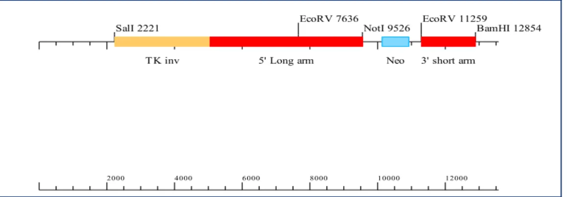
Figure 5.11 - Schematic representation of the targeted deletion of the first exon from ADTK1 gene ... 115
Figure 5.12 - 3' arm PCR screen for neo band ... 116 Figure 5.13 - 3' arm PCR screen for wt band ... 116 Figure 5.14 - Southern Blot Image using an external 3‟ probe ... 117 Figure 5.15 - Southern Blot Image using an internal probe for the neo cassette .. 118 Figure 5.16 - ADTK +/- ES cells ... 119 Figure 5.17 - ADTK1 male chimeras ... 120 Figure 5.18 - ADTK1 genotyping ... 121 Figure 5.19 - Newborns form ADTK intercrosses ... 122 Figure 5.20 - Wild-type and ADTK1 KO kidney comparison ... 123 Figure 5.21 - Histological analyses of kidneys from ADTK1 intercrosses... 124 Figure 5.22 - Skeletal analysis of ADTK1 null mutants and wild type littermates 125 Figure 9.1 - Southern assembly ... 165
XXV µL - Microliter µm - Micrometer 3‟ - 3 prime 5‟ - 5 prime aa - Amino acid Ab - Antibody
ADE - Anterior Definitive Endoderm ADTK - Antero Distal Tyrosine Kinase
AER - Apical Ectodermal Ridge
AP - Anterior-posterior
ASE - Asymmetric Intronic Enhancer AVE - Anterior visceral endoderm
BMP . Bone Morphogenetic Protein
bp - Base pair
cDNA - Coding DNA
CDS - Coding Sequence
cerl - Cerberus-like
Chd - Chordin
CNS - Central Nervous System
Dhh - Desert hedgehog
DIG - Digoxigenin
Dkk . Dickkopf
DLHP - Dorsolateral hinge point
DMEM - Dulbecco‟s Modified Eagle Medium DNA - Deoxyribonucleic acid
dpc - Days post coitum
DV - Dorso-Ventral
DVE - Distal Visceral Endoderm
E - Embryonic day
EGO - Early Gastrula Organizer
XXVI
FBS - Fetal Bovine Serum
FD - Faraday
FGF - Fibroblast growth factor
Fw - Forward
Fz - Frizzled
h - Hour
HPRT hypoxanthine guanine phosphoribosyl transferase
ICM - Inner Cell Mass
Ihh - Indian hedgehog
JNK - Jun-N terminal Kinase
LPM - Lateral Plate Mesoderm
L-R - Left-Right
LRP - Low-density-lipoprotein Receptor-related Protein MAP - Mitogen-Activated Protein
MEF - Mouse Embryonic Fibroblast
MGO - Mid Gastrula Organizer
MHP - Medium hinge point
min - Minute
mL - Milliliter
NTD - Neural tube defect
Oep - one eye pinhead
ON - Overnight
Otx2 - Orthodenticle-related homeobox PBS - Phosphate-buffered saline PCP . Planar Cell Polarity
PCR - Polymerase Chain Reaction
P-D - Proximal-Distal
PE - Primitive Endoderm
PFA - Paraformaldehyde
PKC - Protein Kinase C
XXVII
Rev - Reverse
RNA - Ribonucleic acid
RT - Reverse Transcription
RT - Room Temperature
RTK - Receptor tyrosine kinases SDS - Sodium dodecyl sulfate
SFRP - Secreted Frizzled Related Protein
Shh - Sonic hedgehog
SMART - Simple Modular Architecture Research Tool
Smo - Smoothed
TE - Trophoectoderm
TGF-β - Transforming Growth Factor-beta
UTR - Untranslated Region
UV - Ultraviolet
V - Volt
VE - Visceral Endoderm
WISH - Whole mount in situ hybridization ZPA - Zone of Polarizing Activity
2
The origin of life is still poorly understood. As for the origin of life concerning the animal kingdom, much is already known, but much still remains to unravel.
Embryonic development has been fascinating the human being for a very long time. Aristotle, in 384 B.C., was the first person ever to write about embryology, suggesting the two theories that would prevail for long. At that time, the debate was whether the embryo was a preformed miniature individual (a homunculus) or an undifferentiated form that gradually became specialized when fertilized: preformation versus epigenesis. According to the preformation theory, an embryo or miniature individual preexists in either the female's egg or the male's sperm and begins to grow when properly stimulated. Some preformationists believed that all the embryos that would ever develop had been formed by God at the Creation. From the analysis of chick embryos Aristotle preferentially supported the theory of epigenesis.
The subject of embryo development was then left forgotten until the 13th century, when Albertus Magnus, a scholastic, started to study fish development. This reappraisal opened the way to the scientific movement of the Renaissance, when in the 15th century Leonardo da Vinci played an essential role, approaching the development of the human embryo with a series of sketches that supported the preformation theory (Fig. 1.1) (da Vinci 1510-1513).
Figure 1.1 - The dwarf embryo as imagined by Leonardo da Vinci in the 15th century
3
With the use of the microscope, and the discovery of sperm in 1677 by Hartsoeker and Leeuwenhoek, the theory of epigenesis proved the more difficult to defend: Hartsoeker proposed a spermatozoid structure where a tiny human being was concentrated (Fig 1.2) (Hartsoeker 1694). However a question remained, how could complex organisms, as human beings, develop from such simple organisms?
Figure 1.2 - The sperm as miniature human being by Hartsoeker from the 17th century
Adapted from Hartsoeker 1694.
It wasn‟t until two centuries later, in 1827, when Karl Ernst von Baer discovered the mammalian egg by identifying a yellowish spot within the follicle, which was only visible with a microscope (von Baer 1827), that Aristotle‟s theory of epigenesis was again supported. In the late 19th century, in 1890, Wilhelm Roux and Hans Driesch founded the experimental embryology. Roux tried to prove the preformation theory when, in a two-cell stage frog embryo, he destroyed half the embryo with a hot needle and obtained just half an embryo after letting the embryo develop. He believed that the fertilized egg receives substances that represent different characteristics of the organism, which, as cell division occurs, are asymmetrically distributed to daughter cells. This qualitative division fixes the fate of the cells and their descendants, because some of the determinants are lost at each division. The destruction of a single cell in an embryo with a hot needle was a revolutionary technique that would be the base of several experiments many years later (Kirschner 2003).
Driesch refuted this theory when he himself, instead of destroying one of the cells, separated a two cell stage of a sea urchin embryo into two individual cells, and allowed
4
them to develop: both cells developed into complete sea urchins. Driesch concluded, then, that each cell retains all the developmental potential of the zygote (Oppenheimer 1970).
Later on, in the 20th century, Hans Spemann and Hilde Mangold performed a series of experiments on the newt that opened a new field towards the understanding of the embryonic development, namely the organization and the process of axis formation. These experiments led to the beginning of developmental biology, an area of science where much is still to discover and understand (Spemann and Mangold 1924, Spemann 1931).
During the last fifty years, since the finding of DNA in 1953 by Watson and Crick (Watson and Crick 1953), and then the discovery of the translation system which gave a Nobel Prize in 1968 to Holley, Khorana and Nirenberg, there were many breakthroughs towards understanding cell signaling and molecular biology. Furthermore, much has been achieved with the sequencing of the genome. The first draft of the human genome was published in 2001 and was completed in 2006, whereas the complete mouse genome was published in 2002. This only came to unravel the large amount of work that remains to be done with the about 24.000 genes identified in mouse; nowadays, the true work lies on interpreting the proteins translated and the interactions between them, understanding therefore the signaling cascade processes that occur outside and inside the cells.
1.1 M
OUSEE
MBRYOGENESISThe mouse is a widely used model when studying developmental biology and inherent molecular processes. However, mouse embryos are small and are not produced in large numbers in comparison to Xenopus or chick; furthermore, mouse embryos develop relatively slowly, complete mouse gestation takes about three weeks, and the embryos are inaccessible to experimental manipulation because development takes place in utero. In spite of all these drawbacks, mice are mammals, easy to handle, and of low maintenance cost, when compared with other mammalian model organisms.
5
Being mammals ourselves, we have a particular interest in understanding mammalian development. Furthermore, comparing with Xenopus and chick, the mouse genome has a higher homology to the human genome and proteins share similar functions.
1.2 E
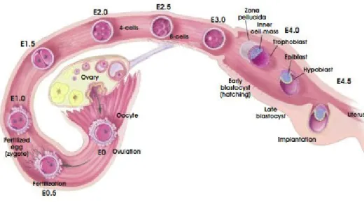
ARLY MOUSE DEVELOPMENTThe fertilized egg divides and develops as it travels along the oviduct to the uterus. In mice this process takes about 4 to 5 days (Fig 1.3). The egg has a protective membrane called the zona pellucida, which stops it from implanting in the oviduct wall (Beddington and Robertson 1999).
Figure 1.3 - Development of the pre-implantation blastocyst in mice
Schematic view of the processes which the embryo undergoes until it is implanted into the uterine wall. E-embryonic day. Adapted from Regenerative Medicine 2006.
By the time it reaches the uterus, the egg has undergone many cell divisions. Cleavage culminates in blastulation right after the morula stage, by day 3.5 to 4.5 (Beddington and Robertson 1999). The blastocyst hatches from the zona pellucida to implant into the uterine wall. The morula stage is reached at 16 cell stage when the outside cells increase contact with each other through a process known as compaction
6
(Zernicka-Goetz 2005). With blastulation two distinct types of tissues are formed: extraembryonic and embryonic. The extraembryonic tissue consists of the trophectoderm (TE) and is called extraembryonic because it does not contribute with any descendant cells in the future body. The embryonic tissue is called the inner cell mass (ICM) (Beddington and Robertson 1999, Zernicka-Goetz 2002). The ICM is said to be pluripotent, as each cell can be differentiated into any cell type. These pluripotent cells are characterized by expressing three transcription factors, Oct4, Nanog and Stat3 that are essential for maintaining the pluripotency of the ICM and for the formation of the embryo. The trophectoderm is characterized by the expression of the T-box transcription factor eomesodermin and the caudal-like transcription factor Cdx2, which are responsible for the downregulation of Nanog and Oct4 (Fig 1.4) (reviewed by Zernicka-Goetz, et al. 2009).
Figure 1.4 - Transcriptional circuitry of cell fate decisions
TE- trophectoderm, ICM- inner cell mass, EPI- epiblast, PE- primitive endoderm. ICM-specific gene expression (yellow; such as Nanog, Oct4, Sox2) represses TE-specific genes (green; such as Tead4, Cdx2) that in turn could repress ICM genes. The ICM then differentiates into EPI (blue; for example, Nanog) and PE (red; such as Gata6), where there is similar reciprocal antagonism of gene expression. Adapted from Zernicka-Goetz, 2009.
7
Later on after implantation, the ICM gives rise to the epiblast (EPI) and the primitive endoderm (PE), which produces the visceral endoderm lining the extraembryonic yolk sac (Tam and Loebel 2007).
Immediately after hatching, the blastocyst is caught by the endometrium where a network of collagen, laminins and fibronectin form an extracellular matrix that, along with the trophoblast enzymes, allow the embryo to attach and bury in the uterine wall (Gilbert 2006).
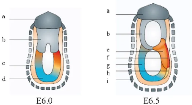
When the mouse embryo implants, it changes its form and size rather dramatically; it starts being a vesicular structure consisting of ICM inside the trophectoderm and, within 3 days, the polar trophectoderm and the ICM develop into an elongated structure that is made up of the ectoplacental cone and the extraembryonic ectoderm (Fig 1.5). The proliferating polar trophectoderm will originate the extra-embryonic ectoderm that seems to „push‟ the proliferating ICM complex into the blastocyst cavity. It is this inward growth, which is characteristic of rodent embryos, that transforms the embryo into the elongated cylindrical structure called the egg cylinder (Zernicka-Goetz 2002). It is at this stage, when the extraembryonic and embryonic regions are well defined, that the polarized proximo-distal (P-D) axis is delineated. More, the prospective dorso-ventral (D-V) axis can also be detected at this stage, the surface of the epiblast that faces the proamniotic cavity corresponds to the future dorsal side and the outer surface of the VE marks the future ventral side of the embryo (Beddington and Robertson 1999).
Figure 1.5 – Implanting blastocyst to pre-DVE stage
a. Polar trophectoderm; b. Inner Cell Mass; c. Primitive endoderm; d. Parietal endoderm; e. Mural trophectoderm; f. Extraembryonic ectoderm; g. Epiblast; h. Visceral endoderm; i. Trophoblast. Adapted from Tam and Loebel 2007.
8
The primitive endoderm, also called hypoblast, is a transitory cell layer that appears along the blastocoelic surface of the embryo between E4.0 and E5.0 dpc (days post coitum) (Fig 1.5) (Tam and Loebel 2007). These cells are of great importance, as they are the origin of two important and distinct extraembryonic cell types, the parietal endoderm and the visceral endoderm. The first, along with trophectoderm, is responsible for the synthesis of the extracellular matrix type IV proteins, collagen and laminins. These proteins will then assemble into Reichert‟s membrane, a specialized membrane that surrounds the embryo and passively filters nutrients (Bielinska, et al. 1999). The second, the visceral endoderm (VE), is associated with the epiblast. The VE cells have microvilli and contain numerous phagocytic and pinocytic vesicles that will grant the absorption and digestion of maternal nutrients. Another feature of VE is that these cells still retain the ability to differentiate into parietal endoderm (Bielinska, et al. 1999).
Curiously, the morphology of VE cells are different along the endoderm layer: the ones overlying the lower or distal pole of the egg cylinder have a squamous morphology, while more proximal visceral endoderm cells are cuboidal (Bielinska, et al. 1999); furthermore, it is also known that the function of these cells also varies. For instance, in the distal tip of the embryo several Nodal antagonists responsible for axis formation are present (Belo, et al. 1997), whereas in posterior proximal region of the VE genes related with hematopoietic commitment such as wnt3 and bmp2 can be detected (Baron 2005).
Asymmetry generation begins very early during embryonic development, even before gastrulation, with the generation of polarity, mostly from maternal information inheritance in the extra embryonic ectoderm (Zernicka-Goetz 2005).
1.2.1 Gastrulation
Gastrulation is a stage in which the embryo faces massive reorganization due to a wide cell movement, from a simple spherical ball of cells, the blastula, into a multi-layered organism. Although the details of gastrulation differ between various groups of animals, the cellular mechanisms involved in gastrulation are common to all animals. Gastrulation involves changes in cell motility, cell shape, and cell adhesion ( reviewed in Gilbert 2006, Wells and Melton 1999).
9
In the early post implantation mouse embryo, the process of gastrulation converts the epiblast into the three primary germ layers: endoderm, mesoderm and ectoderm, from which all the fetal tissues will develop (Fig 1.6). Briefly, from these layers, the ectoderm will generate skin and the central nervous system; mesoderm will give rise to the blood, bone and muscle and the endoderm will be the origin of the respiratory and digestive tracts (reviewed by Wells and Melton 1999).
Figure 1.6 - Early post-implantation development in the mouse: Post DVE to early streak stage
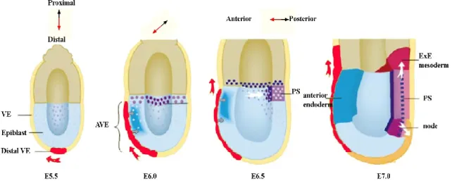
a. Ectoplacental Cone; b. Extraembryonic Ectoderm; c. AVE; d. Epiblast; e. Mesoderm; f. Primitive Streak; g. Ectoderm; h. Definitive Endoderm; i. Parietal yolk sac and Reichert‟s membrane. Adapted from Tam and Loebel 2007.
Gastrulation begins at 6.5 dpc with the formation of a characteristic transient structure, the primitive streak (PS), at the posterior end of the epiblast (Fig 1.7). Embryos that fail to form PS, also fail to proceed with gastrulation and are not viable (Conlon, et al. 1994). During this critical step of embryonic development, several morphogenetic cell movements occur in order to transform a two germ layer into a three germ layer epiblast. In the proximal region of the epiblast, the mesendodermal cells ingress and, as the PS extends, mesoderm, definitive endoderm, and axial mesendoderm precursors arise in the streak by 7.0 dpc, and undergo coordinated morphogenetic movements that lead to their eventual placement in posterior and anterior positions (Tam and Loebel 2007). Mesodermal cells move immediately adjacent to the overlying epiblast, while definitive endoderm cells constitute the outer-most layer (Tam and Loebel 2007, Tam, et al. 2006). The anterior and proximal movement of definitive endoderm cells displaces visceral endoderm to extraembryonic locations. On the other
10
hand, the mesodermal cells that arise in the anterior end of the primitive streak at 7.0 dpc, except for the node, constitute the presumptive heart mesoderm, and have moved to the anterior-most embryonic region by 7.5 dpc where the heart will be formed (Baron 2005).
The PS is marked by the expression of several genes such as Nodal, brachyury,
cripto and wnt3 among others (Rivera-Pérez and Magnuson 2005, Conlon, et al. 1994,
Barrow, et al. 2007). It is involved in cell fate specification, whereby endoderm and mesoderm precursors migrate through the PS in the process of differentiating. It is known that the generation of the primitive streak is regulated by multiple pathways, including Nodal, Wnt and Bmp (Conlon, et al. 1994, Barrow, et al. 2007).
Figure 1.7- Cell Movements and Molecular Signals Controlling Axis Formation
AVE- Anterior Visceral endoderm, ExE- Extraembryonic; PS- Primitive streak,VE- Visceral endoderm. At E5.5, distal VE cells (red), marked by Hex expression, give rise only to anterior progeny, which populate the AVE and eventually move into the extraembryonic region (red arrows). The AVE induces anterior character in the underlying epiblast. Transcripts of future markers of the PS such as Cripto are ubiquitously expressed in the epiblast at E5.5 but restricted to the proximal rim of epiblast at E6.0, where other markers of the PS, such as brachyury (deep purple) start to be expressed. Caudal cell movement in the proximal epiblast (white arrow) results in the PS forming opposite the AVE at E6.5. By E7.0 extraembryonic mesoderm is produced from the posterior aspect of the streak while anteriorly the node forms. Adapted from Beddington and Robertson 1999.
11
Also during gastrulation, a transient structure named node is formed (Fig 1.7) (Beddington 1994, Lee and Anderson 2008, McGrath, et al. 2003). This structure is formed from the mesoderm, at the most anterior part of the PS. As the streak elongates, the subsequent mesendodermal cells that pass through the node will differentiate into anterior definitive endoderm (ADE), prechordal mesendoderm and notochord. (Beddington 1994). By the end of gastrulation, the prechordal plate is preceded posteriorly by the notochord and followed anteriorly by the definitive endoderm. At this time, convergent extension, a combined movement of mesoderm and definitive endoderm, is required for the midline tissues anterior to the node to extend. By this time, the endoderm consists of one thick cell layer of approximately 500 cells, which covers the mesoderm and ectoderm of the embryo (Zernicka-Goetz 2005).
As said before, the definitive endoderm constitutes the precursor of the embryonic gut: the visceral endoderm during gastrulation is replaced by definitive endoderm which ingresses through the anterior segment of the PS; the visceral endoderm only makes a minor contribution to the foregut (Tam, et al., 2003).
Before gastrulation, although it is difficult to identify the anterior-posterior (A-P) axis of the embryo by morphology, the expression of some genes is already asymmetrically established on one side of the VE. This side corresponds to the future anterior end of the embryo, hence its name, the anterior visceral endoderm (AVE).
1.2.2 The origin of body axis and the organizer
Early studies by Spemann and Mangold showed that in amphibian embryos the dorsal blastopore lip, a region that was subsequently named the “Spemann organizer” or “Organizer”, could induce a complete secondary axis, when grafted in the ventral side of an embryo (Spemann and Mangold 1924).In addition, it was also observed that this transplanted tissue could induce the surrounding cells to acquire a new fate, organizing this way the formation of a novel central nervous system (CNS) and notochord, and also contributing to the dorsalization of mesoderm and generation of somites (Spemann and Mangold 1924).
Studies of body axis formation led to the discovery of similar inducing centers in other vertebrates, which were called Hensen‟s node in chick (Waddington 1933), the
12
embryonic shield in zebrafish (Ho 1992), and the node and AVE in mouse (Beddington and Robertson 1999). While grafts of the early Spemann organizer can induce a complete, head-containing secondary axis, grafts of late organizer tissue give rise to the trunk region without a head (Spemann 1931).
The node was the first organizing center proposed in mouse; however, node transplantation in the mouse embryo resulted in the formation of incomplete axis without anterior structures, suggesting then the existence of another organizer for the anterior structures (Beddington 1994).
Until quite recently, anterior patterning of the developing central nervous system in the mouse had been attributed solely to the influence of axial mesoderm or definitive endoderm derived from the node during gastrulation (Beddington 1981, Blum, et al. 1992). Over the past few years it has become apparent that murine AVE, which overlies the future anterior embryonic region, plays an active role in the specification of the A-P axis, as well as in heart formation and positioning of the yolk sac (Thomas and Beddington 1996, Belo, et al. 1997). Accordingly, the AVE was suggested to be an early organizer that specified anterior identity prior to and early in gastrulation (Belo, et al. 1997, Beddington and Robertson 1998).
The gastrula stage has been associated to head formation for a long time. During gastrulation, the AVE organizer assures anterior gene expression, which is essential to the development of the early head, and the beginning of the formation of neural tissues (Anderson 2002)
This way, two distinct organizing centers for the head and trunk have been postulated for the development of the A-P axis in mammals (Belo, et al. 1997). These head and trunk inducing activities are suggested to reside within the AVE and node/primitive streak, respectively. These structures are clearly separated in mouse, contrary to the amphibian embryos where the head and trunk organizer are spatially close, residing in the Spemann organizer (Beddington 1994, Spemann and Mangold 1924, Belo, et al. 1997).
The process of normal left-right (L-R) patterning of the vertebrate embryo can be described in three different stages. The first is the establishment of L-R asymmetry with respect to the D-V and A-P axes in the embryo. This results in a global L-R axis for the embryo such that asymmetries are consistently oriented in the body. Subsequently, this global L-R patterning information is transmitted to developing organ primordia, through
13
signaling molecules. And finally, the organ primordia must correctly interpret the positional cues and execute appropriate morphogenetic responses. When perturbations of L-R development occur, numerous defects result including bilateral symmetry, isomerism, heterotaxia, situs inversus.
1.2.3 The Node
The mouse node is a structure that is formed at the distal tip of the E7.0 embryo, when some groups of columnar cells with small apical surfaces become visible (Beddington 1994). The node is composed of two columnar epithelial layers with adjoining basal surfaces: the dorsal node is adjacent to the surrounding epiblast, whereas the ventral node is contiguous with adjacent endodermal epithelium (Lee and Anderson 2008). There are about 250 cells in a mature node. The node pit consists of a dorsal layer of ectoderm over a ventral layer of cells each with a single monocilium on their apical surface; these cells present apical basal polarity (Lee and Anderson 2008, McGrath, et al. 2003).
At this structure, a leftward fluid flow of great importance to the establishment of the L-R axis, will be generated. This flow depends on two different cellular processes: motility of the cilia and morphogenesis of the node itself (Lee and Anderson 2008). Mutant mouse embryos, that completely lack cilia, or have immotile cilia, such as iv mutants, show an abnormal L-R patterning (McGrath, et al., 2003). Inv mutants have 20% of the cilia pointed anteriorly rather than posteriorly and a nodal flow that moves slowly and not in a polarized way. These mutants do not show a randomization but a complete inversion of the L-R pattern, which is called situs inversus. Moreover, when an artificial flow with reversal of the nodal flow direction is imposed, this leads to the reversal of L-R patterning, suggesting that the flow per se is responsible for subsequent L-R patterning events. (reviewed in Shiratori and Hamada 2006; Lee and Anderson 2008). It is important to mention the presence of an increased influx of Ca2+ ions on the left periphery of the node, which appears to be linked with the nodal flow. This asymmetry in the Ca2+ influx is correlated with the asymmetric activation of nodal expression in the left lateral plate mesoderm (McGrath et al., 2003; Tanaka et al., 2005). This nodal flow is quite conserved between vertebrate. In zebrafish, motile cilia which generate a unidirectional flow have been detected in Kupffer‟s vesicle; whereas
14
in Xenopus, there are cilia present in the gastrocoel roof plate, which also generate a leftward unidirectional flow (reviewed in Lee and Anderson 2008).
In the HNF-3β null mutants, absence of the node allows limited neural induction in some embryos (Levine and Brivanlou 2007). Despite the loss of the node, and with it the loss of the genes that are normally expressed in the node (for instance noggin and
chordin), chordin is still expressed briefly in the mid gastrula organizer in these
mutants, which may account for the limited neural induction (Klingensmith, et al. 1999). Also in fgf8 and cripto mutants, which are again characterized by lacking the node, there is anterior neural induction (Ding, et al. 1998, Sun, et al. 1999). Consequently, the node itself is not required for neural induction. However, the same cannot be said of its predecessor: the node precursors are located at the advancing anterior tip of the primitive streak, as it elongates during gastrulation. This precursor of the node is more potent than the node itself in promoting early anterior neural development (Tam and Steiner 1999). At mid-streak stage, when the primitive streak is elongated midway, grafts of the anterior primitive streak are able to induce anterior brain markers. On the other hand, grafts from earlier or later stages induce more posterior markers (Yang and Klingensmith 2006). These tissues are considered the early gastrula organizer (EGO) and mid gastrula organizer (MGO).
The node of the late-gastrula embryo has been shown to possess organizing activity by virtue of its ability to induce axis formation following heterotopic transplantation. When the node of the mouse is tested for its organizing activity by transplantation, the induced axis is typically made up of the graft-derived tissues in the notochord and somites, and neural tissues that are derived from the host, are morphologically characteristic of the trunk neural tube (Beddington 1994).
Although structurally different, the mouse node shares signaling properties with Hensen‟s node in chick, Kupffer‟s vesicle in zebrafish and the Spemann‟s Organizer in amphibians (Gilbert 2006, Lee and Anderson 2008).
Organizer cell properties, through different vertebrate species, are conserved. In vertebrates such as Xenopus, chicken and mouse, one can detect in these cells the presence of transcription factors such as goosecoid and HNF3β (Zhu, et al. 1999, Belo, et al. 1998), and secreted molecules such as noggin and chordin (Anderson 2002). Furthermore, these cells share similar fates as they become notochord, prechordal mesoderm and gut mesoderm. Moreover, both chicken, Xenopus and mouse organizer
15
cells can induce secondary axis when transplanted, although the mouse node, as said before, induces an incomplete axis, lacking anterior structures (Beddington 1994, Beddington and Robertson 1998).
1.2.3 Anterior visceral endoderm
The VE, which surrounds the epiblast and the extraembryonic endoderm, forms at the distal tip of the egg cylinder at the embryonic day 5.5. The following unilateral polarized movement of these cells towards the proximal region of the embryo establishes the future anterior pole and imparts anterior identity upon the underlying epiblast, becoming the AVE (Fig 1.7) (Rodriguez 2005, Srinivas 2004, Thomas, et al. 1998, Tam and Loebel 2007).
This movement is accompanied by asymmetric gene expression and is the first phenomenon that marks the end of the radial symmetry in the mouse embryo (Zernicka-Goetz 2002). However, recently, it has also been suggested that the intrinsic information within the embryo is involved in the orientation of AVE migration and therefore the orientation of the AP axis (Torres-Padilla 2007). Nevertheless, these changes in cell movement and gene expression mark the conversion of the P-D axis of the embryo into the A–P axis of the embryo.
From the moment it is established, the AVE performs an essential role in the primordial induction of the anterior neuroectoderm.
During mid-gastrulation, as the AVE emerges from the anterior primitive streak and intercalates into the outer VE layer, a population of anterior mesendoderm cells that expresses a similar subset of genes, signals to the anterior epiblast, to pattern the neuroectoderm (Lu, et al. 2001).
This early patterning is processed and transduced into cascades of asymmetric gene expression that will characterize the subsequent phases of dorso-ventral and left-right asymmetry generation in the embryo (Hamada, et al. 2002). It sets off with the expression of some genes in the AVE, encoding for transcription factors like lim1, the homeobox containing genes such as hex, goosecoid, otx2 and Hesx1/Rpx (Thomas and Beddington 1996, Thomas, et al. 1998), and several repressors of Nodal and Wnt signaling such as cer1, lefty1, dkk1, sfrp5 and sfrp1 (Belo, et al. 1997, Perea-Gomez 2002, Yamamoto, et al. 2004). These Nodal antagonists force the initial homogenous
16
expression of Nodal, to the posterior part of the embryo, beginning in this step an asymmetry in the embryo (Belo, et. al 1997, Perea-Gomez 2002).
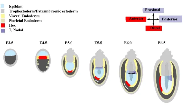
The study of Hex gene was the first experiment to show how the AVE is formed. Initially hex is expressed in the PE, but by 5.5 dpc it starts being expressed only in the VE at the distal tip of the embryo. Subsequently, it marks the unilateral movement of these cells, as its expression shifts towards the anterior side of the embryo (Thomas, et al. 1998).
Initially there were doubts regarding the origin of AVE population, and whether the VE might be the first tissue to acquire A-P polarity in the mouse conceptus; in order to confirm that the VE cells are indeed the precursors of the AVE, DiI was injected at the distal tip of the endoderm, and tracing confirmed VE as the origin of the AVE cells (Thomas, et al. 1998).
Figure 1.8 - A model for anteroposterior axis formation in the mouse embryo
Hex expression (red) is initiated in the nascent PE of the 4.5 dpc blastocyst and at 5.0 dpc is restricted to a
few VE cells at the distal tip of the egg cylinder. At 5.5 dpc this P-D asymmetry is converted into A-P asymmetry by the unilateral movement of the distal Hex-positive cells (white arrow). At this time, T and
Nodal expression patterns (purple) are symmetrical in the proximal epiblast. Subsequently, T and Nodal
expression resolve to the opposite side of the egg cylinder from the AVE (black arrow). Adapted from Thomas, et al. 1998.
17
Before gastrulation, expression markers are already expressed asymmetrically. The distal tip of the embryo is patterned with anterior genes, whereas the proximal region of the embryo expresses genes that are characteristic markers of the primitive streak, such as brachyury, Nodal and fgf8. Furthermore, it is shortly after the VE cells shift towards the anterior region, that a movement from the proximal region and towards the posterior side of the epiblast starts (Thomas, et al. 1998) (Fig 1.8).
There are considered three organizing centers besides the AVE, that are responsible for the correct formation of the head (Anderson 2002). The prechordal plate, an organizing center derived from the mesendodermal cells of the node, confers dorso-ventral patterning to overlying tissue through its secreted molecules; among these molecules is sonic hedgehog (Shh) (Chiang, et al. 1996). The other organizers for head development are the anterior neural ridge and the isthmic organizer; they act in the neural ectoderm (Rubenstein and Beachy, 1998). The first induces and promotes forebrain character, whereas the second is responsible for neural tissues contiguous to the midbrain/hindbrain boundary. These organizers also express morphogens such as FGF8 (Crossley and Martin 1995).
It has been shown the importance of these three organizing centers in head development: when they and their effector molecules are removed, the result is a mispatterning and hypoplasia of the developing forebrain (reviewed by Beddington and Robertson, 1998).
Previous studies suggest an important role in forebrain induction. AVE alone is insufficient to induce forebrain in grafting experiments, but can do so in concert with posterior epiblast fragments (Tam and Steiner 1999). The AVE functions to inhibit the expression of posterior genes (Yamamoto et al., 2004), but much still remains unknown about how it promotes forebrain fates.
Although the AVE is not sufficient to induce forebrain character in explants from the early streak stage, it does promote forebrain gene expression in explants from the mid and late streak stages. This is consistent with findings from other species: the chick equivalent of the AVE, the hypoblast, induces transient neural character.
18
In addition, surgical ablation of the AVE at early gastrulation stages E6.5 leads to the loss of expression of forebrain markers such hesx1 (Thomas and Beddington 1996). Null alleles of genes encoding transcription factors expressed in the AVE such as otx2 and lim1 lead to loss of the forebrain and midbrain (Perea-Gomez, et al. 2001, Shawlot, et al. 1999).
1.3 T
HE IMPORTANCE OF SIGNALING PATHWAYSRight from the onset of cell division, several molecules from different cascades and signaling pathways, act simultaneously in order to obtain the correct development of an embryo. Although it is often referred one or other pathways, none of the signaling pathways exists completely by itself; development is an entanglement of several pathways, which are translated into signal transduction cascades.
The most common target of signaling in development is transcription. Different pathways activate or repress different genes at distinct times and places in the embryo (Gilbert 2006).
All signaling pathways are important at a certain point during development, being sometimes more, sometimes less significant, according to the stage of gestation and signals required at that time.
In development, there are five major signaling pathways, which are grouped on the basis of their structure: the fibroblast growth factor (FGF), the Hedgehog, Notch, Wnt family, and the TGF-β superfamily (Gilbert 2006).
To date, the FGF family consists of twenty three members, all of which contain a conserved 120 amino acid (aa) core region. The members of this family act extracellularly through four tyrosine kinase FGF receptors: the FGF receptor binds to a FGF dormant kinase, which will then be activated and phosphorylate some other proteins (Powers, et al. 2000). FGF signaling, although it stands for fibroblast growth factors, is not limited to cell growth. This family of cytokines, even though induces fibroblast proliferation, is also known to play important roles in several critical
19
processes such as hematopoiesis, angiogenesis, cancer and development (Vasiliauskas and Stern 2004). During development, FGF signaling members play a role in numerous key events, such as gastrulation and organogenesis including somite, kidney, brain and limb development (Perantoni, et al. 2005). One important member of this family is fgf8 which is first expressed in the pre-gastrulation epiblast and then in the primitive streak (Crossley and Martin 1995). Null mutants for fgf8 fail to gastrulate (Sun, et al. 1999), whereas fgf8 hypomorphic alleles or tissue-specific gene inactivation result in several defects such as aberrant cell death of the prospective midbrain and cerebellum (Chi, et al. 2003), abnormalities in cardiovascular and smooth muscle development (Abu-Issa, et al. 2003), defects in limb development and the onset of nephrogenesis (Crossley, et al. 1996). fgf8 is not the only important member of the FGF signaling pathway, FGF receptors are of major importance in signaling transduction, as other member as fgf10 and fgf4, that are involved in limb development (Capdevila and Izpisúa Belmonte 2001).
There are three members of the vertebrate Hedgehog family: Desert hedgehog (Dhh), Indian hedgehog (Ihh) and Sonic hedgehog (Shh) (Cohen 2003). Hedgehog proteins undergo autocatalytic processing and modification that is critical for their signaling activity. The precursor protein is cleaved and gives rise to an N-terminal domain and a C-terminal domain. The auto-processing of Hedgehog causes the covalent attachment of cholesterol to the C-terminal side of the N-terminal domain; it is this attachment with the cholesterol molecule that transforms the Hedgehog protein into a completely functional protein (Jiang and Hui 2008, Ma et al. 2008). The Hedgehog reception system consists of a 12-span transmembrane protein, Patched (Ptc), as the Hedgehog receptor and a 7-span transmembrane protein, Smoothened (Smo), as the obligatory signal transducer across the plasma membrane (Cohen 2003).
These three members all contribute to correct development during embryogenesis: it is already known that Shh is a major player during embryonic development, being involved in several processes such as the development of the CNS, in establishing lateral asymmetry and the anterior posterior limb axis (Chiang 2001), and in the process of nephrogenesis; it is of major importance as it is one of few morphogens known (Cohen 2003). As for Ihh signaling, it is involved in bone development, especially the
20
cartilage, regulating chondrocyte proliferation and differentiation. Dhh is essential for the development of spermatogenesis and the Schwann cells in peripheral nerves (Ma, et al. 2008).
Briefly, Notch signaling is known to be important for specifying cell fates, regulating pattern formation, and defining boundaries between different cell types. Notch proteins are transmembrane receptors for the Jagged and Delta transmembrane ligands. Notch, Delta, and Jagged proteins have EGF domains in their extracellular structures. Notch can act as both a ligand-binding receptor and a nuclear factor that regulates transcription. This pathway has been implicated in several processes such as neural development, apoptosis, and hematopoiesis, among others (Shi and Stanley 2006). One important feature is the ability of the Notch signaling to activate Nodal in the node through its downstream targets which will be a critical step in breaking the symmetry of the embryo (Krebs, et al. 2003). It is also of major importance regarding the epithelial to mesenchymal transition, which is essential, for example, in processes as PS cell migration (Takeuchi, et al. 2007).
After the interaction of Notch with its ligands, the signal induced by ligand binding is transmitted intracellularly, where nuclear translocation of the intracellular domain of the Notch family protein will occur. Once in the nucleus, the Notch intracellular domain will form a complex with the RBP-J protein (Krebs, et al. 2003). This complex activates transcription of downstream target genes such as Nodal, a member of the TGF-β superfamily that is of major importance in embryonic development and will be further discussed ahead.
The Wnt signaling pathway is a highly conserved signal transduction cascade that has a central role in embryonic development. Three different pathways are believed to be activated upon Wnt receptor activation: the canonical Wnt/β-catenin cascade, the non-canonical planar cell polarity (PCP) pathway, and the Wnt/Ca2+ pathway (Clevers 2006, Qian, et al. 2007). Target cell populations respond to secreted Wnt morphogens in a concentration dependent manner, such that the gradient of Wnt concentration determines the resulting gene expression and cellular differentiation. These actions
21
make Wnt molecules central to the signal transduction pathways which underlie cell proliferation, survival and differentiation.
The Wnt ligands are a family of nineteen molecules that are secreted, vary in length between 350 and 400 aa, possess 22 to 24 conserved cysteines, and show 20%-85% aa identity within the family (Bejsovec 2000, Bejsovec 2005).
As this signaling pathway is so important for the correct development of an embryo, it will be discussed more thoroughly ahead.
The TGF-β superfamily is of enormous importance during embryonic development. Several key genes reside within this family as, for example, Smads, BMPs, activin receptors and Nodal signaling pathways (Gilbert 2006). These ligands are synthesized as pre-propeptides. The N-terminal pro-region is cleaved prior to secretion, and the secreted C-terminal mature segment has six or seven spatially conserved cysteines, which form a cysteine knot structure in the monomer. The mature segments will then form dimers via disulfide links (with some exceptions, such as lefty1), which will subsequently serve as the active ligand.
1.3.1 Nodal signaling
During embryonic development, an important step is the establishment of the L-R axis. It has been shown that Nodal signaling is essential for this asymmetry to be processed: it is required the asymmetric activation of the Nodal signaling cascade in the left side of the body wall ( reviewed in Hamada, et al. 2002), the left Lateral Plate Mesoderm (LPM).
Nodal starts being expressed very early. Transcripts can be detected in ES cells in vitro, however, in vivo it is not until shortly after implantation, at E5.5, that Nodal can
be seen in the VE and epiblast (Varlet, et al. 1997). Also at this time, the AVE is formed and starts to dislocate towards the anterior side of the embryo. In the AVE several
Nodal antagonists are expressed, such as cerl-1 and lefty1 (Fig 1.8, Fig 1.9) (reviewed
by Yamamoto, et al. 2004). Later on, at E6.0 Nodal is strongly detected in the proximal ring of the epiblast, while in the VE low levels of Nodal can be detected. As the primitive streak starts to be formed, at E6.5, Nodal can be seen pushed towards the
22
posterior side of the embryo, where the mesoderm is to be formed (Meno, et al. 1999) (Fig 1.8, Fig 1.9), a feature of the responsibility of Nodal‟s antagonists expressed in the AVE. At E7.5, when the primitive streak is completely formed, Nodal transcripts can be detected in the perinodal region. This early expression is necessary for Nodal posterior activation in the left LPM later on, at E8.0 (Lowe, et al. 1996), activating its downstream targets and also leading to an asymmetric expression in the node (Brennan, et al. 2001).
Figure 1.9 - Model for A-P determination by Nodal antagonists
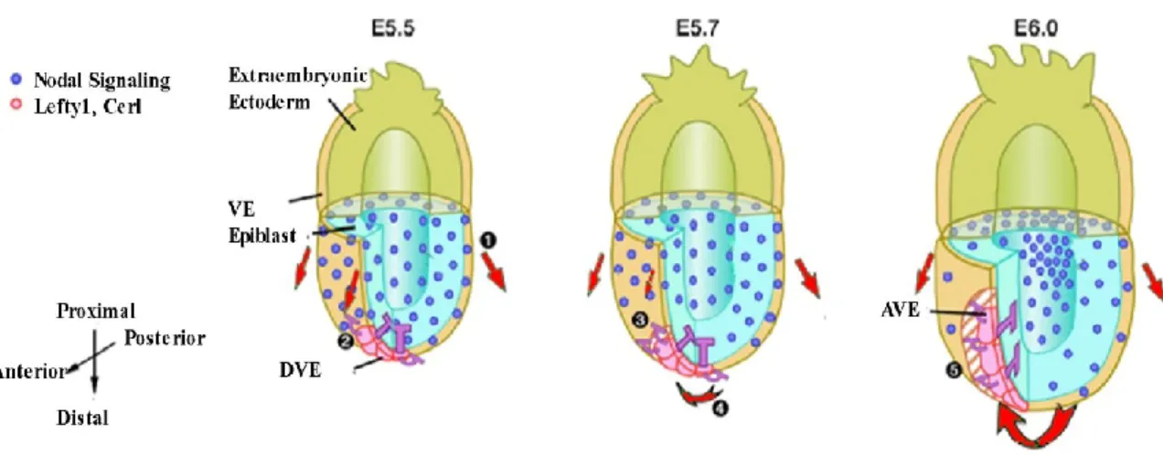
In the wild-type embryo at E5.5, Nodal signals in the epiblast and overlying VE regulate cell proliferation of VE in a symmetric manner (Step 1; red arrows represent putative migration force generated by cell proliferation). Nodal antagonists (Lefty1 and cerl) in DVE whose expression domains are already inclined toward the prospective anterior side start to inhibit the Nodal signals in the region adjacent to the DVE (step 2). Cell proliferation will be inhibited in the VE regions that have received the Nodal antagonists (step 3). This would generate higher migration force on the posterior side and induce the DVE to migrate toward the anterior side (step 4). Migration of the DVE toward the anterior side further establishes A-P asymmetries in Nodal signaling and cell proliferation (step 5). Adapted from Yamamoto, Saijoh, et al. 2004.
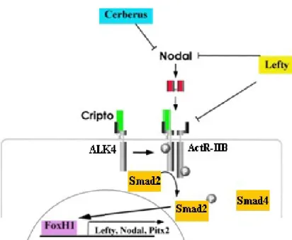
Very briefly, as a member of the TGF β superfamily, Nodal is first a proprotein; this proprotein is processed extracellularly by convertases PACE4 and Furin. Nodal signaling is activated by the interaction of mature Nodal ligand with activin receptors (ActRIIB, ALK4) and EGF-CFC co-receptors, and it is inhibited by leftys and Cerberus family genes (Cheng, et al. 2004, Belo, et al. 1997, Belo, et al. 2000). Nodal signaling is