UNIVERSIDADE DA BEIRA INTERIOR
Ciências da Saúde
Design and production of new nanodevices for
future application in cancer therapy
Ana Sofia Matias da Silva
Thesis dissertation for MsC in Master degree thesis
Biomedical Sciences
(2
ndcycle of studies)
Supervisor: Ilídio Correia, PhD
UNIVERSIDADE DA BEIRA INTERIOR
Ciências da Saúde
Desenho e produção de novos nanodispositivos
para futura aplicação na terapia do cancro
Ana Sofia Matias da Silva
Dissertação para obtenção do Grau de Mestre em
Ciências Biomédicas
(2º ciclo de estudos)
Orientador: Prof. Doutor Ilídio Correia
iv
“Tenho pensamentos que, se pudesse revelá-los e fazê-los viver,acrescentariam nova
luminosidade às estrelas, nova beleza ao mundo e maior amor ao coração dos
homens”. Fernando Pessoa
vi
Dedication
I would like to dedicate this Master Thesis to two of the most important people that ever crossed my life: my Grandmother Deolinda and my ‗second‘ father Fernando. Because I know that both of you would like to be next to me in this moment of my life and because I do believe that in ‗Every step I take, you‘ll be watching me‘.
viii
Acknowledgments
I would like to thank my supervisor Professor Ilídio Correia for the unconditional support, guidance and help during my master‘s degree. Specially, I feel the need to acknowledge him for having believed in my capabilities and for making me believe in myself. It has been a privilege working with him.
Furthermore, I would like to thank Professor António Mendonça for all the help and for the recipe to produce gold nanoparticles.
I also acknowledge to Professor Ana Aguiar and Professor Vasco Bonifácio from the Universidade Nova de Lisboa, for providing us oligoaziridine biosensor and for the support to continue this project.
Moreover, I would like to thank to Eng. Ana Paula from the Optics department of Universidade da Beira Interior for the help in acquiring scanning electron microscopy images of the nanoparticles produced.
I would also like to thank to Dr. Catarina Ferreira for the help in the acquisition of immunofluorescence and confocal laser microscopy images.
In addition, I would like to thank to all of my group colleagues for all the teaching, for having supported me whenever I was feeling down, for all the laughs that made me feel good and for all the experience shared during these months. Their friendship was, without any doubt, very important to overcome the difficulties faced.
From the bottom of my heart I thank to my family and closest friends, from the youngest to the elderly for the love, support and patience not only during the execution of this master thesis, but also through my entire academic course. They were truly important in all of my victories and they are definitely ‗the only love that doesn‘t make me weak‘.
I could not end these acknowledgments without a special thanks to my boyfriend for having appeared in my life when I needed the most. For all the love, support, care and patience during the good and the bad days and for all the advices, I do thank him a lot. Specially, I thank him for having shown me that I‘m an amazing woman who deserves to be loved.
x
Abstract
Nanotechnology is a multidisciplinary area of research that involves different knowledgements from, like life sciences, engineering and medicine. It has been used for different applications such as molecular imaging, molecular diagnosis and also targeted therapy. So far, ddifferent nanoscale devices have been produced, among them, inorganic nanoparticles, dendrimes, lipossomes, polymeric micelles, polymeric nanoparticles, nanotubes and nanofibers are some of the examples. Some of these particles exhibit unique optical and electrical properties allowing their course identification and precise location in the body. Gold nanoparticles are an example of inorganic particles with exceptional physico-chemical properties that demonstrate a huge potential for biomedicine applications.
The present study aimed to produce gold nanoparticles by two different methods: the citrate reduction method developed by Frens in 1973 (method 1), and its functionalization with oligoaziridine, developed by the colleagues from Universidade Nova de Lisboa, as a capping agent (method 2). This second method relies on the fact that gold nanoparticles can be prepared in water directly by the complexation of the alkylamine molecules that act as reducing agents and consequently stabilizes gold nanoparticles.
Moreover, gold nanoparticles produced by method 1 were also grafted with homofunctional maleimide poly(ethylene glycol) and then capped with oligoaziridine and the same parameters mentioned above were also evaluated.
The cytotoxicity and cell internalization of the different nanoparticles herein produced, was evaluated through in vitro studies.
The use of this new biosensor allow us to confirm the entry of the produced nanoparticles into cells opening new sights for the use of these particles as drug/gene delivery agents and/or as a new method for optimal imaging when methodologies like X-ray computed tomography or magnetic resonance cannot be used.
Keywords
Nanotechnology; Gold nanoparticles; Oligoaziridine; Homofunctional Maleimide Poly(ethylene glycol)
xii
Resumo
A nanotecnologia é uma área de investigação multidisciplinar que abrange conhecimentos das ciências da vida, da engenharia e da medicina. Esta área do conheciemnto tem contribuido para melhorar as tecnologias de imagiologia, diagnóstico molecular e na terapia direccionada. Nos últimos anos têm sido produzidos diferentes dispositivos à nanoescala, entre eles destacam-se as nanopartículas inorgânicas, dendrímeros, lipossomas, micelas poliméricas, nanopartículas poliméricas, nanotubos e nanofibras. As nanopartículas de ouro são um exemplo de partículas inorgânicas, e apresentam propriedades físicas e químicas excepcionais que lhe conferem um elevado potencial para aplicações biomédicas.
O presente estudo teve como objectivo produzir nanopartículas de ouro por dois métodos diferentes: o método de redução de citrato desenvolvido por Frens em 1973 (método 1); e o da funcionalização das aminas através da adição de oligoaziridina, um biosensor desenvolvido pelos colegas da Universidade Nova de Lisboa, como agente de revestimento (método 2). Este segundo método envolve a preparação das nanopartículas de ouro directamente em água através da complexação com moléculas acilaminas, que actuam como agentes redutores, estabilizando as nanopartículas de ouro.
Numa segunda fase, as nanopartículas de ouro produzidas pelo método 1 foram revestidas com polietilenoglicol maleimida homofuncional e, em seguida, adiciounou-se oligoaziridine. A citoxicidade e a capacidade de entrarem nas células foi tambem avaliada para estas nanopartículas. Os resultados obtidos demonstram que o polimero polietilenoglicol maleimida homofuncional se liga de uma forma efectiva às nanopartículas de ouro. Por outro lado, provou-se que o oligoaziridine se liga tanto ao polietilenoglicol como às nanopartículas isoladas.
Após a sintese das nanopartículas pelos dois métodos foi avaliada a sua toxicidade e a capacidade de entrarem nas células eucarióticas.
A utilização deste novo biosensor permite confirmar a entrada das nanopartículas nas células, o que possibilitará o uso destas partículas como agentes de entrega direccionada de fármacos, genes ou como um novo método para a obtenção de imagens quando metodologias como Tomografia Computadorizada por raios X ou Ressonância Magnética não poderem ser usadas.
Palavras-chave
Nanotecnologia; Nanopartículas de ouro, Oligoaziridine, Polietilenoglicol Maleimida Homofuncional
xiv
Index
Chapter I - Introduction ... 1
1. Introduction ... 2
1.1. Cancer – A brief discussion of the disease ... 2
1.1 1. Events that lead to cancer disease ... 3
1.1.2. Cancer treatments ... 5
1.2. Nanotechnology ... 7
1.2.1. Nanoparticles as anti-cancer agents ... 8
1.2.2. Nanoparticles surface constituents for a target delivery ... 9
1.2.2.1. Polymers for nanoparticles‘ coating ... 9
1.2.2.2. Extracellular matrix targeting ligands for coating nanoparticles . 11
1.2.2.3. Other molecules used for nanoparticles targeting ... 12
1.2.3. Different types of nanoparticles ... 14
1.2.3.1. Amphiphile based particles ... 15
1.2.3.1.1. Liposomes ... 15
1.2.3.1.2. Polymersomes ... 17
1.2.3.1.3. Micelles ... 17
1.2.3.2. Dendrimers ... 17
1.2.3.3. Carbon nanotubes ... 18
1.2.3.4. Inorganic nanoparticles ... 18
1.2.3.4.1. Ceramic nanoparticles ... 19
1.2.3.4.2. Metallic nanoparticles ... 19
1.2.3.4.2.1. Gold nanoparticles ... 20
1.3. Oligoaziridine for coating nanoparticles ... 22
Chapter II – Material and Methods ... 23
2. Materials and Methods ... 24
xv
2.2. Methods ... 24
2.2 1. Preparation of gold nanoparticles by the citrate reduction method -
Method 1 ... 24
2.2 2. Preparation of gold nanoparticles by oligoaziridine reduction method -
Method 2 ... 24
2.2 3. Coating of gold nanoparticles produced by method 1 with oligoaziridine 25
2.2 4. Preparation of gold nanoparticles coated with homofunctional maleimide
poly(ethylene glycol) ... 25
2.2 5. Coating AuNP‘s_PEG with oligoaziridine ... 26
2.2 6. Scanning Electron Microscopy analysis ... 26
2.2 7. Ultraviolet-Visible spectroscopy analysis ... 27
2.2 8. Fourier Transform Infrared spectroscopy analysis ... 27
2.2 9. X - ray Diffraction analysis ... 27
2.2 10. Proliferation of A549 small lung carcinoma cell line in the presence of
the produced nanoparticles ... 27
2.2 11. In vitro transfection of cells with the nanoparticles produced ... 28
2.2 12. Qualitative evaluation of in vitro transfection ... 28
2.2 13. Evaluation of the cytotoxic profile of the produced nanoparticles ... 28
2.2 14. Statistical analysis ... 29
Chapter III – Results and Discussion ... 30
3. Results and Discussion ... 31
3.1. Particles Morphology ... 31
3.2. Ultraviolet Visible analysis ... 34
3.3. FTIR – analysis of the nanoparticles ... 38
3.4. X ray Diffraction analysis ... 44
3.5. Qualitative evaluation of in vitro transfection ... 45
xvi
Chapter IV – Conclusion and Future Perspectives ... 51
Bibliography ... 54
xviii
List of Figures
Chapter I – Introduction
Figure 1 – Cancer evolutive model ... 2
Figure 2 – Illustration of the metabolic changes that occur in cancer cells ... 3
Figure 3 – Framework of genetic events on cancer development ... 4
Figure 4 – Schematic representation of different mechanisms by which
nanocarriers can deliver drugs into tumors ... 9
Figure 5 – Illustration of a multifunctional nanoparticle ... 14
Figure 6 – Different types of nanoparticles for drug delivery ... 15
Chapter III – Results and Discussion
Figure 7 – SEM images of the produced nanoparticles ... 33
Figure 8 – UV-VIS spectra of the produced nanoparticles and the compounds used
for their production ... 36
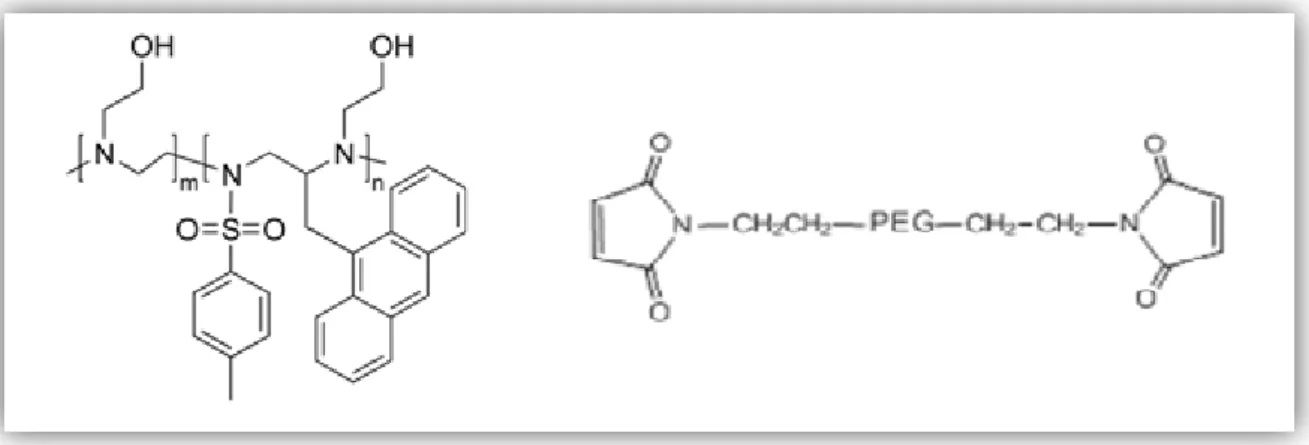
Figure 9 – Molecular structures of oligoazidirine and MAL-PEG-MAL ... 38
Figure 10 – FTIR spectra of the produced nanoparticles and the compounds used
for their production ... 41
Figure 11 – XRD spectra of AuNP‘s, MAL-PEG-MAL and AuNP‘s_PEG ... 44
Figure 12 – Immunofluorescence images of A549 cells transfected with
oligoaziridine and the produced nanoparticles ... 45
Figure 13 – Confocal Laser Scan Microscopy images of A549 cells transfected with
oligoazirine and the produced nanoparticles ... 47
Figure 14
-Inverted Light Microscope images of A549 cells in contact with
materials after 24 and 48 hours of incubation ... 49
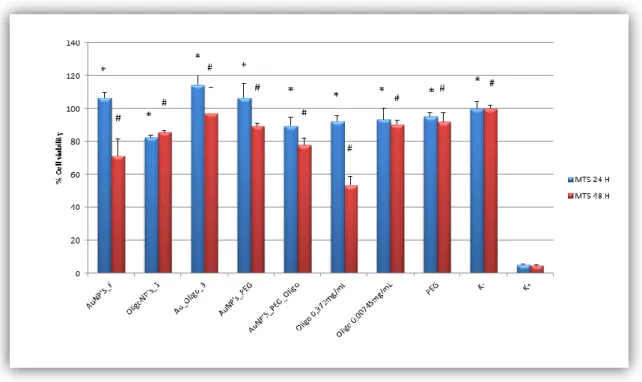
Figure 15 – Cellular activities measured by MTS assay after 24 and 48 hours in
contact with the materials... 50
xx
List of Tables
Table 1 – Selection of FDA approved antibodies and molecules that are clinically
used for the treatment of tumors and that can be conjugated with nanoparticles . 12
Table 2– Examples of clinically approved tumor target nanoparticle ... 16
xxii
List of Acronyms
ABC ATP Binding Cassette transporters
ATP Adenosine Triphosphate
Au_Oligo Gold nanoparticles produced by method 1 capped with oligoaziridine AuNP‘s Gold nanoparticles
AuNP‘s_F Gold nanoparticles produced by method 1
AuNP‘s_PEG Gold nanoparticles (produced by method 1) capped with MAL-PEG-MAL AuNP‘s_PEG_Oligo Gold nanoparticles (produced by method 1) capped with MAL-PEG-MAL
and Oligoaziridine
CEA Carcinoembryonic Antigen
CLSM Confocal Laser Scan Microscopy
EGF Epidermal Growth Factor
EPR Enhanced Permeability and Retention effect ErbB2 Human Epidermal Growth Factor receptor 2
EtOH Ethanol
FBS Fetal Bovine Serum
FDA Food and Drug Administration
Frs Folate receptors
FTIR Fourier Transform Infrared
K- Negative control
K+ Positive control
mAB Monoclonal Antibody
MAL-PEG-MAL Maleimide-PEG-Maleimide
MDR Multidrug Resistance
MPS Mononuclear phagocytic system
MR Magnetic ressonance
MRI Magnetic Resonance Imaging
MTS 3-(4,5-dimethylthiazol-2-yl)-5-(3-carboxymethoxyphenyl)-2-(4-sulfophenyl)-2H-tetrazolium
NaBH4 Sodium Borohydride
NIR Near-Infrared
NLS Nuclear Location Signal
OLA Oleyl amine
OligoNP‘s Gold nanoparticles produced by method 2
PAAm Poly(acrylamide)
PAMAM Polyamidoamine
PBS Phosphate-Buffered Saline
PEG Poly(ethylene glycol)
PEG:AuNP‘s Poly(ethylene)glycol: Gold nanoparticles ratio
PEG-b-PPO-b-PEG Poly(ethylene glycol)-b-poly(propylene oxide)-b-poly(ethylene glycol) PHSRN Proline – Histidine – Seronine – Arginine – Asparagine
PMS Phenazine Methosulfate
pNIPAAM Poly(N-isopropylacrylamide) PPE Palmar-plantar erythrodyesthesia Pr_b Fibronetic-mimic peptide
xxiii
PVA Poly(vinyl alcohol)
RGD Arginine – Glycine – Aspartic acid SEM Scanning Electron Microscopy
SPR Surface Plasmon Resonance
TEM Transmission Electron Microscopy
Tf Transferrin
TFrS Transferrin receptors UV-VIS Ultraviolet-Visible
VEGF Vascular endothelial growth factor X Ray TC X ray Computed Tomography
YIGSR Tyrosine – Isoleucine - Glycine – Seronine – Arginine YIGSR-NPS YIGSR attached to nanoparticles surface
Chapter I – Introduction
2
1. Introduction
1.1. Cancer – A brief discussion of the disease
Cancer is the leading cause of death worldwide accounting for 7.9 million of deaths in 2007 (Dong and Mumper 2010) and World Wide Health Organization predicts that in the year of 2030 this value will rise up to 20 million (Bode & Z. Dong 2009). Cancer may develop from different cell types in the human body and is characterized by relatively uncontrolled proliferation of cells that can invade healthy tissues and metastasize into remote organs (Stratton et al., 2009). The first insights in cancer development emerged in the late 19th and
early 20th centuries through the examination of cancer cells by optical microscopy (Stratton
et al., 2009). Later on, the observation of peculiar chromosomal aberrations led to the proposal that cancer are abnormal clones of cells, that appear due to the occurrence of modifications in the hereditary material (Stratton et al., 2009). Natural selection, acting on the resulting phenotypic diversity, eliminates cells which have acquired deleterious mutations and foster the ones that have acquired proliferation capabilities (cancer cells) (figure 1) (Visvader and Lindeman 2008; Stratton et al., 2009).
Figure 1 - Cancer evolutive model. All cells possess similar tumorigenic capacity. Mutant tumor cells with growth advantage prevail over normal cell types thus increasing tumorigenic tissue (Adapted from Visvader and Lindeman 2008).
Nevertheless, cancer development is accepted as a multistep process, during which genetic alterations promote abnormal cell growth, which leads to the progressive transformation of normal and healthy cells into to cancer ones (Hanahan 2000). Figure 2 summarizes the key event changes that characterize the appearance and growth of a cancer cell.
Chapter I – Introduction
3 Figure 2 – Illustration of the metabolic changes that occur in cancer cells. Malignant growth is characterized by several key events as self-sufficiency in growth signals, insensitivity to anti-growth signals, cell avoids apoptosis, deregulated proliferation potential, enhanced angiogenesis, ability to invade tissues and metastasize. (Adapted from Hanahan 2000)
1.1.1. Events that lead to cancer disease
The genetic alterations in cancer must be understood in a context of cellular organization, differentiation, tissue organization, host response and susceptibility to angiogenesis. Several factors can influence the evolution of a cancer, like different events that occur inside of the cell (as gain or loss of function events), epigenetic events, environmental exposures (e.g., radiations or smoke), systemic effects like hormones or growth factors and paracrine interactions with neighbouring cells (figure 3) (Hanahan 2000; Ponder 2001).
Chapter I – Introduction
4 Figure 3 – Framework of genetic events on cancer development. Horizontal arrows represent the pathway of successive genetic or epigenetic events through which the cell may acquire the cancer phenotype. Vertical arrows indicate the influences in this pathway that affect the probability of a specific event to occur ( Adapted from Ponder 2001).
The three major classes of genes in which genetic variations occur and subsequently cancer cells arise are oncogenes, tumor suppressor genes, and caretaker genes (Vogelstein and Kinzler 2004). Oncogenes encode proteins that control cell proliferation, apoptose or both. These types of genes can be activated by structural modifications that result from either mutations or gene fusion, or by juxtaposition to enhancer elements (Croce 2008). Mutations in this type of genes are usually associated with gain of function events, i.e., events that confer enhanced activity to the expressed proteins (Collins 1997). Tumor suppressor genes or anti-oncogenes regulate cell division. This type of genes reduces the probability of the cell, in a certain multicellular organism, becomes a tumorigenic one. Mutations in tumor suppressor genes cause a loss or reduction in its function, leading a malignant cell to progress to cancer (Ponder 2001; Vogelstein and Kinzler 2004).
Inactivation of caretaker genes (genes that control genomic integrity) leads to genomic instability and thus contributes for the increase of the probability of alterations in the oncogenes and in the tumor suppressor genes (Ponder 2001; Vogelstein and Kinzler 2004).
Chapter I – Introduction
5 Epigenetic events, gene expression alterations that do not alter nucleotide sequences like DNA methylation and histones modification, occur in all stadiums of tumor formation, including premature phases. Due to the genetic alterations caused by these events, they have been recognized as the main mechanisms involved in aberrant silencing of genes, that are important to avoid the initiation and progression of tumors (Ponder 2001). The huge importance of these epigenetic variations is due to the fact that they can be reverted, by applying small molecules like inhibitors of DNA methylation (5-azadeoxycytidine) and inhibitors of histone deacetylase (Moradei et al., 2005; Ting et al., 2006; Momparler, 2003).
Genetic variations acting either inside or outside the cancer cell may determine the outcome of interaction with exogenous carcinogens (Ponder 2001). For instance, sun exposure in individuals with fair skin, chemical exposures and genetic variations in the metabolic pathways may account for substantial differences in cancer susceptibility within the population (Ponder 2001). Moreover, gene-environment interaction may also provide new sights for cancer prevention, since it is possible to categorize subpopulations in terms of genetic risk. This will increase the effectiveness to detect cancers environment causative exposures. This type of information may also be helpful for epidemiologic studies in a way to categorize populations and thus be able to detect the main causes of cancer disease (Ponder 2001). Variations in the circulating levels of hormones or growth factors are also associated with cancer development. For example, high levels of oestrogen might have carcinogenic effects, either through direct stimulation of cancer growth or as a product of mutagenic metabolites (Ponder 2001). Paracrine interactions between inflammatory cells and healthy ones, may also have an important role in cancer development (Ponder 2001). For example, the production of the matrix metalloproteinase 9 by inflammatory cells, has been implicated in the development of squamous cell carcinomas in an HPV-16 transgenic model (Ponder 2001).
1.1.2. Cancer Treatments
A single cancer cell enclosed by healthy tissue will replicate at a higher rate than the normal ones, placing a strain on the supply of nutrients and removal of metabolic wastes. Often tumor cells undergo apoptosis in environments with low nutrients concentrations (such as oxygen, glucose and amino acids) (Nie et al., 2007; Pathak et al., 2007).
Current diagnostic do not reflect the whole clinical heterogeneity of tumors and are insufficient to make predictions for the successful treatment of the disease and patient outcome (Nie et al., 2007). Cancer is often diagnosed in an advanced stage, when cancer cells have already been metastasized and invaded other parts of the body, leading to a loss of effectiveness of the different therapies (Nie et al., 2007).
The usual cancer treatments are limited to chemotherapy, radiation and surgery (Dong and Mumper 2010; Cho et al., 2008). The most recurrent and most active first line of
Chapter I – Introduction
6 chemotherapeutic agents are anthracyclines (such as doxorubicin, epirubicin and daunorubicin), and taxanes (like docetaxel and paclitaxel) (Dong and Mumper 2010).
One of the drawbacks of the existent cancer therapies include nonspecific systemic distribution of the antitumor agents, which leads to body toxicity. The majority of drugs available lack of selectivity for cancer cells resulting in healthy cells death (Brewer et al., 2011; Dong and Mumper 2010; Nie et al., 2007; Peer et al., 2007; Cho et al., 2008). Moreover, the rapid clearance of the therapeutic molecules from blood circulation requires larger doses for the treatment to be effective. Such doses can be responsible for side effects (Brewer et al., 2011; Nie et al., 2007; Peer et al., 2007; Cho et al., 2008). Others obstacles of the current therapies are the inadequate drug concentrations reaching the tumor and the limited ability to monitor therapeutic efficiency (Cho et al., 2008).The biological properties of the solid tumor, that limit drug arrival into neoplasic cells, include abnormal heterogeneous tumor vasculature, interstitium, interstitial fluid pressure and cell density (Dong and Mumper 2010; Peer et al., 2007).
Drug resistance is the major obstacle that limits the therapeutic efficacy of chemotherapeutic agents. Multidrug resistance (MDR) is the phenomenon in which exposure of tumor cells to a single cytotoxic agent accounts for cross-resistance to other structurally unrelated classes of cytotoxic agents (Dong and Mumper, 2010). This is related to the broad spectrum resistance of cancer cells to chemotherapy (Dong and Mumper, 2010). MDR can be caused either by non-cellular mechanisms such as physiological barriers or by cellular ones, like modifications in the biological and biochemical features of the cancer cells (Davis et al., 2008). Non cellular drug resistance may include poorly vascularized tumor regions, high interstitial pressure and low microvascular pressure (Davis 2008). These features appear to significantly reduce drug access to tumor region (Dong and Mumper 2010; Cho et al., 2008; Davis et al., 2008). On the other hand, cellular mechanisms involve changes in specific enzyme systems for drug metabolism, mutations in the drug delivery target and increased drug efflux from tumor cells (Davis et al., 2008). The most widely investigated mechanism of MDR is the change in the membrane transporters that act as drug efflux pumps (Davis et al., 2008). MDR transporters actively pump chemotherapeutic drugs out of the cell reducing intracellular doses to nontherapeutic levels. Not every cancer cell express this type of transporters and chemotherapy drugs are only able to kill cells that do not express MDR transporters or present only a residual expression of these membrane transporters (Peer et al., 2007). One of the most described membrane transporters is glycoprotein P(P-gp), a member of the ABC superfamily (transmembrane proteins that use the energy of ATP hydrolysis to shuttle various substrates across the cell membrane), that is overexpressed in the plasma membrane of tumor cells (Dong and Mumper 2010; Nie et al., 2007; Davis 2008). This glycoprotein is capable of effluxing several anticancer drugs like doxorubicin and paclitaxel out of the cells (Dong and Mumper 2010; Nie et al., 2007). To overcome this problem different inhibitors for this P-gp have been investigated. However, some inhibitors used do not only block the P-gp of tumor cells, but also those from healthy cells, and thus
Chapter I – Introduction
7 contributes for the increase of toxicity (causing side effects in different tissues such as bone marrow suppression, cardiomyopathy and neurotoxicity). This limits the drug concentration that reaches the target tissue (Dong and Mumper 2010; Nie et al., 2007).
Enhanced targeting selectivity and delivery efficiency are two of the foremost goals in the development of therapeutic agents. Ideally, a therapeutic drug would be selectively enriched in the tumor with minimal damage to the surrounding healthy tissues, and would have no degradation before reaching the target cells (Dong and Mumper 2010; Cho et al., 2008). In order to overcome both non-cellular and cellular mechanisms of resistance, and to increase drugs selectively towards cancer cells, nanosized devices are currently being developed (Brigger et al., 2002).
1.2. Nanotechnology
Nanotechnology is a multidisciplinary area of research in life sciences, engineering and medicine with broad band applications for molecular imaging, molecular diagnosis and targeted therapy (Misra et al., 2010). The American Nanotechnology Institute defines nanotechnology as a science in which 10−100 nm size structures are developed (Nair et al., 2010; Liu et al., 2007; Farokhzad and Langer 2009). Overtime, the scope of this definition was expanded in terms of its upper limit (Davis et al., 2008), since nanodevices must be large enough to prevent their fast escape into blood capillaries, but small enough to escape from the capture from macrophages, which are lodge in the reticuloendothelial system, like the liver and the spleen. Considering this, the size of the sinusoids in the spleen and fenestra of the Kupffer cells, which varies between 150 to 200 nm, and the size of gap junction between endothelial cells of the leaky tumor vasculature ranges from 100 to 600 nm, nanoparticles size should be at least 100 nm in order to achieve tumour tissues (Cho et al.,, 2008). However, due to sieving coefficients measurements for the glomerular capillary wall, and based on the threshold for the first-pass elimination by the kidneys, the lower limit of 10 nm was maintained (Davis 2008).
Nanometer scale devices are mainly built up from their basic constituents since they can be formed by chemical synthesis, spontaneous self-assembly of molecular clusters (molecular self-assembly), biological molecules (e.g., DNA) used as building blocks for production of three-dimensional nanostructures, and quantum dots (nanocrystals) of arbitrary diameter (about 10–105 atoms) (Bhushan 2010). Delivery vehicles at nano or micro scale
consisting on aggregates of macromolecules have been designed to release the drugs via local injection or systemic release (Baldwin and Kiick, 2010). Methods of imparting biological activity to materials involve the use of polysaccharides derived from natural or synthetic sources. These materials present low immunogenicity, ionic charge and other properties that are fundamental for their use in biomedical applications (Baldwin and Kiick 2010).
Chapter I – Introduction
8
1.2.1. Nanoparticles as anti-cancer agents
Nanoparticles were produced 39 years ago as vaccine carriers and chemotherapeutic agents for cancer (Pathak et al., 2007). Cancer nanotherapies have been implemented to undertake several limitations of the conventional drug delivery systems, which are: nonspecific for distribution and targeting, low water solubility, poor oral bioavailability and lower therapeutic efficacy (Yih and Al Fandi 2006).
In contrast to normal cells, tumor anatomical defectiveness along with functional abnormalities such as tumor blood vessels with irregular shape, dilated, leaky or defective and endothelial cells disorganized with large fenestrations, results in extensive leakage of blood plasma components into the tumor. These features help the retention of nanoparticles in tumor site, long enough, to allow local nanoparticle disintegration and release of the drug into tumor‘s vicinity (Wang and Thanou 2010; Iyer et al., 2006; Liu et al., 2007). Venous return to tumor tissue is slow, lymphatic clearance is poor, but the extravasation into the tumour interstitium continues, leading to an accumulation of macromolecules in the tumor (Iyer et al., 2006). Nanoparticles can penetrate into leaky and hyperpermeable tumor vasculature, accumulate in its proximity using the enhanced permeability and retention effect (EPR). They can also deliver more than one therapeutic agent for combinatory therapy (Danhier et al., 2010; Jain and Stylianopoulos, 2010). This type of approach is currently denominated by passive targeting (figure 4) (Danhier et al., 2010; Cho et al., 2008). However, EPR is not applicable to low molecular weight drugs due to their quick diffusion into the circulating blood, followed by renal elimination (Iyer et al., 2006).
On the other hand, active targeting overcome permeability limitations of the passive targeting since it allows tissue penetration and cellular uptake by cancer cells (Pathak et al., 2007; Peer et al., 2007; Cho et al., 2008). This active targeting involves the functionalization of a carrier system containing the chemotherapeutical agents, which are selectively recognized by the overexpressed receptors existing at the surface of the interest cancer cells, a feature also represented in figure 4 (Pathak et al., 2007; Peer et al., 2007; Cho et al., 2008). Since ligand-receptor interactions can be highly selective, the functionalization of the nanoparticle surface will allow a more precise targeting for tissues of interest and will reduce of the toxic effects in the surrounding normal tissues (Pathak et al., 2007; Peer et al., 2007; Cho et al., 2008). As a result, the potential benefits of such delivery vehicles include: controlled and long-term release rates, extended bioactivity, reduced side effects, decreased administrated frequency to the patient, and the ability to co-deliver multiple drugs with synergistic effects to the same target site (Brewer et al., 2011; Pathak et al., 2007; Farokhzad and Langer 2009).
Chapter I – Introduction
9 Figure 4 – Schematic representation of different mechanisms by which nanocarriers can deliver drugs into tumors. In the passive mode, nanoparticles accumulate preferential at the tumors sites through EPR , as in the active targeting, ligand molecules such as antibodies and peptides are often used to recognize specific tumor antigens (Adapted from Dong and Mumper 2010; Nie et al., 2010; Farokhzad and Langer 2009)
1.2.2. Nanoparticle surface constituents for a target delivery
The biophysico-chemical features of the carrier such as size, charge and surface hydrophilicity, can all affect both circulating time of the particles and their biodistribution (Farokhzad and Langer 2009). Structures such as antibodies, antibody fragments, small molecules, aptamers (nucleic acids) and peptides, vitamins and carbohydrates have all demonstrated abilities to induce nanoparticle-targeting to cancer cells (Peer et al., 2007).
1.2.2.1 Polymers for nanoparticles’ coating
Nanoparticles present a high surface-to-volume ratio when compared with larger particles (Davis, 2008). As a result, the control of nanoparticles surface characteristics is crucial for their administration into human body (Davis, 2008).
The outcome of nanoparticles within the human body can be determined by nanoparticles interactions with their local environment, which depends on a combination of size and surface features (Davis, 2008). Nanocarriers sterically stabilized by polymers like poly(ethylene glycol) (PEG), and with surface charges either slightly negative or slightly positive tend to have minimal self-interactions (Davis, 2008).
The coating of hydrophobic nanoparticles with hydrophilic polymers include common strategies like ligand exchange, micelle encapsulation and covalent bonding (Fang et al.,
Active targeting
Chapter I – Introduction
10 2009). Synthetic polymers used for coating biomaterials include hydrophilic and non-hydro degradable materials like PEG, poly(vinyl alcohol) (PVA) currently used for coating nanoparticles for oral chemotherapy (Win and Feng 2005), and poly(acrylamide) (PAAm) usually used for coating magnetic nanoparticles (Sun et al., 2006). On the other hand, hydrophobic and hydro degradable polymers are used as well. These include poly(α-esters) good for coating drug delivery systems (Chan-Seng et al., 2009), amphiphilic block polymers like PEG-b-PPO-b-PEG and thermally sensitive polymers such as poly(N-isopropylacrylamide) (pNIPAAM) that have been used for cancer targeting in conjugation with hyperthermia (Baldwin and Kiick, 2010; Win and Feng 2005).
PEG has been used by different researches as an effective coating material for nanoparticles, due to its ability to resist protein fouling and provide steric hindrance, preventing nanoparticles to aggregate (Fang et al., 2009). PEG is a neutral, crystalline, thermoplastic polymer with a high solubility in water and organic solvents. PEG layers have been added to nanoparticles (liposome, dendrimer or inorganic nanoparticles) that are aimed to be administered intravenously in order to accumulate into tumors (Allen et al., 2002). This approach has been widely applied to form ‗stealth‘ micelles in order to enhance circulation time, by preventing their interaction with plasma proteins, and inhibit the accumulation of opsonises and their uptake by macrophages (Brewer et al., 2011; Dong and Mumper, 2010; Wang and Thanou, 2010). It has been reported that PEG‘s size and density are the key features that control the circulation times and accumulation in tumors (Dong and Mumper 2010; Wang and Thanou, 2010). PEG is placed on the outmost exterior of the nanoparticle delivery system and targeting ligands are generally presented on top of its layer (Brewer et al., 2010). The enlarged residence time increases the probability of interaction between the receptor and the target cancer cells (Brewer et al., 2011). Akyyama and collaborators studied the effects of PEG grafting level and injection dose on gold nanorod biodistribution in the tumor-bearing mice (Akiyama et al., 2009). This investigation reported that as PEG concentration increases, the amount of gold retained in the spleen decreases, in contrast to the concentrations within the liver and cancer cells (Akiyamai et al., 2009). However, for PEG: gold molar ratios of 1.5 or above, this concentration was higher within the tumor tissue (Akiyama et al., 2009). A decrease in splenic accumulation by increasing the amounts of PEG grafted onto gold nanorods surface can be explained by the ability that particles, larger than 200 nm, have to avoid the reticuloendothelial system and to escape from splenic physical filtration (Akiyama et al., 2009). Gold nanorods that escaped from the spleen were able to reach the tumor tissue and enter within it due to its EPR effect (Akiyamai et al., 2009). Nevertheless, a large amount of PEG may also lead to hepatic accumulation due to the uptake by hepatic parenchyma (Akiyama et al., 2009). This undesired hepatic accumulation of PEGylated nanoparticles might result in an inflammatory response triggered by the liver as described by Cho and colleagues (Cho et al.,, 2009).
Chapter I – Introduction
11
1.2.2.2. Extracellular matrix targeting ligands for coating nanoparticles
As cancer cells often present similar characteristics to its surrounding healthy tissue, ligands can be designed in order to promote the specific recognition of cancer cells. These molecules have specificity to bind receptors that are overexpressed on cancerous cells. In healthy cells, these nanoparticles should not present capability to induce receptor mediated endocytosis (Brewer et al., 2011). An important aspect is the type of ligand–receptor interaction which modifies the rate of cellular internalization. This rate can affect the accumulation of nanoparticle inside tumor cells (Brewer et al., 2011).
Extracellular matrix (EMC) is mainly composed of proteoglycans and proteins such as collagen, laminin, fibronectin and integrins (Choi et al., 2010). EMC derived short peptides act as receptors binding motifs that can be immobilized or incorporated onto several surfaces, including polymers, hydrogels and nanodevices (Choi et al., 2010).
Sarfati and collaborators performed an investigation that aimed to studied nanoparticles‘ targeting to lung metastasizes via interactions with the laminin receptor, whose expression is upregulated in metastatic cells (Sarfati et al., 2011). The laminin receptor binding peptide Tyr-Ile-Gly-Ser-Arg (YIGSR) was attached to nanoparticles surface (YIGSR-NPs) to facilitate targeting (Sarfati et al., 2011). When intravenously administered into B16 melanoma tumor – bearing mice, YIGSR-NPs reached the cancerous metastatic cells in lungs, with nearly no binding to the healthy lung cells, which lead to the conclusion that YIGSR-targeted nanoparticles have a potential to be used for systemic delivery of chemotherapeutic drugs for the treatment of metastatic lung cancer (Sarfati et al., 2011) .
Peptides containing the Arg-Gly-Asp (RGD) can be recognized by αβ integrins which are overexpressed in tumor cells (Sugahara et al., 2009). RGD sequences have been widely attached to nanoparticles in order to enhance their penetration into tumor vasculature (Sugahara et al., 2009). Kang‘s group described the ability of gold nanoparticles conjugated with RGD sequence to be recognized by cancer cells (Kang et al., 2010).
Besides integrin receptors, the adhesive glycoprotein fibronectin also appears to play important roles in the progression of cancer disease (Akiyama et al., 1995). Pro-His-Ser-Arg-Asn (PHSNR), the peptide fragment of fibronectin that is recognized by α5 1 receptor of cancer cells, is known for its ability to enhance drug entrapment (KoKKoli 2011). Demirgo and collaborators proposed the incorporation of a fibronectin-mimic peptide into PEGylated lipossomes with the aim of targeting α5 1 which is overexpressed on prostate cancer cells (Demirgo et al., 2008).
There is also a need to optimize ligand density at the drug delivery vehicle surface. Targeting ligands are generally presented on the top of the stealth layer which is attached to the nanoparticles (Wang and Thanou, 2010). This is responsible for the tissue penetration and cellular uptake of the drug delivery systems in order to enhance therapeutic efficacy (Wang and Thanou, 2010; Farokhzad and Langer 2009).
Chapter I – Introduction
12
1.2.2.3. Other molecules used for nanoparticles targeting
Antibodies are a group of serum proteins produced by plasma cells that bind specifically to antigens. Regarding targeting, they can be either used in their native state or only as a fragment (Woof and Burton 2004; Peer et al., 2007). Targeting cancer with monoclonal antibodies (mAb) was first described in 1981 (Warenius et al., 1981; Woof and Burton 2004; Peer et al., 2007). Since then, different studies reported their use. In 1997 mAb Rituximab (Rituxan), which binds to antigen CD-20, was approved for the treatment of non-Hodgkin‘s lymphoma, a cancer originating from lymphocytes. In 1998 Trastuzumud (Herceptin) was approved for breast cancer therapy, binding to human epidermal growth factor Receptor 2 (HER-2) (Peer et al., 2007). Six years later, Bevacizumad (Avastin), an anti-vascular endothelial growth factor (VEGF) mAb, was developed in order to inhibit the factor responsible for the growth of new blood vessels. This antibody was approved for treating colorectal cancer (Peer et al., 2007). Table 1 shows the antibodies and molecules approved by the Food and Drug Administration (FDA) that are often conjugate with nanoparticles to be used in cancer therapy (Cherukuri et al., 2010).
Tabela 1 FDA approved antibodies and molecules currently being used for tumor treatment (Adapted from Adams 2004; Cherukuri, 2010; Ciardiello et al.,, 2000; Clemons, 2002; Takimoto and Awada, 2008)
Name Brand Name Type Target Cancer Type
Bevacizumab Avastin Antibody VEGF receptor Colorectal,
non-small cell, lung, breast
Bortezomib Velcade Multicatalytic
enzyme Protein complex proteasome 26 s Myeloma, lymphoma
Trastuzumab Herceptin Antibody Human epidermal
receptor-2 (HER-2)
Breast
Gefitinib Iressa Quinazoline
derivate; Tyrosine kinase inhibitor Epithelial growth factor receptor (EGFR) Non-small cell lung
Sorafenib Nexavar Multi kinase
inhibitor Vascular endothelial growth factor receptor (VEGFR) and Platelet derived growth factor receptor Kidney, liver
Tositumomab Bexxar Antibody Antigen CD20 Lymphoma
Tamoxifen Nolvadex Selective estrogen
receptor modulator
Estrogen receptor Breast
Rituximab Rituxan Antibody Antigen CD20 Lumphoma
All antibody molecules possess a similar basic structure comprised of two identical heavy chains as well as two identical light chains. Such chains are arranged to form two antigen binding portions (Fab portion) and one portion that binds to other elements of the immune system like macrophages or other complement proteins (Fc portion) (Woof and
Chapter I – Introduction
13 Burton 2004; Peer et al., 2007). In order to have a higher binding activity it is suitable to use the intact antibody. In addition, the binding of immune cells to the Fc portion of de mAb will start a cascade of processes in order to kill the cancer cells. Yet, this mAb portion can also react with the Fc receptors of healthy cells, initiating the immunogenic process (Peer et al., 2007).
In order to increase antibodies efficacy it is possible to conjugate them directly with therapeutic drugs for targeting delivery. However these therapies appear to have some lethal side effects due to nonspecific binding between the targeting agent and non-target receptors on the cell surface (Peer et al., 2007).
The creation of new technologies like fusion proteins (combination of two or more genes producing a new protein with the desired properties) and engineering proteins that are detected by a specific conformation of a target receptor, may improve targeting and selectivity of the drug delivery systems (Peer et al., 2007). Small protein domains like affibodies, a new class of targeting molecules are engineered to bind specifically to different target proteins in a conformational-sensitive manner (Peer et al., 2007). Other small proteins, avimers are used to bind selectively to target receptors through multivalent domains. Heavy-chain antibodies engineered to one tenth of the size of an intact antibody (nanobodies) have been used to bind to carcinoembryonic antigen (CEA), a protein used as a tumor marker (Peer et al., 2007).
Growth factor or vitamin interactions are also used in the targeting strategy, as cancer cells often overexpress the receptors for nutrients to maintain their fast-growing metabolism (Peer et al., 2007). EGFR is overexpressed in a variety of tumor cells such as tongue cancer, epidermal growth factor (EGF) has been shown to block and reduce tumor expression of this receptor (Peer et al., 2007). Folic acid (folate) has also been used in cancer therapy, since folate receptors (FRs) are quite overexpressed in tumors like ovarian, endometrial as well as kidney cancer (Peer et al., 2007). Another example is transferrin (Tf), that interacts with Tf receptors (TFrS), receptors overexpressed in different types of tumors, such as pancreatic, colon, lung, and bladder cancer (Peer et al., 2007). Direct coupling of these targeting agents to nanoparticles containing chemotherapies agents has improved intracellular delivery and therapeutic outcome in animal tumor models (Peer et al., 2007).
Nanomedicine efficacy can be slightly improved by producing nanoparticles that respond to chemical or physical features in the tumor microenvironment (Brewer et al., 2011; Jain and Stylianopoulos, 2010). For instance, nanomaterials can take advantage of cancers lower pH, in order to promote drug delivery (Brewer et al., 2011; Jain and Stylianopoulos, 2010). Hyperthermia is another feature that has been widely investigated as a method for trigger drug release (Brewer et al., 2011). The heat increases microvessel permeability in tumors, which causes the accumulation of the respective thermosensitive nanocarrier in the tumor site, where they release their encapsulated drugs (Brewer et al., 2010). Moreover, hyperthermia also increases cell permeability which induces drug diffusion through tumor walls and sensitivity to thermal injury (Brewer et al., 2011). Nanoparticles can also be design
Chapter I – Introduction
14 in order to be activated by the proteinases in tumors (Jain and Stylianopoulos, 2010). In addition, the targeting of nanoparticles to tumors can also be achieved through the use of external sources like magnetic or electric fields, ultrasound and light (Jain and Stylianopoulos, 2010).
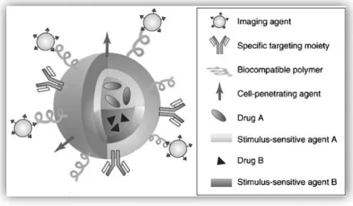
By exploiting the varied chemistry of the nanocarriers it is possible to conjugate ligands, polymers and imaging contrast agents all in one type of nanoparticle, as well as make it suitable to accommodate multiple drugs (Park et al., 2009). This multifunctional nanoparticle type is illustrated in figure 5, and is a great promise for the field of individualized medicine (Park et al., 2009).
Figure 5 - Illustration of a multifunctional nanoparticle. Multifunctional nanoparticle can combine a specific targeting agent (usually with an antibody or peptide), a cell-penetrating agent, a stimulus-selective element for drug release, a stabilizing polymer to ensure biocompatibility and the therapeutic compound (Adapted from Park et al., 2009)
1.2.3. Different types of nanoparticles
According to the process used to produce nanoparticles it is possible to obtain nanospheres or nanocapsules. Unlike nanospheres (a system in which the drug is dispersed), nanocapsules are vesicular systems in which the drug is confined to an aqueous or oily cavity surrounded by single polymeric membrane. Nanocapsules can then be regarded as a reservoir system (Brigger et al., 2002).
Most of the researchers agree that nanocarriers or nanovectors are nanosized systems that can carry multiple drugs and/or imaging agents. These systems are produced with polymers, ceramics and biological with several shapes, where drugs and contrast agents are attached to. They also possess a corona of polymeric material that improves biokinetics and biodistribution, and a ligand that adds specificity to the particle in order to recognize and attach to cancer cells (Wang and Thanou, 2010; Yih and Al Fandi 2006; Farokhzad and Langer 2009).
Chapter I – Introduction
15 The family of nanocarriers includes polymer conjugates, polymeric nanoparticles, lipid-based carriers such as liposomes and micelles, dendrimers, carbon nanotubes, and organic nanoparticles, like ceramic and metallic ones (figure 6) (Peer et al., 2007). These nanocarriers have been explored not only for gene and drug delivery, but also for imaging, photothermal ablation of tumors, radiation sensitizers, detection of apoptosis, and sentinel lymphnode mapping (Peer et al., 2007).
1.2.3.2. Amphiphile-based particles
Amphiphile molecules are made of two different fractions or ‗blocks‘, one hydrophilic and other hydrophobic (Discher and Ahmed 2006). Liposomes, polymersomes, and micelles represent a class of amphiphile-based particles (Wang and Thanou 2010).
1.2.3.2.1. Liposomes
Liposomes are considered the most intensively researched colloidal drug delivery systems (Wang and Thanou 2010). These particles have a size in the nanoscale range and consist of a lipid bilayer surrounding a water core loading the drug. Since they are made of natural materials, liposomes are safe drug carriers that can circulate in the bloodstream for a long period of time (Wang and Thanou 2010). Nowadays, liposomes are approved by regulatory agencies to be used for chemotherapeutics since they can transport anticancer Figure 6 – Different types of nanoparticles for drug delivery: Polymeric nanoparticles (A); polymeric micelles (B); Dendrimers (C); Liposomes (D); Viral-based nanoparticles (E); Carbon nanotubes (F) (Adapted from Cho et al., 2008)
Chapter I – Introduction
16 drugs without affecting normal cells. Liposomes can be loaded during its production with hydrophilic or hydrophobic drugs. However, when adding charged hydrophilic drugs, there is a need to use pH-gradient between the internal and external aqueous domains. This gradient drives the drug into the interior of liposomes by partitioning through the membrane. On the other hand liposomes have poor loading capacity for hydrophobic drugs (Dong and Mumper 2010; Wang and Thanou 2010; Cho et al., 2008). Liposomes were first developed in order to improve the pharmacokinetics and biodistribuition of the drug doxorubicin (Wang and Thanou 2010). This drug appears to induce cardiotoxicity which limit the administered dose. In this perspective, doxorubicin was encapsulated into anionic liposomes leading to an improvement in drug accumulation in tumors and decreasing its side effects. Liposomal doxorubicin has also shown to be efficient and save in the treatment of ovarian and breast cancer (Wang and Thanou 2010). Another approved nanoparticle for cancer therapy was Abraxane (an albumin Taxol) that was produced to overcome Taxol insolubility. Abraxane is an improvement of Cremophor EL that used solvents (polyethoxylated castor oil) to solubilise Taxol (Table 2). Endogenous hydrophobic molecules bound to albumin through non covalent interactions, which makes albumin a natural carrier for this type of molecules (Wang and Thanou 2010). Therapeutic nanocarriers based on this strategy have been improved and approved for a wider use (Peer et al., 2007). Liposomal systems have shown preferential accumulation in tumors via EPR effect and also present reduced toxicity, being therefore very suitable for clinical practice. On the other hand, long-circulating liposomes may lead to extravasation of the drug in unexpected locals (Peer et al., 2007). This can cause palmar-plantar erythrodysesthesia (PPE) also denoted as hand-foot syndrome, a dermatologic toxicity reaction seen when high doses of chemotherapy are used and that can be solved by changing the doses and scheduling of the treatment (Peer et al., 2007). However, liposomes lack controlled release mechanisms and present some limitations like pharmacokinetic shortcomings (Discher and Ahmed 2006). Table 2 – Examples of clinical approved tumor target nanoparticles (Adapted from Jain and Stylianopoulos; Davis et
al., 2008; Lammers et al., 2008)
Compound Name Cancer Indication
Liposomal doxorubicin Myocet,Caelyx(Doxil) Breast, Ovarian
Liposomal daunorubicin Daunoxome Kaposi Sarcoma
Liposoaml vincristine Onco- TCS Non-Hodgkin Lymphoma
Albumin-paclitaxel Abraxane Breast
90Yttrium- Ibritumomab tiuxetan (α-CD20) Zevalin Non-Hodgkin Lymphoma
DTA- IL2 fusion protein (α-CD25) Ontak T-cell Lymphoma
Chapter I – Introduction
17 1.2.3.2.2. Polymersomes
Polymersomes are self-assembled shells composed of a block copolymers amphiphiles that have been widely engineered to perform some of the functions as viral capsids (Ahmed et al., 2006; Discher and Ahmed 2006; Meng et al., 2009). This type of particles have also shown remarkable applications in medicine and biotechnology, since they are able of transporting hydrophilic as well as hydrophobic species such as anticancer drugs, genes, proteins and diagnostic probes (Ahmed et al., 2006; Discher and Ahmed 2006; Meng et al., 2009).
Unlike liposomes, polymersomes are in general prepared from macromolecular amphiphiles of various architectures including amphiphilic blocks, graft and dendritic copolymers (Ahmed et al., 2006; Discher and Ahmed 2006; Meng et al., 2009). Incorporation of biomolecules as well as a broad spectrum of functionality suggests that these synthetic carrier systems offer a truly generic approach for drug delivery (Meng et al., 2009).
1.2.3.2.3. Micelles
Micelles are self-assembling lipid monolayers with a hydrophobic core and hydrophilic shell, that have been used with success in the transport of water-insoluble drugs (Phillips et al., 2010; Peer et al., 2007). As liposomes, micelles belong to the group of amphiphilic colloids that can be formed spontaneously under certain concentrations and temperatures from amphiphilic or surface-active agents (Peer et al., 2007).
Polymeric micelles are non-biodegradable, stable in biological environments and, due to its small size (<100nm) and flexibility to incorporate the drug, can effectively reach the solid tumors since they are able to take advantage of the EPR effect of cancer cells(Dong and Mumper 2010; Cho et al., 2008).
1.2.3.3. Dendrimers
Dendrimers are the main polymeric architectures that are constructed from a series of branches around a core, providing products of different generations with a nearly perfect 3D geometry pattern (Wang and Thanou 2010). Dendrimers are versatile particles, with special features like size, functionality and chemistry, that makes them good candidates for several modifications in certain imaging modalities (Wang and Thanou 2010). They are more expensive than other nanoparticles and require many repetitive steps for synthesis, which is a challenge for large-scale production (Peer et al., 2007). This type of nanodevices can be synthesized with either divergent methods (outward from the core) or convergent ones (inwards towards the core) (Peer et al., 2007). An example of this type of nanocarries is Polyamidoamine (PAMAM) synthesis using divergent methods. This agent can be easily conjugated with targeting molecules, imaging agents and drugs, since they present high water
Chapter I – Introduction
18 solubility and well defined chemical structures, are biocompatible, and present good clearance due to their small size (<5nm) excluding the need of biodegradability (Wang and Thanou 2010).
Dendrimer-methotrexate conjugates are used in in vivo as drug delivery systems resulting in a tenfold reduction in tumor size compared with that achieved with the same molar concentration of free systemic methotrexate (Peer et al., 2007).
This type of nanodevices can either carry drugs as complexes or as conjugates. However, controlling the rate of drug release from dendrimer is a difficult assignment since encapsulated drugs tend to be released before reaching the target site. The conjugated ones have their release controlled by the chemical linkage between the dendrimer and the drug (Dong and Mumper 2010; Phillips et al., 2010; Wang and Thanou 2010; Peer et al., 2007; Haley and Frenkel 2008).
Gadolinium has also been complexed with dendrimers in order to improve conventional magnetic resonance (MR) images and it has shown better images then that obtained with the usual diethylenetriaminepentaacetic acid Gd (III) chelate (Wang and Thanou 2010).
1.2.3.4. Carbon nanotubes
Carbon nanotubes are extremely small tubes with one or more carbon sheets (Wang and Thanou 2010; Peer et al., 2007). They are completely insoluble in all solvents which may lead to some toxicity. To solve this, carbon nanotubes can be chemically modified in order to make them water soluble and be able to be conjugated with a wide variety of active molecules such as peptides, proteins, nucleic acids and therapeutic agents, like doxorubicin and paclitaxel. Carbon nanotubes have been applied in biology as sensors for detecting DNA and proteins, diagnostic devices for the discrimination of different proteins from serum samples, and carriers to deliver vaccines or proteins (Wang and Thanou 2010; Peer et al., 2007). The multiple covalent functionalizations on the sidewall or tips of carbon nanotubes allows them to carry several molecules at once which is advantageous for cancer therapy (Cho et al., 2008).
Several studies have been made in order to understand the type of blood stream circulation of carbon nanotubes and if they do accumulate in target tissues or cells. Drugs conjugated with this nanodevice have been shown a more effective internalization into cells when compared to the free drug alone (Cho et al., 2008). Some studies concerning carbon nanotubes have also demonstrated the existence of a high antifungal activity (Cho et al., 2008).
Chapter I – Introduction
19
1.2.3.5. Inorganic nanoparticles
Inorganic nanoparticles are mainly metal based and have been widely investigated for MRI as well as high-resolution superconducting quantum interference devices (Peer et al., 2007; Rotello 2008). The inorganic component of the particles imparts exceptional features to the resultant systems, providing capabilities for both delivery and imaging applications (Peer et al., 2007; Rotello 2008). Since inorganic nanoparticles are usually smaller and do not present the flexibility of polymeric nanoparticles and liposomes, the conjugation between inorganic ones and polymers is suitable in order to enhance nanoparticles blood circulation time as well as tissues targeted uptake (Wang and Thanou, 2010). Some examples of inorganic nanoparticles are briefly described below.
1.2.3.5.1. Ceramic nanoparticles
Ceramic nanoparticles are one example of nanoparticles produced with inorganic materials and are known for their porous structure that can be easily produced with a desired size and porosity. These features have great interest to its use as drug vehicles (Wang and Thanou, 2010). Some of the ceramic nanoparticles used in cancer therapy are produced with alumina, silica and titania (Zhang et al., 2007). These nanoparticles possess high physiological stability and can be loaded with DNA to destroy, for instance, cancerous liver cells (Yih and Al Fandi 2006). Hydroxyapatite nanoparticles have been used for insulin release, presenting promising results regarding to repeated injections (Yih and Al Fandi 2006).
1.2.3.5.2. Metallic nanoparticles
Another type of inorganic nanoparticles are those produced with metallic ions. They can be synthesized with small sizes (<50nm) but their high surface area allows them to transport high doses of drugs. Metallic nanoparticles can also release the drug to the target site using for this purpose a source of external excitation, such as infrared light or a magnetic field (Gunasekera et al., 2009). Specific types of recently developed inorganic nanoparticles include iron oxide nanoparticles, nanoshells and gold nanoparticles (Wang and Thanou 2010; Peer et al., 2007). As mentioned above, magnetic nanoparticles composed of a magnetic core and biocompatible polymeric shell have been widely used in tumor target delivery and magnetic resonance imaging (MRI) (Chertok et al., 2008; Sun et al., 2008). Iron oxide magnetic nanoparticles are known to be strong enhancers of proton spin–spin relaxation, which makes them suitable for a noninvasive detection of MRI signal (Chertok et al., 2008). Due to the reduction in signal intensity (negative contrast) at the spatial location of magnetic nanoparticles it is possible to visualize them at the MRI images collected under in vivo conditions (Chertok et al., 2008).
Chapter I – Introduction
20 Nanoshells with size ranging from 100–200 nm may be used both for imaging and therapy (Wang and Thanou 2010; Peer et al., 2007). They present optical resonances that can be adjusted to absorb or scatter anywhere in the electromagnetic spectrum, including the near infrared region (NIR), where transmission of light through tissue is optimal (Peer et al., 2007). Absorbing nanoshells are suitable for hyperthermia-based therapeutics, where nanoshells absorb radiation and heat up the surrounding cancer tissue, as scattering nanoshells are desirable as contrast agents for imaging applications (Peer et al., 2007).
A similar approach involves the use of gold nanocages, which are smaller (<50 nm) than nanoshells, to generate heat in response to NIR light and thus may also be useful in hyperthermia-based therapeutics (Peer et al., 2007).
1.2.3.5.2.1. Gold nanoparticles
Healthy tissue toxicity caused by non-specific interactions may hold back the use of many types of nanoparticles (Peer et al., 2007). If inorganic particles are used for photo-ablation, they are going to avoid non-specific toxicity, since light is locally directed (Peer, et al., 2007). Gold nanodevices seem to exhibit low toxicity (Huang and El-Sayed 2010; Nelson and Rothberg 2011; Chen et al., 2008). However it is extremely important to take into account that cytotoxicity is strongly dependent on the exact nature of the gold nanoparticles (AuNP‘s) (Schlehe et al., 2011).
AuNP‘s are relatively easy to synthesize and manipulate (Peer et al., 2007) and monodisperse nanoparticles can be formed with core sizes ranging from 1 nm to 150 nm, with chemically active surfaces that bind to amine and thiol groups (Nelson and Rothberg 2011; Selvakannan et al., 2003; Ghosh et al., 2008). The most popular technique to produce AuNP‘s is by the chemical reduction of gold salts using citrate as a reducing agent. This method was used for the first time in 1973 by Frens (Frens 1973; Chen et al., 2008). Very small AuNP‘s can be misrecognized by cellular barriers as biological compounds, leading to undesired cellular entry which can compromise normal cellular function (Schleh et al., 2011; Chen et al., 2008). These particles can be incorporated into the groves of DNA molecules and thereby induce oxidative stress (Schleh et al., 2011). In 2007, Pan and colleagues demonstrated that AuNP‘s display a size dependent cytotoxicity with AuNP‘s with sizes ranging from 1 to 2 nm being considered toxic, and 15 nm AuNP‘s as nontoxic, confirming the results previously reported in several studies, in which AuNP‘s presented a size dependent toxicity (Chen et al., 2008). This characteristic can be taken into consideration in order to induce DNA damage and therefore cellular apoptosis of cancer cells. Recent studies report the functionalization of AuNP‘s with specific sequences like RGD sequences and nuclear localization signal (NLS) peptide in order to improve the delivery of these nanoparticles into cancer cells (Kang et al., 2010).
Another feature that appears to interfere with AuNP‘s cytoxicity is their surface charge. Nevertheless this particular aspect is still raising some conflicts and as some authors