Arq Neuropsiquiatr 2009;67(4):1106-1108
1106
Letter
Subarachnoid hemorrhage in iSolated
cortical Vein thromboSiS
A rare presentation of an unusual condition
Leonardo Kayat Bittencourt
1, Fernando Palma-Filho
2,
Romeu Côrtes Domingues
3, Emerson Leandro Gasparetto
4hemorragia Subaracnóide na tromboSe iSolada de Veia cortical: apreSentação rara de uma condição incomum
Clínica de Diagnóstico por Imagem (CDPI), Rio de Janeiro RJ, Brazil: 1Médico Radiologista, CDPI e Multi-Imagem, Rio de Janeiro RJ, Brazil. Pós-Graduando
em Radiologia, Universidade Federal do Rio de Janeiro (UFRJ), Rio de Janeiro RJ, Brazil; 2Médico Radiologista, CDPI e Multi-Imagem; 3Médico Radiologista
e Diretor Médico, CDPI e Multi-Imagem; 4Médico Radiologista, CDPI e Multi-Imagem, Professor Adjunto do Departamento de Radiologia, UFRJ.
Received 3 March 2009, received in inal form 1 July 2009. Accepted 18 July 2009.
Dr. Leonardo Kayat Bittencourt – Rua Cinco de Julho 142 / 1101 - 22051-030 Rio de Janeiro RJ - Brasil. E-mail: [email protected]
Cerebral vein thrombosis (CVT) is a dificult diagno-sis to establish, due to its widely variable clinical man-ifestations1. Among those, subarachnoid hemorrhage
(SAH) is regarded as one of the rarest presentations and of relatively recent recognition2,3. Isolated cortical vein
thrombosis (ICoVT), i.e. without concomitant venous si-nus thrombosis, is an extremely rare presentation of CVT, being mainly reported as sparse case reports or small se-ries of cases1,4,5 .
We describe a case of ICoVT presenting with SAH, in-cluding clinical features, MR imaging indings and imag-ing follow-up.
caSe
A 31 year-old female patient presented with a 12-day histo-ry of worsening “thunderclap” pulsatile headache since she un-derwent an ankle surgery with spinal anesthesia. The symptom-atology was then attributable to the procedure, until the patient
developed syncope and transient right hemiparesis on the sev-enth day post-surgery. The patient reported use of oral contra-ceptives and denied family history of coagulopathies or deep vein thrombosis.
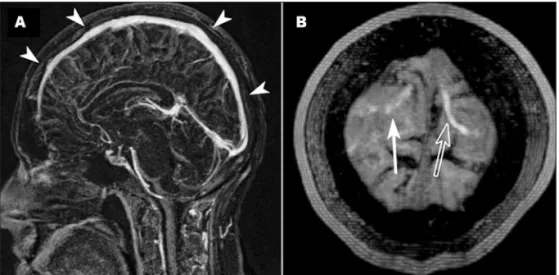
She underwent an MR imaging scan and luid-attenuated in-version-recovery (FLAIR) sequence showed linear hyperinten-sities illing in the cortical sulci of the parietal lobes bilateral-ly, suggesting SAH (Fig 1A). Mild hyperintense lesions on FLAIR were also seen on the adjacent gyri (Fig 1B). In addition, tubular serpiginous structures, hyperintense on T1-weighted images and converging to the superior sagittal sinus (SSS) were noted adja-cent to the areas suspected of SAH (Fig 1C,D). An MR venogra-phy showed no evidence of underlying sinus venous thrombo-sis (Fig 2A,B). The indings were compatible with a diagnothrombo-sis of isolated cortical venous thrombosis associated with subarach-noid hemorrhage.
The patient was managed conservatively, with withdrawal of the oral contraceptives, physical rest, and anticoagulation
Arq Neuropsiquiatr 2009;67(4)
1107
Subarachnoid hemorrhage: cortical vein thrombosis Bittencourt et al.
measures. The symptoms improved, and the follow-up MR im-aging 10 days later showed resolution of the aforementioned indings (Fig 3).
diScuSSion
Cerebral vein thrombosis accounts for 1–2% of strokes in young adults4. There are more than 100 etiologic factors
associated with the condition6, among which the use of
oral contraceptives, recent surgery, coagulopathies, dehy-dration and malignancy are the most prevalent. The clin-ical manifestations are highly variable, most frequent-ly being presented as headache (95%), seizures (47%), fo-cal motor deicits (43%), papilledema (41%), altered con-sciousness state (39%), intracranial hypertension (20%), or coma (15%)7.
Subarachnoid hemorrhage is regarded as an extreme-ly rare manifestation of CVT, being found in the literature only through isolated case reports3,4. Worthy of mention,
though, is one series with 32 patients that reported 50% of them with more than 100 erythrocytes per cubic mil-limeter of CSF, although no MR imaging scans were avail-able, and only two patients presented with sudden onset headache8. The exact cause of its association with CVT is
object of speculation, with most of the theories suggest-ing rupture of small cortical veins secondary to hemor-rhagic infarction or to venous hypertension9. The main
distribution is along the cortical sulci at the convexities, with typical sparing of basal cisterns3.
In patients with CVT, the dural sinuses are affected in as much as 98% of the time5, being the involvement of
the cortical veins usually secondary to retrograde prop-agation of the primary thrombus. Isolated cortical vein thrombosis is rather unusual, and also conined to case reports or small series1. The clinical manifestations and
risk factors are not well established, but reviewed cases
suggest that they are analogous to dural sinus thrombo-sis. When symptomatology is mild, the diagnosis may be often missed or underestimated, due to frequent anatom-ical variations in cortanatom-ical vein distribution.
The concomitance of ICoVT and SAH is even rarer, and to our knowledge there are only 3 reports describing a to-tal of 5 cases of this association4,10,11. Another article
men-tioned a case of ICoVT where the CT revealed “thin hyper-densities between frontal sulci”, but this inding was inter-preted as “cortical venous stasis and dilatation”5.
We reported a case of ICoVT associated with SAH in a post-operative female patient in use of oral contra-ceptives. The presenting complaint consisted of
“thun-Fig 3. Axial FLAIR (9000/2500/80) image, 10 days after the irst ex-amination, showing complete resolution of the indings.
Arq Neuropsiquiatr 2009;67(4)
1108
Subarachnoid hemorrhage: cortical vein thrombosis Bittencourt et al.
derclap” headache, and there were transient focal dei-cits during the evolution of the case. Once the imaging diagnosis was established and proper therapeutic mea-sures were taken, rapid resolution of the symptoms was achieved. Our case is in accordance with the available re-ports in the literature regarding the risk factors13,
symp-tomatology and outcomes4,11, and provides another
exam-ple of this rare association.
In conclusion, SAH is a readily recognizable condition, most frequently urging invasive procedures or surgical measures, while ICoVT is related to a more subtle imaging inding, generally requiring only medical treatment. In the setting of SAH, CVT can be suspected whenever the cor-tical sulci are affected and the basal cisterns are spared. The imaging diagnosis is based on FLAIR, T1-weighted im-ages and MR venography indings. Care should be taken to evaluate the cortical veins, for there may be thrombosis even though the sinuses are pervious. This case is thus an example of an unusual manifestation of a common ind-ing – SAH – helpind-ing to establish the diagnosis of a rare condition – ICoVT.
referenceS
1. Boukobza M, Crassard I, Bousser MG, Chabriat H. MR Imaging features of isolated cortical vein thrombosis: diagnosis and follow-up. AJNR 2009;30:344-348.
2. Sztajzel R, Coeytaux A, Dehdashti AR, Delavelle J, Sinnreich M. Sub-arachnoid hemorrhage: a rare presentation of cerebral venous throm-bosis. Headache 2001;41:889-892.
3. Oppenheim C, Domingo V, Gauvrit JY, et al. Subarachnoid hemorrhage as the initial presentation of dural sinus thrombosis. AJNR 2005;26: 614-617.
4. Chang R, Friedman DP. Isolated cortical venous thrombosis present-ing as subarachnoid hemorrhage: a report of three cases. AJNR 2004;25: 1676-1679.
5. Miranda H, Mellado P, Sandoval R, Huete L. [Isolated cortical throm-bosis: report of two patients] – original in Spanish. Rev Med Chile 2007; 135:1313-1317.
6. Bousser MG. Cerebral venous thrombosis: nothing, heparin or local thrombolysis. Stroke 1999;30:481-483.
7. Kimber J. Cerebral venous sinus thrombosis. QJM 2002;95:137-142. 8. de Bruin S, de Haan R, Stam J. Clinical features and prognostic factors
of cerebral venous sinus thrombosis in a prospective series of 59 pa-tients. J Neurol Neurosurg Psychiatry 2001;70:105-108.
9. Sakaki T, Matsuyama T, Nagata K, Nakase H, Hirabayashi H, Morim-oto T. Delayed intracerebral haemorrhage after intracranial surgery. J Clin Neurosci 1999;6:54-57.
10. Spitzer C, Mull M, Rohde V, Kosinski CM. Non-traumatic cortical sub-arachnoid haemorrhage: diagnostic work-up and aetiological back-ground. Neuroradiology 2005;47:525-531.
11. Wang YF, Fuh JL, Lirng JF, Chang FC, Wang SJ. Spontaneous intracra-nial hypotension with isolated cortical vein thrombosis and subarach-noid haemorrhage. Cephalalgia 2007;27:1413-1417.
12. Leach JL, Strub WM, Gaskill-Shipley MF. Cerebral venous thrombus signal intensity and susceptibility effects on gradient recalled-echo MR imaging. AJNR 2007; 28:940-945.