C
a s eR
e p o Rt4 1 4 Arq Bras Oftalmol. 2016;79(6):414-6 http://dx.doi.org/10.5935/0004-2749.20160117
CASE REPORT
A 21-year-old female presented with decreased vision in her right eye (OD), which already had a low vision, for the past 4 days. She had undergone several surgeries in both eyes because of congenital glaucoma and had lost light perception in her left eye (OS) several years prior. For treatment of both her eyes, she was on topical anti-glau comatous medication, including dorzolamide hydrochloride and timolol maleate combination and brimonidine tartrate.
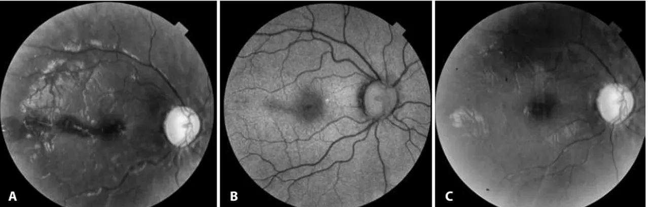
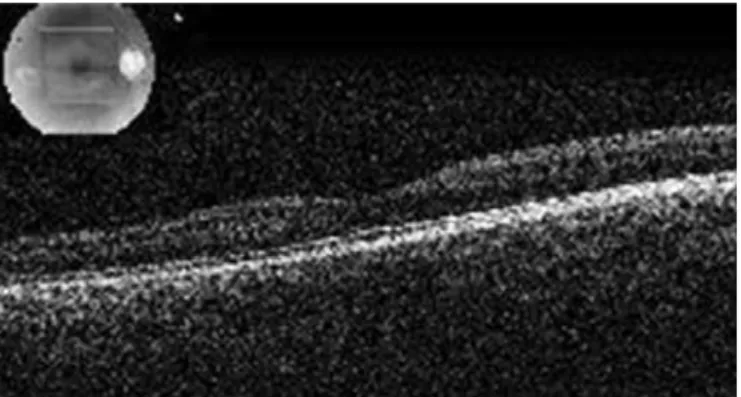
Ophthalmological examination revealed 0.15 Snellen visual acuity OD and no light perception in OS. Intraocular pressure was 14 mmHg OD and 15 mmHg OS, as evidenced using applanation tonometry. Anterior segment findings included megalocornea and temporally located posterior embryotoxon bilaterally. Fundoscopy revealed nearly total cupping and pallor of the optic disc and multiple retinal hemorrhagic foci in the macula with decreased autofluores-cence corresponding to the hemorrhagic areas (Figures 1 A and B). Spectral domain optical coherence tomography (SD-OCT; Topcon 3D OCT-2000, Topcon Inc., Japan) revealed thickening and increased reflectivity of the outer retinal layers at the fovea with disturbance of the IS/OS layer (Figure 2 A). There was a full-thickness loss of retinal integrity along with subretinal hyper-reflectivity competent with he-morrhage, temporal to the macula. Hyperreflectivity over the retina in the vitreous because of hemorrhage and increased hyper-reflectivity of the internal limiting membrane (ILM) over the patchy hemorrhagic area were also noted (Figure 2 B).
The patient did not have any previous or current systemic disease, did not use any systemic drugs, and had no recent history of acci-dent, trauma, or Valsalva maneuver. When asked to help identify any etiological factor that may have caused the retinal hemorrhage, she recounted riding a roller coaster at an amusement park 5 days prior.
Hematological evaluation revealed iron deficiency anemia, and she was prescribed iron supplements. We followed up with her weekly during the first month and monthly thereafter up to the sixth month. One week after her initial evaluation, her visual acuity in creased to 0.3 in OD. After 2 months, the hemorrhages had com-pletely resorbed (Figure 1 C), and SD-OCT sections were found to be normal without any evidence of disturbance of the retinal layers. Only the hyper-reflectivity of the ILM at the fovea remained stable. At the six-month visit, visual acuity was still 0.3, with stable SD-OCT findings (Figure 3).
DISCUSSION
We considered this retinal hemorrhage to be caused by gravita-tional forces during a roller coaster ride. The excitement of roller coas-ter rides partly comes from complex and abrupt changes in motion. Roller coaster riding causes rapid acceleration and deceleration in all three dimensions with positive and negative g-forces. The complex and abrupt changes in motion have the potential to cause sudden hyperextension, hyperflexion, or rotation of the neck, causing injury
Macular hemorrhage after roller coaster riding in a single-eyed patient with
congenital glaucoma
Hemorragia macular após passeio em montanha russa em paciente monocular com glaucoma congênito
Dilek Guven1, Zeynep AcAr1, MehMet DeMir1, yektA SenDul1, AtillA Gokce DeMir1, erDeM erGen1
Submitted for publication: June 1, 2015 Accepted for publication: January 7, 2016
1 Ophthalmology Department, Sisli Hamidiye Etfal Education and Research Hospital, Istanbul, Turkey.
Funding: No specific financial support was available for this study.
Disclosure of potential conflicts of interest: None of the authors have any potential conflict of interest to disclose.
Corresponding author: Zeynep Acar. Vardar sok. 16/5 - Caddebostan, Istanbul - Turkey E-mail: [email protected]
ABSTRACT
A 21-year-old female presented with a 4-day history of decreased vision in her only functional eye (right eye, OD). She had a history of multiple ocular surgeries in both eyes because of congenital glaucoma and had lost light perception in her left eye several years prior. Ophthalmological examination revealed 0.15 Snellen visual acuity, and fundoscopy revealed nearly total cupping and pallor of the optic disc and multiple retinal hemorrhagic foci in the macula in OD. Lesions spontaneously resolved over a few months. Gravitational forces during a roller coaster ride may have caused this macular hemorrhage.
Keywords: Eye hemorrhage; Tomography, optical coherence;
Glaucoma/con-genital
RESUMO
Uma paciente de 21 anos de idade se apresentou com perda de visão há quatro dias em seu único olho com visão útil. Ela tinha uma história de cirurgias oculares múltiplas nos dois olhos devido a um glaucoma congênito e perda de percepção lu-minosa em olho esquerdo há muitos anos. O exame oftalmológico revelou acuidade visual de Snellen de 0,15 e na fundoscopia foi observada escavação do nervo óptico quase total e palidez de papila, assim como focos hemorrágicos múltiplos na região macular. As lesões se resolveram espontaneamente em alguns meses. Acreditamos que essas hemorragias maculares tenham sido causadas pelas forças gravitacionais gera das durante o passeio na montanha russa.
Gu v e n D, e ta l.
4 1 5
Arq Bras Oftalmol. 2016;79(6):414-6 to the carotid or vertebral arteries through the creation of intimal
tears(1). Individuals with connective tissue disorders are more
vulne-rable to these injuries; however, phenotypically normal people may also have ultrastructural connective tissue abnormalities, which may increase the risk of injury.
Riding roller coasters can cause systemic complications, inclu-ding headache, intracranial hemorrhage, vascular dissection (verte-bral artery and internal carotid artery), cere(verte-bral aneurysm, and cardiac complications such as arrhythmia and myocardial infarction. Repor-ted ocular complications because of indirect trauma include retinal
Figure1. A) Grey-scale fundus image of the right eye showing foveal and extrafoveal retinal hemorrhage 5 days after the roller coaster ride. B) De-creased autoluorescence in the areas where retinal hemorrhage was noted. C) Grey-scale fundus image of the right eye 2 months after admission, showing resolution of the hemorrhages.
A B C
A
B
Ma c u l a rh e M o r r h a g ea f t e rr o l l e rc o a s t e rr i d i n gi nas i n g l e-e y e d pat i e n tw i t h c o n g e n i ta lg l au c o M a
4 1 6 Arq Bras Oftalmol. 2016;79(6):414-6
Figure 3. Normal spectral-domain optical coherence tomography image of the fovea six months after presentation as seen with the minimal hyper-relectivity of internal limiting membrane (ILM) at the fovea.
macrovessel decompensation as observed in a 19-year-old female(2),
macular hemorrhage as observed in a 26-year-old female(3), and
re-tinal artery occlusion that is associated with ipsilateral ophthalmic artery occlusion secondary to internal carotid artery dissection in a
35-year-old female(4). Our patient developed blurred vision 1 day after
the roller coaster ride. We believe there could be two explanations for the delay in symptoms; either she may not have noticed the blurring in her eye which already had poor visual acuity or the retinal edema and hemorrhage may have migrated towards the fovea.
Mechanical disruption of macular vessels by shearing forces at the interface between the vitreous and retina, increased retinal venous pressure caused by sudden increases in cerebral pressure, high hydrostatic pressure because of g-forces temporarily and inter-mittently disrupting blood flow to the head and eye, or a mechanism similar to shaken baby syndrome (SBS) are possible reasons for the hemorrhage(3).
Roller coasters can accelerate passengers up to 100 km/h in 2.5 s(5). Repositioning of a dislocated intraocular lens during a roller
coaster ride after reaching a height of 73 m and being brought to a speed of 130 km/h exposing passengers to a centripetal force of 4 G
has been reported(6). In SBS, which is primarily caused by repeated
acceleration-deceleration forces, retinal hemorrhage is observed in
50%-100% of patients(7); usually bilaterally but also unilaterally with
asymmetric presentation. Shaking induces shearing forces at the vitreoretinal interface and is the main contributing factor in
multila-yered retinal hemorrhages observed in SBS. Repeated acceleration and deceleration is believed to be necessary for the vitreous to
separate from the retina in cases with retinoschisis(8). Asymmetric
presentation was observed in this case. This may be attributed to the difference in ocular dynamics in these two eyes, which had undergo-ne several major operations in the past.
FAF is a method of evaluating the integrity of the retinal pigment epithelium (RPE), with a normal RPE metabolic activity showing a homogenous pattern and hypo-autofluorescence showing probable photoreceptor or RPE cell impairment. A report on FAF imaging in patients with blunt ocular-trauma demonstrated hypo-autofluores-cence of recent subretinal hemorrhage with no degradation of red blood cells and hyper-autofluorescence of old subretinal
hemorrha-ge on degradation of blood cells(7). In our case, the area of
hypo-au-tofluorescence because of blockage of hemorrhage resolved, and iso-autofluorescence was observed during follow-up.
With this case report, we wish to attract attention to a completely and spontaneously recovered sub- and intraretinal macular hemor-rahge in a patient went for a roller coaster ride. This patient has the risks of probable ocular tissue alterations because of congenital glau-coma and previous surgeries. As ophthalmologists, we should warn our younger patients with predisposing factors against the possible risks of injury on riding roller coasters.
REFERENCES
1. Lascelles K, Hewes D, Ganesan V. An unexpected consequence of a roller coaster ride. J Neurol Neurosurg Psychiatry. 2001;71(5):704-5.
2. Beatty S, Goodall K, Radford R, Lavin MJ. Decompensation of a congenital retinal macrovessel with arteriovenous communications induced by repetitive rollercoaster rides. Am J Ophthalmol. 2000;130(4):527-8.
3. Asefzadeh B, Connell N. Macular hemorrhage after repetitive roller coaster riding. Clin Exp Optom. 2009;92(5):447-8.
4. Ozkan Arat Y, Volpi J, Arat A, Klucznik R, Diaz O. Bilateral internal carotid artery and vertebral artery dissections with retinal artery occlusion after a roller coaster ride - case report and a review. Ulus Travma Acil Cerrahi Derg. 2011;17(1):75-8.
5. Kumar A, Sinha A, Al-Waa AM. Resolution of sudden sensorineural hearing loss fol lowing a roller coaster ride. Indian J Otolaryngol Head Neck Surg. 2011;63(Suppl 1):104-6. 6. Bosch MM, Landau K, Thiel MA. Repositioning of a dislocated intraocular lens during
a roller-coaster ride. N Engl J Med. 2003;349(11):1094-6.
7. Forbes BJ.Clues as to the pathophysiology of retinal hemorrhages in Shaken Baby syndrome determined with optical coherence tomography. Am J Ophthalmol. 2008; 146(3):344-5.
8. Sturm V, Landau K, Menke MN. Optical coherence tomography findings in Shaken Baby syndrome.Am J Ophthalmol. 2008;146(3):363-8.