Chemical and morphological analysis of the
human dental enamel treated with argon
laser during orthodontic bonding
Glaucio Serra Guimarães*, Liliane Siqueira de Morais**, Carlos Nelson Elias***, Carlos André de Castro Pérez****, Ana Maria Bolognese*****
Introduction: The main utilities of the argon laser in orthodontics are the high speed curing process in orthodontic bonding and the caries resistance promotion of the tooth enamel.
Objective: To evaluate the chemical and morphological changes in the tooth enamel
treat-ed with the argon laser in the orthodontic bonding parameters. Methods: Fifteen sound human irst premolars, removed for orthodontic reason, were selected and sectioned across the long axis in two equal segments. One section of each tooth was treated and the other remained untreated. A total of thirty samples was analyzed, creating the laser (n =15) and the control groups (n =15). The treatment was done with 250 mW argon laser beam for 5 seconds, with energy density of 8 J/cm2. Results: The X-ray analysis demonstrated two
different phases in both groups, the apatite and the monetite phases. The reduction of the monetite phase was signiicant following laser treatment, suggesting higher crystallinity. The EDS analysis showed an increase in the calcium-phosphorus ratio in the laser group, linked with the decrease of the monetite phase. The surface morphology was smoother after the laser exposure. Conclusion: The results of high crystallinity and supericial enamel smooth-ness in the laser group are suggestive of the caries resistance increase of the tooth enamel.
Abstract
Keywords: Argon laser. Tooth enamel. Orthodontic bonding.
* Assistant Professor, UFF-NF. PhD in Science Materials, MSc in Orthodontics. ** PhD in Science Materials IME / UCSD. MSc in Orthodontics UFRJ.
*** Professor of Mechanical Engineering and Materials Science Department - IME.
on these interactions, the argon laser has ive main utilities in dentistry: early caries detection by luo-rescence,7 soft tissue cutting,21,27 bleaching agent
activator,27 laser curing of dental materials,2,6 and
promotion of tooth enamel resistance against de-mineralization.9,10
High-speed polymerization and enamel re-sistance promotion are the most signiicant clinical properties in orthodontic treatment that justiies laser application. In 1999, Blanke-nau et al5 showed that 5 seconds of argon
la-ser exposure created a composite with higher compressive strength than 20 seconds of visible light curing. Losche16 reported a greater
conver-sion rate of canphoroquinone with the argon laser when compared with visible light. Many authors tested the different properties of dental materials cured with argon laser or visible light. Better or equal results in argon laser polymer-ization were found in these studies.3,12,21 Pulpal
histology from “in vivo” tests conirm that the argon laser used at the energy levels used in re-storative dentistry creates neither short-term nor long-term pulpal pathology.21
Sedivy et al24 tested the argon laser in the
bond-ing of orthodontic metallic brackets. They con-cluded that with 1 W power, the argon laser took 87% less time to obtain similar bond strength than conventional light cure. In a similar study, Lalani et al13 conirmed that 5 seconds polymerization using
argon laser produced bond failure loads compara-ble to 40 seconds of conventional light cure. Wein-berger et al29 investigated the bonding of ceramic
brackets and showed that it can be done with 231 mW power for 10 seconds of argon laser.
Another important effect of the laser on human enamel is related to a prevention characteristic.
resistance. In a clinical study, Anderson et al1
dem-onstrated that argon laser radiation with 325 mW for 60 seconds reduced in 90% the depth and the area of caries lesion.
Using the argon laser in the orthodontic bracket bonding, the enamel around the bracket is modiied. The effects of the argon laser therapy in tooth enamel vary with the several combina-tions of power and time curing described, al-though most of them are for the caries resistance treatment. Nevertheless, the power and the cur-ing time for bondcur-ing and for resistance promo-tion are different.
The purpose of the present study was to inves-tigate the chemical and morphological effects of argon laser irradiation on human enamel treated in a protocol of high speed curing of orthodontic brackets.
MAtERIAL And MEtHOdS
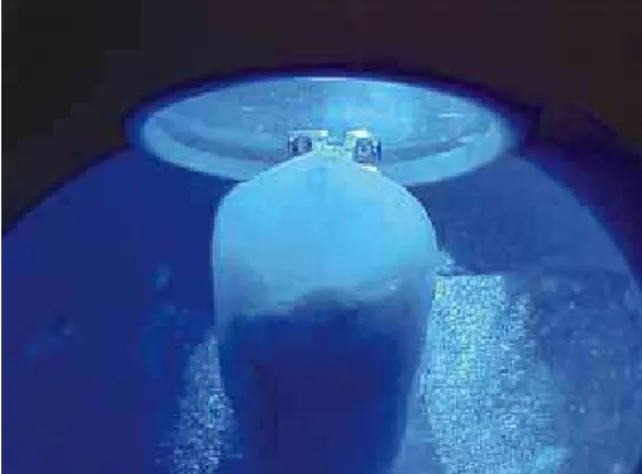
Fifteen human irst premolars extracted for orthodontic purposes were selected for this “in vitro” study. Following the extractions, the soft tis-sues were removed and the teeth were evaluated using a halogen light,32 only sound elements were
selected (Fig 1).
The dental elements were stored in a 0.1% thymol waterish solution and kept in a tempera-ture of 361ºC.4,8 All the teeth underwent
prophy-laxis, using pumice, water, and brush in low speed for 10 seconds.23 It was followed by washing with
water for 10 seconds and drying with a hair dryer for 15 seconds, so the surface became free from oil contamination.17 In order to produce uniform
The dental elements were sectioned in two equal segments, cut across the long axis with a carburundum disc in low rotation and water refrigeration. Each tooth had one half treated and the other half remained untreated, thereby creating a laser group (n =15) and a control group (n =15).
The treatment was done with an argon laser (Accucure 3000®, Laser Med, Salt Lake City,
USA) with 250 mW power for 5 seconds during each cycle, delivering an energy density of 8 J/cm2
(Fig 2). The laser power was checked with a cali-bration meter built into the laser before its use on each sample.
Energy dispersive Spectroscopic analysis (EdS)
The EDS analysis was done in a 4000 µm2
enamel area of the buccal surface (Jeol 5800 LV®,
Tokyo, Japan). The relative calcium-phosphorus ratio was compared in both the treated and un-treated samples, using the technique of least squares it.
Descriptive statistics were performed on the data to obtain means and standard deviations for each group and the groups were analyzed for sig-niicant differences using a paired-sample T test, at 5% signiicance (SPSS for Windows Release
11.0 SPSS software Corp., Munich, Germany). Following this analysis, the sample was divided and 10 pairs were submitted to X-ray diffraction analysis and the other 5 pairs were submitted to scanning electron microscopy (SEM).
X-ray diffraction analysis
The sample was laid out with the buccal enam-el surface tangent to the diffraction plane and analyzed using an X-ray diffractometer (Rigaku, Dmax 2200, Osaka, Japan) with monochroma-tized CuKradiation (wavelength λ = 1.540 Å) at 40 kV and 40 mA. The diffractograms were col-lected in the angular interval of 5º ≤ 2θ ≤ 80º us-ing 0.05º steps. The ixed time was two seconds per step and the diffractogram of each group was obtained by mean peaks.
The phase identiication was done by a match-ing process usmatch-ing the International Center for Dif-fraction Data (ICDD) database. The cell reine-ment report and the crystallinity evaluation were done with the Materials Data Inc Jade® program,
version 5.0, California, USA.
Scanning electron microscopy analysis
Five pairs of the sample received a gold layer for 3 minutes in the coater (Pollaron SC 500®,
Sputter, VG, Microtec) at 20 mA current and FIGURE 1 - Enamel quality evaluation by halogen light evaluation. FIGURE 2 - Argon laser treatment with 250 mW power continuously
10 20 30 40 50 60 70 80 90 200 mTorr vacuum. The enamel surfaces were
evaluated by secondary electron detection (Jeol 5800 LV®, Tokyo, Japan) at 500X, 1000X and
1500X original magniication.
RESuLtS
Energy dispersive Spectroscopic Analysis
The paired-sample T test showed signiicant differences between the relative calcium and phosphorus ratio after the treatment with the ar-gon laser (p<0.05). The results indicated higher relative calcium rate and lower relative phospho-rous rate after the laser exposure (Table 1).
X-ray diffraction analysis
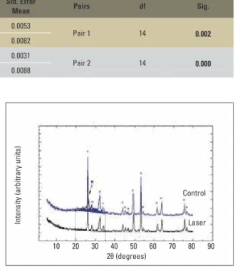
The phase identiication showed a princi-pal and a secondary phase in both groups. The principal phase was the apatite (database card # 09-0432) and the secondary phase was the mon-etite (database card # 09-0080). The original dif-fractogram of the control group showed a broad peak between 20º and 35º, which is characteristic phase of amorphous materials (Fig 3).
No new peaks were observed in the laser group when compared with the control group. Neverthe-less, the diffractogram of the laser group showed narrower peaks and reduction of the amorphous phase. Furthermore, the monetite phase was sig-niicantly decreased, indicating higher crystallinity of the treated enamel surface (Fig 3).
In the cell reinement analysis, both a- and c-ax-is of the apatite structure showed signiicant differ-ences between the control and laser groups. After the laser treatment, the a-axis showed a contraction of 0.064 Å and the c-axis an expansion of 0,016 Å.
These values obtained from the laser group came close to hydroxyapatite values (Table 2).
Scanning Electron Microscopic analysis (SEM)
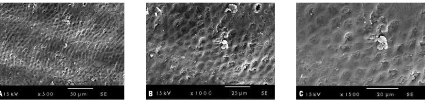
Untreated enamel surfaces from the control group showed voids and microvoids, representing the normal enamel prism end markings (Fig 4). In contrast, following argon laser irradiation, the surface morphology was substantially changed, becoming smoother (Fig 5).
FIGURE 3 - Diffractogram of control and laser groups. (*) Apatite phase peaks,(•)monetitephasepeaks.Reductionofamorphousphase(blue area above the diffractogram).
TABLE 2 - Values of experimental and hydroxyapatite cell parameters (database card #09-0432).
Pair 2 14 0.000
Laser P 15 0.1872 0.0341 0.0088
Groups Axis Mean Std deviation
Control a-axis 9.530 Å 0.003 Å c-axis 6.861 Å 0.006 Å
Laser a-axis 9.466 Å 0.006 Å c-axis 6.877 Å 0.002 Å
Hidroxyapatite a-axis 9.418 Å
--c-axis 6.884 Å
--2θ (degrees)
Control
Laser
A B C
A B C
dISCuSSIOn
The irst laser application in dentistry was done with a ruby laser, which increased enamel resistance to decalciication.26 Since then, some
authors reported this same effect after the enamel treatment with different types of lasers.
The main explanation for the acid resistance of the enamel tissue is less permeability and re-duction of carbonate content,19,20 water and
or-ganic substances in the treated enamel20.
Blankaneau et al5 reported “in vivo” argon
laser radiation effects on human enamel resis-tance against decalciication. This study de-scribed reduction of 29.1% on average lesion depth in a laser treatment with a 250 mW beam for 10 seconds. Anderson et al,1 using a 325 mW
beam for 60 seconds, found reduction of 91.6%. In this way, we could expect similar result on the enamel around the brackets during the
orth-odontic bonding. Although, the time exposure and the laser power for that are different (250 mW beam for 5 seconds).13,25,28
The energy density (ED) could be calculated by the division of the energy (E) by the spot area (S). The energy is expressed by the prod-uct of the power (P) and the exposure time6 (t)
(Equation 1).
ED = E = P x t
S S
Nelson et al19 investigated the effects of
pulsed, infrared laser radiation on human dental enamel with an energy density varying between 10 to 50 J/cm2. They concluded that the laser
radiation resulted in a melted surface and the heat delivery was limited to 10-20 µm depth. A new phase of tetracalcium diphosphate monox-ide was monox-identiied in the treated surface with a FIGURE 4 - Enamel surface morphology in control group: A) SEM at 500X original magnification; B) SEM at 1000X original magnification; C) SEM at 1500X original magnification (SE detection).
oka20 did not ind any new phases in the argon
laser treatment with 67 J/cm2.
The difference among these studies can be attributed to the effect of infrared19 and the
argon laser20 radiation on enamel surfaces. The
higher energy absorption of the infrared spec-trum by the enamel results in higher thermal energy conversion7 and more signiicant changes
in comparison with the argon laser changes. An interesting finding of this work was the correlation between the EDS and X-ray dif-fraction results. The EDS analysis showed the increase of the calcium-phosphorus ratio in the laser group. This result was brought into relation with the decrease of the monetite phase found in the x-ray diffraction analysis. In the diffractograms of the control and laser groups, the main phases observed were the apatite and the monetite phase. The apatite phase (Ca10(PO4)6(OH)2) had a calcium-phos-phorus ratio of 1.67 and the monetite phase (CaPO3(OH)) had a ratio of 1.0. Hence, the decrease of the monetite phase in the laser group, in theory, this should result in an in-crease of the calcium-phosphorus rate. Actu-ally, the increase of the calcium-phosphorus ra-tio following the laser treatment was observed, sustaining the change in the enamel surface.
The diffractogram analysis showed the de-crease of the amorphous phase after the laser treatment. This result added up to the reduc-tion of the monetite phase and to the narrower apatite peaks in the laser group indicated high-er crystallinity in the treated enamel. These re-sults are supported by Oho and Morioka20 and
Nelson et al19 findings, where a best
arrange-ment of ions in the crystal lattice of the laser
ditional studies are needed to determine the influence of these factors in the orthodontic bonding protocol.
The changes in the apatite structure ar-rangement were analyzed by the cell refine-ment of the X-ray analysis. The most signifi-cant change found in the present work was the apatite a-axis contraction of 0.064 Å. Based on previous studies, reductions of water and carbonate in the apatite phase11,15 affected
the length of the a-axis of the apatite enamel crystal. The argon laser treatment with energy density of either 11.5 or 100 J/cm2 induced
a contraction of the a-axis of apatite and this result was linked with the reduction of lesion depth and the caries resistance increase of the enamel 9,10,30,31. In this manner, due to the fact
that the argon laser treatment with energy density of 8 J/cm2 induces an a-axis
contrac-tion of 0.064 Å. It is possible that a similar resistance mechanism occurs in the parameters of this study. In such case, it could be suggest-ed that this contraction indicates rsuggest-eduction of water and carbonate, resulting in enamel resis-tance. However, additional studies are needed to prove this mechanism.
COnCLuSIOnS
1. Argon laser treatment with 250 mW for 5 seconds modiied the enamel surface result-ing in the increase of the enamel
crystallin-ity, suggesting a higher caries resistance. 2. The enamel surface morphology became
smoother after the argon laser treatment in the orthodontic bonding parameters.
1. Anderson AM, Kao E, Gladwin M, Benli O, Ngan P. The
effects of argon laser irradiation on enamel decalciication:
an in vivo study. Am J Orthod Dentofacial Orthop. 2002
Sep;122(3):251-9.
2. Arai S, Hinoura K, Ando S, Kuruda T, Onose H. Comparison of curing between activator light argon laser ion. J Dent Res.
1989;68:342.
3. Aw TC, Nicholls JI. Polymerization shrinkage of restorative resins using laser and visible light. J Clin Laser Med Surg.
1997;15(3):137-41.
4. Bishara SE, Fehr DE, Jakobsen JR. A comparative study of
debonding strengths of different ceramic brackets, enamel conditioners and adhesives. Am J Orthod Dentofacial
Orthop. 1993 Aug;104(2):170-9.
5. Blankenau RJ, Powell G, Ellis RW, Westerman GH. In vivo caries-like lesion prevention with argon laser: pilot study. J Clin Laser Med Surg. 1999 Dec;17(6):241-3.
6. Brugnera AJ, Pinheiro AL. Lasers na Odontologia moderna. 1ª ed. São Paulo: Pancast;1998.
7. Featherstone JDB. Caries detection and prevention with laser energy. Dent Clin North Am. 2000 Oct;44(4):955-69. 8. Guimarães GS, Pacheco N, Chevitarese O. Resistência ao
cisalhamento da colagem do aço inoxidável austenítico ao esmalte bovino utilizando Transbond XTTM e o primer MIP.
RGO. 2001;5(1):57-62.
9. Hicks MJ, Flaitz CM, Westerman GH, Berg JH, Blankenau
RL, Powell GL. Caries-like lesion initiation and progression
in sound enamel following argon laser irradiation: a study in vitro. ASDC J Dent Child. 1993 May-Jun;60(3):201-6. 10. Hicks MJ, Flaitz CM, Westerman GH, Blankenau RJ, Powell
GL, Berg JH. Enamel caries Initiation and progression
following low luencies (energy) argon laser and luoride treatment. J Clinic Pediatr Dent. 1995;20(1):9-13.
11. Holcomb DW, Young RA. Thermal decomposition of human tooth enamel. Calcif Tissue Int. 1980;31(3):189-201. 12. James JW, Miller BH, English JD, Tadlock LP, Buschang PH.
Effects of high-speed curing devices on shear bond strength and microleakage of orthodontic brackets. Am J Orthod
Dentofacial Orthop. 2003 May;123(5):555-61.
REfEREnCES
13. Lalani N, Foley TF, Voth R, Banting D, Mamandras A.
Polymerization with the argon laser: curing time and shear bond strength. Angle Orthod. 2000 Feb;70(1):28-33. 14. LeGeros RZ, Bonel G, Legros R. Types of "H2O" in human
enamel and in precipitated apatites.. Calcif Tissue Res. 1978 Dec 8;26(2):111-8.
15. LeGeros RZ. Effects of carbonate on the lattice parameters of apatite. Nature. 1965 Apr;206:403-4.
16. Losche GM. Color measurement for comparison of campheroquinon conversion rate. J Dent Res. 1990;69:232. 17. McCarthy MF, Hondrum SO. Mechanical bond strength
properties of light-cured and chemically glass ionomer
cements. Am J Orthod Dentofacial Orthop. 1994 Feb;105(2):135-41.
18. Miller M, Trure T. Lasers in dentistry: an overview. J Am Dent Assoc. 1993;124(2):32-5.
19. Nelson DG, Wefel JS, Jongebloed WL, Featherstone JD.
Morphology, histology and crystallography of human dental enamel treated with pulsed low-energy infrared laser
radiation. Caries Res. 1987;21(5):411-26.
20. Oho T, Morioka T. A possible mechanism of acquired acid resistance of human dental enamel by laser irradiation.
Caries Res. 1990;24(2):86-92.
21. Powell GL, Blankenau RJ. Laser curing of dental materials.
Dent Clin North Am. 2000 Oct;44(4):923-30.
22. Powell LG. Lasers in the limelight: what will the future bring? J Am Dent Assoc. 1992 Feb;123(2):71-4.
23. Pus MD, Way DC. Enamel loss due to orthodontic bonding with illed and unilled resins using various clean-up techniques. Am J Orthod. 1980 Mar;77(3):269-83. 24. Sedivy M, Ferguson D, Dhuru V, Kittleson R. Orthodontic
resin adhesive cured with argon laser: tensile bond strength. J Dent Res. 1993;72:176.
25. Shanthala BM, Munshi AK. Laser vs. visible light cured composite resin: An in vitro shear bond study. J Clin Pediatr Dent. 1995 Winter;19(2):121-5.
26. Sognaes RF, Stern RH. Laser effect on resistance of human
dental enamel to demineralization in vitro. J South Calif
Contact address Gláucio Serra Guimarães
Avenida Nossa Senhora de Copacabana, 647/1108 CEP: 22.050-000 - Copacabana - Rio de Janeiro / RJ, Brazil E-mail: [email protected]
Feb;124(2):30-1.
Submitted: July 2007
Revised and accepted: November 2007
1997;67(3):173-8.
30. Westerman GH, Hicks MJ, Flaitz CM, Berg JH, Blankaneau RJ, Powell GL. Argon laser irradiation in root surface caries:
in vitro study examines laser’s effect. J Am Dent Assoc.