637
CLINICS 2007;62(5):637-40
LETTER TO THE EDITOR
Department of Gastroenterology, São Paulo Univesity Medical College, São Paulo/SP, Brazil
Email: [email protected]
PLASMOCYTOMA OF THE JEJUNUM: DIAGNOSIS BY
DOUBLE-BALLOON ENTEROSCOPY
Adriana Safatle-Ribeiro, Thiago Souza, Elisa Baba, Eduardo Oppitz, Osmar Kenji, Paulo Sakai
INTRODUCTION
Multiple myeloma (MM) or Kahler’s disease is a ma-lignant neoplasia of the lymphoplasmocyte lineage, char-acterized by uncontrolled and progressive proliferation of an abnormal plasmocyte clone within the bone marrow (BM) with subsequent production of monoclonal immu-noglobulin.
On the other hand, focal plasmocyte tumors (plasmo-cytomas) with formation of tumorous masses without med-ullary involvement may rarely affect the gastrointestinal tract (GIT) and are denominated solitary extramedullary plasmocytomas. The main extramedullary site of plasmo-cytoma is the jejunum. We report a patient who presented with jejunal plasmocytoma and emphasize the importance of the doubballoon endoscope for diagnosis of the le-sion.1, 2
CASE REPORT
A 72 year-old white man presented with a history of abdominal discomfort and intermittent diarrhea in addition to weight-loss (4 kg in 5 months) and iron deficiency anemia. Physical examination revealed the presence of a non-fixed, slightly hardened mass at the mesogastrium.
Upper digestive endoscopy and colonoscopy did evi-dence no alterations. Abdominal computed tomography scan revealed thickness of small intestine (Figure 1).
Double-balloon enteroscopy detected at mid jejunum an ulcerated and infiltrative lesion with imprecise limits, cov-ered with fibrin and necrotic areas, involving 2/3 of the lu-men and measuring approximately 5 cm of extension (Fi-gure 2).
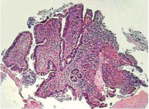
Histopathologic examination showed enteric infiltration by plasmocytoma – Kappa chain producing multiple my-eloma (Figure 3). Immunohistochemistry was positive for CD31, CD20, CD138, and Kappa with rare Lambda cells (Figure 4). Hematological evaluation did not confirm med-ullary and/or systemic disease, i.e. peripheral blood elec-trophoresis without M protein, negative Bence-Jones pro-tein and aspirative bone marrow exam without abnormali-ties.
The patient underwent laparotomy, with enterectomy involving 30 cm of the jejunum and right hemicolectomy due to the presence of an infiltration in the wall of the
as-Figure 1 – Abdominal computed tomography scan evidences thickening of jejunal wall.
Figures 2a and 2b – Double-balloon enteroscopy reveals an ulcerated and infiltrative lesion of the jejunum.
638
CLINICS 2007;62(5):637-40 Plasmocytoma of the jejunum
Safatle-Ribeiro A et al.
cending colon (Figure 5). The postoperative period was uneventful and the patient was discharged from the hospi-tal on the 5th postoperative day. Peritoneal gavage
cytol-ogy was negative for neoplasic cells and the histological study of the surgical sample confirmed the diagnosis of plasmocytoma of the jejunum (with free resection margins), including an inflammatory infiltrate in the colonic wall (Figure 6). Thirteen resected lymph nodes were free of dis-ease.
DISCUSSION
MM is an unusual malignant neoplasia of plasmocyte lineage, whose clinical manifestations are based on the sys-temic effects of the monoclonal protein and on the con-comitant humoral immunodeficiency state, as well as on the effects of bone marrow invasion by malignant cells.1
The disease affects patients with a mean age of 60 years (less than 2% of the cases occur before the age of 40) and typically presents with bone pain, mild anemia, fatigue, weight-loss and an elevated sedimentation rate.
Extramedullary MM involvement is more frequent than formerly supposed, affecting spleen, liver, lymph nodes and kidney in up to 65% of the cases.3
The diagnosis of plasmocytoma of the gastrointestinal tract is uncommon (12% of the cases), and it may be seen as a primary or a secondary lesion to the systemic disease, usually with impairment of the jejunal segment.
Clinical suspicion of isolated plasmocytoma is infre-quent, except for the cases with systemic disease. The com-monest clinical presentation is abdominal pain, palpable mass, anemia, weight-loss, nausea and vomiting. Bleeding and intestinal obstruction may also occur.1, 3, 4, 5
In the presence of a palpable mass, imaging tests are the first to be performed. Contrasted study may be used, and should be avoided in the presence of a sub occlusive presentation and is contraindicated in patients with intes-tinal obstruction. The presence of lesions in multiple seg-ments is rare. Abdominal computed tomography scan helps to determine the size, local regional extension and impair-ment of other organs. However, histological diagnosis is obtained through fragments of biopsy performed through endoscopic examination or through surgical procedure. There are reports of utilization of fine needle puncture for the establishment of the diagnosis. The presence of M pro-tein in peripheral blood helps with diagnostic complementation and may occur in cases of solitary plas-mocytomas.1, 6, 7
Double-balloon enteroscopy allows endoscopic study and biopsies of the entire small intestine, and permits his-tological diagnosis throughout. Capsule endoscopy may be used; however, it should be avoided in obstructed patients, and does not have the potential for histological confirma-tion.
Histology demonstrates the involvement of the entire thickness of the wall, with atypical plasma cells and bi-zarre plasmocytes in varied levels of differentiation. In the presence of associated systemic disease, comparisons be-tween the aspirate and tumorous histology may help the diagnostic conclusion. The immunohistochemical study is fundamental to confirm the B cell origin.
Figure 4 – Immunohistochemistry for CD138 shows positive plasmocyte cell lineage (100X, biopsy).
Figure 5 – Surgical specimen showing small intestinal lesion with invasion of the colon.
639
CLINICS 2007;62(5):637-40 Plasmocytoma of the jejunum
Safatle-Ribeiro A et al.
In some cases angiographic studies have been applied, especially in patients with systemic disease, and demon-strate the presence of hypervascular lesions, positive for gallium-67.3
Such tumors should be distinguished from other neoplasias and correlated with clinical presentation and possible systemic disease. The differential diagnosis be-tween intestinal lymphoma, adenocarcinoma and other rare lesions may be difficult, emphasizing the role of histologi-cal confirmation.
Treatment of MM is based on chemotherapy and the use of corticosteroids, with improvement occurring in most patients. Presently, eradication of all tumor cells can only be obtained with bone marrow transplant combined with chemotherapy. Surgical resection remains as the optimal therapeutic modality in cases of solitary extramedullary
plasmocytoma, mainly if related to symptoms, with cure as a possibility when associated with chemotherapy.7,8,10
Prognosis of MM is related to the cell load. Poor prog-nosis is associated with a high cell load, reflected as anemia, reduced renal function, hypercalcemia, extensive bone impairment and high peaks of monoclonal pro-tein.8,9,10,11
In summary, the diagnosis of a solitary plasmocytoma of the small intestine presents difficulties, considering its rarity, unspecific presentation and localization. Double-bal-loon enteroscopy, allowing access and performance of bi-opsies, enables histological diagnosis. Surgical resection remains the main therapeutic modality, but clinical and en-doscopic follow-up is recommended because of the possi-bility of relapse or appearance of another plasmocyte tumor.
REFERENCES
1. Griffiths AP, Shepherd NA, Beddall A, Williams JG. Gastrointestinal tumor masses due to multiple myeloma: a pathological mimic of malignant lymphoma. Histopathology. 1997;31:318-23.
2. Yamamoto H, Sekine Y, Sato Y, Higashizawa T, Miyata T, Iino S, et al. Total enteroscopy with a nonsurgical steerable double-balloon method. Gastrointest Endosc. 2001;53:216-20.
3. Tsuruda T, Ohashi T, Uezono S, Kato J, Nagamoto Y, Sakata J, et al. Extramedullary plasmocytoma of the jejunum. Intern Med. 1996;35:422-6.
4. Curcio CM, Feinstein RS, Humphrey RL, Jones B, Siegelman SS. Computed tomography of entero-enteric intussusception. J Comput Assist Tomogr. 1982;6:969-74.
5. Park IG, Lee YJ, Park do H, Han SY, Lee GH, Myungg SJ, et al. A case of jejunal stricture due to extramedullary plasmocytoma. Korean J Gastroenterol. 2003;42:67-71.
640
CLINICS 2007;62(5):637-40 Plasmocytoma of the jejunum
Safatle-Ribeiro A et al.
7. Okamura S, Tsujigami K, Muguruma N, Ito S, Wakatsuki S, Sano T, et al. Jejunal extramedullary plasmocytoma. Gastrointestinal Endoscopy. 2005;61:107-8.
8. Homma K, Ihzumi T, Nemoto K, Ohnishi Y. Primary extramedullary plasmocytoma of the small intestine. Int J Hematol. 1992;56:179-84. 9. Rygaard-Olsen, Boedker A, Emus HC, Olsen HA. Extramedullary
plasmocytoma of the small intestine: a case report studied with electron microscopy and immunoperoxidase technique. Cancer. 1982;50:573-6.
10. Merle H, Cassuto JP, Laselve L, Blein L, Delmont J. Malignant plasmocytoma of the duodeno-jejunal angle. Apropos of a case. Chirugie. 1976;102:678-82.