Severe episodic memory impairment
after strategic infarct
A case report
Francisco Wilson Nogueira Holanda Júnior1, Katie Moraes de Almondes2, Rodrigo Alencar e Silva3
ABSTRACT. Brain infarcts located in strategic regions often result in cognitive impairment. Based on a case study, this paper describes unusual and specific clinical and neuropsychological features of a strategic ischemic lesion in the left medial temporal lobe (MTL) structures. Taken together with the literature data, the case illustrates that a unilateral strategic infarct in MTL structures may result in severe impairment of episodic memory (EM), which refers to the ability to encode and retrieve personal experiences, including information about the time and place of an event and detailed description of the event itself. The preservation of other cognitive functions, the severe functional impairment, and the type of visual-verbal deficit in a left-sided lesion were identified as singular features of the case. The current case supports the critical role of the MTL structures in EM formation.
Key words: memory, strategic infarct, neuropsychological assessment, stroke.
GRAVE COMPROMETIMENTO DE MEMÓRIA EPISÓDICA APÓS INFARTO ESTRATÉGICO: UM ESTUDO DE CASO
RESUMO. Infartos cerebrais localizados em regiões estratégicas comumente resultam em comprometimento cognitivo. Baseado em um estudo de caso, este artigo descreve características clínicas e neuropsicológicas incomuns e específicas de uma lesão isquêmica estratégica nas estruturas mediais do lobo temporal esquerdo. Tomado em conjunto com os dados da literatura, este caso ilustra que uma lesão isquêmica estratégica unilateral em estruturas do lobo temporal medial resulta no comprometimento da memória episódica, a qual se refere à habilidade de armazenar e recuperar experiências pessoais, o que inclui informações sobre o tempo e o local do evento e a descrição detalhada do próprio evento. A preservação de outras funções cognitivas, o grave comprometimento funcional e a modalidade de déficit visual-verbal em uma lesão à esquerda foram apontados como características singulares do caso. Este estudo de caso corrobora o papel crítico das estruturas do lobo temporal medial na formação da memória episódica.
Palavras-chave: memória, infarto estratégico, avaliação neuropsicológica, acidente vascular encefálico.
INTRODUCTION
T
he occurrence of strategically located isch-emic vascular lesions in brain areas criti-cal for cognition and behavior (e.g., associa-tive, limbic, paralimbic circuitry) is referred to as strategic infarcts (SI).1 Regions such as theangular gyrus, the medial part of the frontal lobes, the medial part of the temporal lobe, the thalamus, and the caudate nucleus are the most common sites of strategic infarcts.2,3 SI
are of great interest to cognitive neurology and clinical neuropsychology due to the pos-sibility of studying the relationship between cognitive impairments and underlying brain damage (i.e., brain structure-function correlation).
SI are often associated with the gradual or abrupt development of mild vascular cognitive impairment and even dementia.4,5
Depending on the location, extent and
sever-This study was conducted at the Neuropsychology of Aging Service, University Hospital Onofre Lopes, Natal, RN, Brazil.
1Master’s student on the Postgraduate Program in Psychology, Federal University of Rio Grande do Norte, RN, Brazil. 2Associate Professor at the Department of Psychology and on the Postgraduate Program in Psychobiology, Federal University of Rio Grande do Norte, RN, Brazil. 3Neurologist at the University Hospital Onofre Lopes, Federal University of Rio Grande do Norte, RN, Brazil.
Francisco Wilson Nogueira Holanda Júnior. Federal University of Rio Grande do Norte / Lagoa Nova / Mailbox 1622 – 59078-970 Natal RN – Brazil. E-mail: [email protected]
Disclosure: The authors report no conflicts of interest.
Received August 24, 2017. Accepted in final form November 13, 2017.
ity of the lesions, patients may present varying degrees and types of cognitive impairment, such as memory deicits.6 Episodic memory (EM), which refers to the
ability to encode and retrieve daily personal experi-ences,7 is one of the most discussed memory systems.
Clinical features of impairment in EM may be observed in lesions that are more limited to the medial temporal lobe (MTL) region.8 However, data concerning
altera-tions in EM associated with SI in the MTL structures are lacking.
his paper reports a case of severe episodic memory impairment as a result of focal ischemic lesion in the left MTL structures, contrasting and discussing unusual and speciic clinical and neuropsychological features: (1) impairment of verbal and visual mnemonic modalities in a left-sided lesion; (2) preservation of other cogni-tive functions; and (3) severe functional impairment. Case reports with these characteristics are uncommon in Brazilian scientiic literature, and highlights the importance of the brain structure-function correlation, a fundamental cornerstone of cognitive neurology and neuropsychology.
CASE REPORT
JE, a 68-year-old right-handed male with 10th-grade
education was referred by a neurologist for neuropsy-chological evaluation in December 2016 due to forget-fulness which emerged after January of the same year. In January 2016, he had sufered an ischemic stroke of cardioembolic etiology. JE had a medical history of hypertension and chronic atrial ibrillation. According to JE and his wife, before the stroke he was normally able to manage his home activities and job tasks as a machine mechanic. However, after the stroke, the patient had severe diiculty organizing his inances and could no longer work or drive. His family perceived memory deicits such as forgetting new events,
commit-ments, asking about people who died recently, and repeatedly asking the same question. JE also presented disorientation in time and new places.
On medical evaluation, laboratory test results were within normal ranges (including blood count, compre-hensive metabolic panel, thyroid-stimulating hormone, vitamin B12 and venereal disease research laboratory). A magnetic resonance imaging scan of the brain per-formed in January 2016 indicated an ischemic lesion afecting the left parahippocampal gyrus, hippocampus, and anterior portion of the middle and inferior tempo-ral gyrus. here were some additional tiny lesions of the white matter not exceeding possible typical age-related changes (Figure 1). At the time of the neurolo-gist’s observation, JE scored 23/30 on the Mini-Mental State Examination (MMSE), with deicits mainly on the subitems of time, place, and delayed recall. his dem-onstrated a marked decline of orientation and EM. On the Montreal Cognitive Assessment (MoCA), JE’s score (18/30) was mainly attributed to errors in memory and orientation subtests. A diagnostic hypothesis of neuro-degenerative dementia (e.g., Alzheimer’s disease demen-tia) was excluded. here was evidence of a stroke tempo-rally related to the onset of memory impairment, as well as lack of insidious onset of the decline. herefore, the diagnosis of probable Alzheimer’s disease dementia and other neurodegenerative dementias was not applicable.9
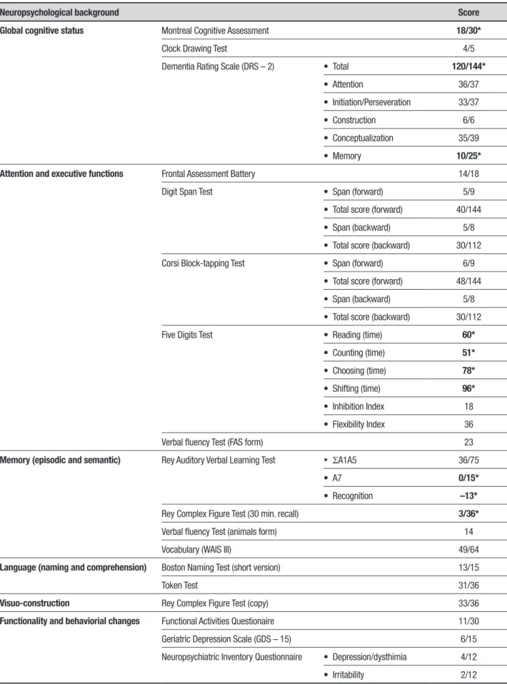
he patient underwent a comprehensive neuropsy-chological assessment to evaluate memory impairment and other functions (Table 1). Overall, JE’s performance showed severe EM impairment in verbal and visual modalities indicated by delayed recall measures of the Rey Auditory Verbal Learning Test (RAVLT) (0/15) and the Rey-Osterrieth Complex Figure Test (RCFT) (3/36) with scores below the 5th percentile. he Memory
Sub-scale from the Dementia Rating Scale (DRS – 2) also indicated impaired performance (10/25, < 5th
Table 1. Patient’s neuropsychological background.
Neuropsychological background Score
Global cognitive status Montreal Cognitive Assessment 18/30*
Clock Drawing Test 4/5
Dementia Rating Scale (DRS – 2) • Total 120/144*
• Attention 36/37
• Initiation/Perseveration 33/37
• Construction 6/6
• Conceptualization 35/39
• Memory 10/25*
Attention and executive functions Frontal Assessment Battery 14/18
Digit Span Test • Span (forward) 5/9
• Total score (forward) 40/144
• Span (backward) 5/8
• Total score (backward) 30/112
Corsi Block-tapping Test • Span (forward) 6/9
• Total score (forward) 48/144
• Span (backward) 5/8
• Total score (backward) 30/112
Five Digits Test • Reading (time) 60*
• Counting (time) 51*
• Choosing (time) 78*
• Shifting (time) 96*
• Inhibition Index 18
• Flexibility Index 36
Verbal fluency Test (FAS form) 23
Memory (episodic and semantic) Rey Auditory Verbal Learning Test • ΣA1A5 36/75
• A7 0/15*
• Recognition –13*
Rey Complex Figure Test (30 min. recall) 3/36*
Verbal fluency Test (animals form) 14
Vocabulary (WAIS III) 49/64
Language (naming and comprehension) Boston Naming Test (short version) 13/15
Token Test 31/36
Visuo-construction Rey Complex Figure Test (copy) 33/36
Functionality and behaviorial changes Functional Activities Questionaire 11/30
Geriatric Depression Scale (GDS – 15) 6/15
Neuropsychiatric Inventory Questionnaire • Depression/dysthimia 4/12
• Irritability 2/12
tile). He underperformed on the RAVLT Word Recog-nition list, which may indicate an inability to encode and consolidate new episodic contents. Category lu-ency (animals form) and Vocabulary (WAIS III) tests indicated preserved semantic memory (50th
percen-tile). JE had an appropriate strategy for visuospatial construction, making few mistakes on the ROCF Copy test. Regarding language processes such as naming and comprehension, there were no indications of impair-ment according to results on the Boston Naming Test (short version) and Token Test, respectively.
here was no evidence of executive impairment in general. On the Five Digit Test, which is a numeric-Stroop paradigm applying four steps (Reading, Count-ing, Choosing and Shifting) that evaluates processes such as processing speed, selective attention, inhibitory control and cognitive lexibility, JE had scores indicative of clinical impairment (< 5th percentile). However,
con-sidering the indices of Inhibition and Flexibility (inter-ference scores that subtract Reading time from Choos-ing and ShiftChoos-ing time, which minimize the inluence of processing speed on executive performance), there was no indication of impairment in these measures (50th
percentile). his discrepancy can be explained by the decreased processing speed observed in the patient, and not by a deicit in superior/executive functioning. his is corroborated by the fact that the patient was slow to perform the tests, but did so without errors, and due to the fact that he had adequate performance in all other tests of executive functioning.
DISCUSSION
he main inding of this report was that an isolated left unilateral irst-ever ischemic stroke conined to the MTL structures manifesting as an anterograde EM impair-ment. JE was unable to consolidate new episodic verbal and visual content, events and information, while other cognitive functions were found to be intact (except for slow processing speed). he capacity to consciously recol-lect information acquired at a certain time and place is referred to as EM.10 Tulving11 described EM as memory
of personally experienced events or remembering what happened, where and when. Normal EM functioning involves a set of interconnected brain circuits, especially the MTL region. MTL represents a system of highly related structures, including the hippocampal forma-tion (the dentate gyrus, CA1, CA2, CA3 subields, and the subiculum) and the collectively denominated para-hippocampal region (adjacent cortical areas, namely, entorhinal, perirhinal, and parahippocampal cortices).7
In the MTL, information originating from
neocor-tical areas is processed through a hierarchical network represented by: (1) the perirhinal and parahippocampal cortices; (2) the entorhinal cortex; and (3) the hippo-campal formation itself. he outputs of hippohippo-campal processing encompass feedback connections successively back (i.e., towards the entorhinal cortex, then perirhinal and parahippocampal cortices) to neocortical areas from which the inputs to the MTL were generated.12 EM is
mainly contingent on hippocampal and parahippocam-pal processing that facilitates consolidation of episodic information (e.g., new verbal and visual contents, spatial context, object recognition) from short-term to long-term memory.13 In this mediation, the hippocampal
sys-tem and related structures then slowly transfer informa-tion into the neocortical storage system. Notably, other areas beyond the MTL, such as the dorsomedial nucleus of the thalamus, mammillary bodies, amygdala, and the basal forebrain also play a role in EM functioning.14 Due
to the complex functioning of the MTL circuit, bilateral or even unilateral lesions in this region usually cause devastating efects on memory and learning.
he current case exhibited some singularities. Although the stroke was unilateral (left-sided lesion), both verbal and visual deicits of EM were observed. his is unusual since memory function specializes with hemi-spheric function: the left hemisphere is more related to verbal components of memory, while the right hemi-sphere mediates visuospatial features of memory. Also, the functional impairment in JE’s everyday life after the stroke was highly severe. his is an uncommon inding because severe functional impairment in MTL lesions is more probable as a consequence of bilateral lesions as opposed to unilateral ones. Moreover, although isolated and stable memory impairment that presents acutely may be permanent, some patients experience changes over time. As expected for typical patients with MTL amnesia, JE also presented preserved insight, mild or absent retrograde amnesia and lack of confabulation. Furthermore, no semantic memory impairment was observed. Although semantic memory is a component of declarative memory along with EM, there has been a debate regarding the extent to which the encoding of new semantic information is dependent solely on the MTL structures, suggesting that semantic memory acquisition may difer in some manner compared to EM.7 Indeed, some evidence based on patients with MTL
lesions has shown acquisition of new semantic informa-tion, in spite of major diiculty.15
severe functional impairment. he case serves as an important example of brain structure-function correla-tion, a fundamental cornerstone of cognitive neurology and neuropsychology.
Author contributions. All authors have contributed signiicantly and all authors are in agreement with the content of the manuscript. FWNHJ, KAM and RAS
were responsible for the study conception and design. FWNHJ was responsible for writing the manuscript. FWNHJ, KAM and RAS were responsible for the critical revision. All authors approved the inal manuscript.
Acknowledgements. he authors wish to thank JE and his wife for their time and collaboration in this study.
REFERENCES
1. Jellinger KA. Morphologic diagnosis of “vascular dementia” – A critical update. J Neurol Sci. 2008;270:1-12.
2. Khan A, Kalaria RN, Corbett A, Ballard C. Update on vascular dementia. J Geriatr Psychiatry Neurol. 2016;29:281-301.
3. Auriacombe S, Amarenco P, Baron JC, et al. Update on vascular demen-tias. RevNeurol (Paris). 2008;164:22-41.
4. Engelhardt E, Tocquer C, André C, Moreira DM, Okamoto IH, Cavalcanti JLS. Vascular dementia: Diagnostic criteria and supplementary exams: Recommendations of the Scientific Department of Cognitive Neurology and Aging of the Brazilian Academy of Neurology. Part I. Dement Neuro-psychol. 2011;5:251-63.
5. Lanna ME, Alves CE, Sudo FK, et al. Cognitive disconnective syndrome by single strategic strokes in vascular dementia. J Neurol Sci. 2012;322:176-83.
6. Hoffmann M, Schmitt F, Bromley E. Vascular cognitive syndromes: relation to stroke etiology and topography. Acta Neurol Scand. 2009;20:161-9.
7. Dickerson BC, Eichenbaum H. The Episodic Memory System: Neuro-circuitry and Disorders. Neuropsychopharmacology. 2010;35:86-104.
8. Squire LR, Wixted JT. The Cognitive Neuroscience of Human Memory Since H.M. Ann Rev Neurosci. 2011;34:259-88.
9. McKhann GM, Knopman DS, Chertkow H, et al. The diagnosis of dementia due to Alzheimer’s disease: Recommendations from the National Institute on Aging-Alzheimer’s Association workgroups on diagnostic guidelines for Alzheimer’s disease. Alzheimers Dement. 2011; 7:263-9. 10. Tulving E. Elements of Episodic Memory. 1988; Oxford: Clarendon Press. 11. Tulving E. Episodic memory and common sense: how far apart? Philos
Trans R Soc Lond B Biol Sci. 2001;356:1505-15.
12. Squire LR, Stark CE, Clark RE. The Medial Temporal Lobe. Annu Rev Neurosci. 2004;27:279-306.
13. Sheldon S, McAndrews MP, Moscovitch M. Episodic memory processes mediated by the medial temporal lobes contribute to open-ended problem solving. Neuropsychologia. 2011;49:2439-47.
14. Nadel L, Hardt O. Update on Memory Systems and Processes. Neuro-psychopharmacology. 2011;36:251-73.