INTRODUCTION
Address to: Dr. Diogo Henrique Saliba de Souza. Rua T-30, 1050/Apto 1501, Residencial Porto Real I, Setor Bueno, 74210-060 Goiânia, GO, Brasil.
Phone: 55 62 8138-1818
e-mail: [email protected]
Received 25 March 2013
Accepted 29 May 2013
Current epidemiological profi le of Chagasic
megaesophagus in Central Brazil
Diogo Henrique Saliba de Souza
[1],
Maria da Glória Merheb Vaz
[1],
Cristiano Rezio Fonseca
[1],
Alejandro Luquetti
[1],[2],
Joffre Rezende Filho
[1],[3]and Enio Chaves de Oliveira
[1],[4][1]. Núcleo de Estudos de Doença de Chagas, Universidade Federal de Goiás, Goiânia, GO. [2]. Curso de Pós-Graduação, Faculdade de Medicina, Universidade Federal de Goiás, Goiânia, GO. [3]. Serviço de Gastroenterologia, Departamento de Clínica Médica, Universidade Federal de Goiás, Goiânia, GO. [4]. Serviço de Cirurgia do Aparelho Digestivo, Departamento de Cirurgia, Universidade Federal de Goiás, Goiânia, GO.
ABSTRACT
Introduction: Chagasic megaesophagus (CM) is the most common digestive manifestation of Chagas disease in Brazil, and the State of Goiás is one of the most affected regions. In recent decades, the Hospital das Clínicas (HC)/Universidade Federal de Goiás (UFG) has been a reference center for the study and treatment of CM. The objective of this study was to characterize
the current epidemiological profi le of patients with CM observed at the HC of the UFG from 1998 to 2010. Methods: In total,
939 patient records were analyzed, and age, gender, place of birth, serology, symptoms and radiological classifi cation according
to Rezende et al. were analyzed. Results: The median patient age was 55 years. Male patients were more (54%) prevalent than female patients. The prevalence of younger patients (less than 31 years of age) was 4.2%, but 82.1% of the younger patients were from State of Bahia. Patients older than 40 years were the majority (85.5%). The radiological groups were distributed as follows: Group I (35.9%), Group II (32.9%), Group III (17%) and Group IV (14.2%). Conclusions: Compared with previous studies by the same group in 1975, 1994 and 1995, the number of younger patients decreased, and the frequency curve has shifted to older patients.
Keywords:Chagasic megaesophagus. Chagas disease. Epidemiology.
Chagas disease (CD) is endemic in Latin America, and approximately 8 million individuals are chronically infected1. This disease may manifest as cardiopathies; digestive disorders, such as esophagopathy and colopathy; and associated cardiodigestive disorders. Regional differences exist regarding the clinical manifestation of the disease, and the State of Goiás, representing the central region of Brazil, has high prevalence of digestive disorders2-4.
Chagasic megaesophagus (CM) is a chronic disease that is characterized by the destruction of the myenteric plexus of
the esophagus by the fl agellate protozoan Trypanosoma cruzi. T. cruzi is the etiological agent of CD and is responsible for functional changes in the esophagus, such as hypercontractility, motor dyskinesia and achalasia of the lower esophageal sphincter. These changes are responsible for dysphagia, the primary symptom noted in this disease5-7. The pathogenesis of
cell destruction by T. cruzi has not been well defi ned. Some studies suggest that this protozoa compromises the muscle cells of the digestive tract and, consequently, that the enteric nerve
plexuses are destroyed by the local infl ammatory response and
immune mechanisms4,8,9.
Vector-borne transmission of CD has been under control in recent decades in Brazil, and triatomine species, such as Triatoma infestans, have been eradicated in some countries as a result of vector control programs conducted in South America10. However, CD remains a challenge to public health in view of the thousands of infected individuals who live in urban areas. The domestic migratory movement from the rural setting to
urban areas and international migration may infl uence the
demographic data of infected individuals8,11,12.
The Nucleo de Estudo de Doença de Chagas (NEDoC) is located at the Hospital das Clínicas (HC), which is the teaching hospital of the Universidade Federal de Goiás (UFG), and has been a reference center for the diagnosis and treatment of CM for several decades. The NEDoC has reported epidemiological series on Chagasic esophagopathy in the State of Goiás since 1975. The last epidemiological survey published was performed from May 1976 to July 1997 by Rezende and Moreira7. In that study, the mean age of patients with CM was reported to have increased in relation to previous studies conducted by Rezende13. A decrease in vector-borne transmission and improvements in the living conditions of the population that have occurred in
METHODS
RESULTS CM patients currently followed in the central region of Brazil.
Therefore, the purpose of this study was to determine the current
profi le of CM patients referred to the NEDoC, from January 1998 to December 2010, and compare the current fi ndings with
previous studies.
A case series study was performed by retrospectively evaluating 1,150 medical records of patients with megaesophagus who were observed at the outpatient clinic of the NEDoC from January 1998 to December 2010. Among these records, 17 records belonged to patients with negative serology for CD, and 194 were patients with CM who had undergone previous esophageal treatment (balloon dilation and surgery) before their
fi rst appointment at the clinic. These records were not included
in the cases chosen for this study. The final sample for this study included 939 medical records of patients presenting with CM who were not previously treated and had positive serology.
Data were organized following a structured questionnaire as follows: a) age: patient age on the occasion of the first appointment; b) gender; c) birth place: state where the patient was born, also considering the division of each state in micro-regions, as established by the Brazilian Institute of Geography and Statistics14; d) serological tests: results of at least 3 tests for antibodies against T. cruzi (ELISA, indirect hemagglutination
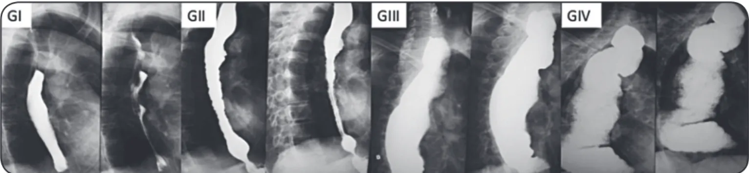
and indirect immunofl uorescence); e) digestive symptoms; and f) CM radiological group, according to the classifi cation defi ned by
Rezende et al.15. The radiological groups were defi ned as follows (Figure 1): Group I – esophagus with an apparently normal diameter, presenting with a small retention of radiological contrast; Group II – esophagus with a moderate increase in diameter,
signifi cant retention of the contrast medium and the presence of
tertiary waves caused by greater uncoordinated motor activity of the organ, commonly associated with a hypertonic lower esophagus;
Group III – hypotonic esophagus with a signifi cantly increased
diameter, exhibiting little wall contractile activity and marked contrast retention; and Group IV – dolico-megaesophagus with an extremely increased diameter that was elongated and tortuous, folded itself over the diaphragmatic dome, retained a great amount of contrast and was without contractile activity.
Microsoft® Excel 2007 software , Microsoft Corporation, Washington, United States of America, was used to tabulate
FIGURE 1 - Chagasic megaesophagus radiological groups according to Rezende et al.15. GI: Group I; GII: Group II; GIII: Group III; GIV: Group IV. the data. SPSS® for Windows®, version 15.0, was used for the statistical analyses. To evaluate and compare gender and mean age among the 4 CM groups, the chi-square test was used for proportions, and Student’s t-test was used to compare means.
The signifi cance level adopted was 5%. Ethical considerations
The Internal Review Board for Research on Humans and Animals of the Hospital das Clínicas, Federal University of Goiás (CEPMHA-HC-UFG), approved the study (Board's Decision No. 052/2011).
A total of 939 medical records of patients observed from January 1998 to December 2010 were selected from the NEDoC
fi les. Patients ranged in age from 15 to 88 years old. The mean
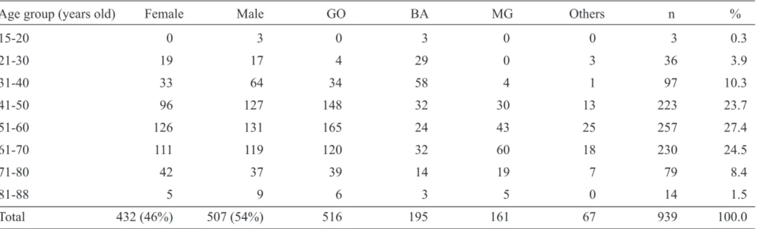
patient age was 54.4 years, and the median age was 55 years. Most (75.6%) patients were in their 5th, 6th and 7th decades of life, and male patients were more prevalent. Patients younger than 31 years old represented 4.2% of the cases. Regarding place of birth, more than half of the patients were born in the States of Goiás and Bahia. See Table 1 for the demographic data.
Younger patients (less than 31 years of age) represented 4.2% of the cases (39 patients). Among these patients, the majority (32 [82.1%] cases) was born in State of Bahia, and 4 (10.2%) cases were born in State of Goiás (Figure 2). Regarding the micro-regions of Bahia, the highest prevalence was found in the areas where Barreiras and Santa Maria da Vitória are located, with 14 (43.7%) cases each.
Regarding the radiological classifi cation of Rezende et al.15,
the esophagogram fi ndings were distributed as follows: Group I – 337 (35.9%) patients, Group II – 309 (32.9%) patients, Group III – 160 (17%) patients and Group IV – 133 (14.2%) patients. The mean age of each CM group is shown in Table 2, and
a signifi cant increase in age was observed relative to the groups
with more advanced CM. When separated by gender, the CM
groups showed a signifi cant prevalence of female patients relative
to males in Group I (58.5% cases). In the other Groups (II, III and IV), males were more prevalent, as demonstrated in Table 2.
DISCUSSION
TABLE 1 - Demographic data for patients with Chagasic megaesophagus at the Hospital das Clínicas, Universidade Federal de Goiás, from 1998 to 2010.
Age group (years old) Female Male GO BA MG Others n %
15-20 0 3 0 3 0 0 3 0.3
21-30 19 17 4 29 0 3 36 3.9
31-40 33 64 34 58 4 1 97 10.3
41-50 96 127 148 32 30 13 223 23.7
51-60 126 131 165 24 43 25 257 27.4
61-70 111 119 120 32 60 18 230 24.5
71-80 42 37 39 14 19 7 79 8.4
81-88 5 9 6 3 5 0 14 1.5
Total 432 (46%) 507 (54%) 516 195 161 67 939 100.0
GO: State of Goiás; BA: State of Bahia; MG: State of Minas Gerais.
82.1% 10.2%
7.7%
State of Bahia
others
n = 39
State of Goiás
FIGURE 2 - Place of birth of patients less than 31 years of age who presented with Chagasic megaesophagus at the Hospital das Clínicas, Universidade
Federal de Goiás, from 1998 to 2010.
This retrospective study evaluated the current epidemiological
profi le of patients with CM observed in a reference center in the
central region of Brazil. Our data show that, currently, patients who are referred to the NEDoC with CM are adults (median
55 years) and predominantly male; were born in Goiás, Bahia or Minas Gerais; and belong to CM Groups I and II.
Chagasic megaesophagus was more frequently found in the 5th, 6th and 7th decades of life. Group I and II patients have a non-advanced form of the disease, which corresponded to 68.8% of the cases included in this study. In an epidemiological series of a general population sample, rather than a hospital sample, Castro et al.16 also noted a prevalence of Group I and II (80.5%) patients with CM. Ceneviva et al.17 compared 2 epidemiological series and found a greater prevalence of Group III patients in a series from the 1960s and Group II patients in a series from the 1990s. In 2 previous studies that were also performed in Goiás, Rezende13 and Rezende and Luquetti18 also found a greater prevalence of CM patients in Groups II and III, as did Oliveira et al.19 in a study performed in Campinas. Therefore, a change in the proportion of patients in each of the CM groups has occurred, and there is a higher prevalence of patients in CM groups with less advanced disease and smaller dilation (Groups I and II). Improved sanitary measures, including the use of insecticides, along with educational campaigns and preventive measures against contamination by the vector, the kissing bug, have reduced the general incidence of the disease throughout the country. Better access to health care at the primary level and referral to the secondary and tertiary levels have also contributed to the adequate control and treatment of CM in its less advanced forms (Groups I and II)20,21.
TABLE 2 - Megaesophagus (ME) groups according to mean patient age and gender. Hospital das Clínicas, Universidade Federal de Goiás, 1998 to 2010.
ME Group I II III IV
Mean age 51.6 (±11.7)a 54.2 (±13.7)b 57.1 (±13.4)b 58.9 (±12.1)b
Female 197 (58.5%)c 132 (42.7%)e 64 (40%)e 39 (29.3%)e
Male 140 (41.5%)d 177 (57.3%)f 96 (60%)f 94 (70.7%)f
b x a (p<0.05); c x d (p<0.001); f x e (p<0.001).
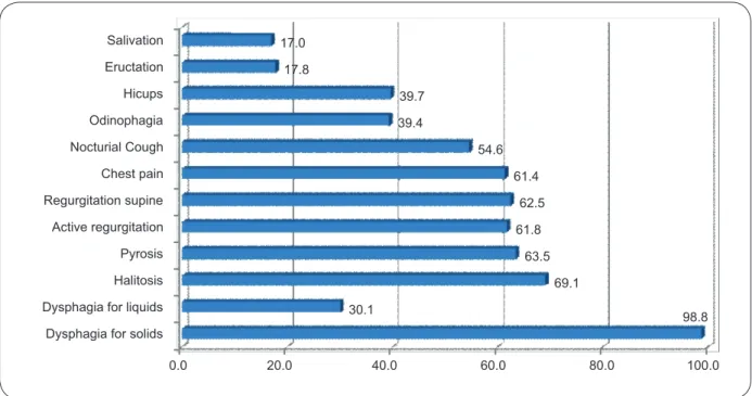
Dysphagia for solids Dysphagia for liquids Halitosis Pyrosis Active regurgitation Regurgitation supine Chest pain Nocturial Cough Odinophagia Hicups Eructation Salivation
98.8 30.1
69.1 63.5 61.8
62.5 61.4 54.6 39.4
39.7 17.8
17.0
0.0 20.0 40.0 60.0 80.0 100.0
FIGURE 3- Digestive symptoms (%) in patients with Chagasic megaesophagus observed at the Hospital das Clínicas, Universidade Federal de Goiás, from 1998 to 2010.
respective prevalences of 12.9% and 38.5% of patients older than 40 years of age, and after comparing his data with the cases in this study and the studies by Rezende and Luquetti18 and Vaz et al.21, we conclude that the population of CM patients in the central region of Brazil has grown older over the last 3 decades.
Studies performed with CM patients at the HC of Ribeirão Preto, São Paulo, an area that is also endemic for CD, have also demonstrated a prevalence of older patients (> 40 years). For example, the median in the study performed by Kamiji and Oliveira20 was 67 years, and the study conducted by Meneghelli et al.23 revealed that a median of 55 years. Thus, the prevalence of older patients (> 40 years old) with CM deserves special attention, not only with regard to esophagopathy but also other comorbidities that are prevalent in the population in this age group, such as cardiovascular and metabolic diseases19.
Additionally, we found that the younger CM patients (< 31 years) comprised 4.2% (39 patients) of the cases. The previous cases investigated in the Rezende13,22 study, performed in 1956 and 1975, and in the Rezende and Luquetti18 study, performed in the same endemic region, which focused on this younger age group, showed prevalences of 71.7%, 37.1% and 18% among the cases, respectively. When comparing these
epidemiological series, a decline in the number of younger patients with CM who have been recently observed at the NEDoC has clearly occurred. This decline is most likely a consequence of better sanitary measures aimed at controlling the transmission of CD. These measures have decreased the general incidence of this disease in the country7,20.
Regarding the place of birth, the States of Goiás (54.9% of the cases), Bahia (20.8%) and Minas Gerais (17.1%) had the
highest prevalences of patients studied, a fi nding that agrees
with the epidemiological series by Vaz et al.21. Therefore, we demonstrated that the central region of Brazil, represented by the State of Goiás, is an endemic region for CD and that the digestive form is highly prevalent1,7. This distribution refl ects
the general profi le of NEDoC patients.
Most of the young patients born in Bahia are from the cities of Barreiras and Santa Maria da Vitória, which had 14 (43.7%) cases each. These 2 cities represent a microregion located in the western-most area of the State of Bahia, bordering the State of Goiás14. For this reason, these patients are frequently referred to the NEDoC for treatment. Young patients from Bahia, which has the highest prevalence among the states, may be related to the delay and disproportionate effectiveness of the sanitary measures implemented in 1975 to combat the vectors, such as spraying insecticides. Some areas of Bahia, including the western area of the state, were not covered by the vector control program during the initial stages. Therefore, the risk of CD transmission among these populations remained high during that period. The control program became widespread only after 1983, when the entire area at risk for vector transmission in Brazil was covered10.
Regarding distribution by gender, similar to the fi ndings of
Rezende13, Rezende and Luquetti18 and Vaz et al.21, this study found that males were generally more prevalent (54% of cases) relative to females (46% of cases). However, studies performed by Kamiji and Oliveira20 and Almeida et al.24 demonstrated a greater prevalence of female patients, and Peñaranda-Carrillo et al.25 and Oliveira et al.19 did not observe any gender differences in their studies. The predominance of CM in a given gender has resulted in many studies with controversial results. Therefore, it does not seem reasonable to attribute longer survival or a
predominance of this disease to a specifi c gender24.
However, separating the patients in this study into CM groups according to Rezende et al.15 demonstrated that female patients were more prevalent relative to males in Group I
(58.5% vs. 41.5%), and the difference was signifi cant (p < 0.05).
In the other Groups (II, III and IV), the prevalence of male
patients relative to females was greater and equally signifi cant
(p < 0.05). The studies performed by Rezende and Luquetti18 and Vaz et al.21 also demonstrated a greater prevalence of female patients in CM Group I. The predominance of females in Group I, which had the less advanced form of the disease, and of males in Groups II, III and IV, which included the more advanced forms (e.g., a dilated esophagus), may be associated with the expression of the disease as it relates to the patient's gender. The suggestion is that females present with higher immunological resistance to aggressive infection by T. cruzi21. This difference may also indicate that the disease evolves more aggressively in males18.
When the CM groups were compared, the mean patient ages were higher in the more advanced Groups III and IV (57.1 and 58.9 years, respectively) relative to patients in the less advanced Groups I and II (51.6 and 54.2 years, respectively). The small difference between the mean ages shows that radiological changes and the magnitude of esophageal dilation are more dependent on the intensity of myenteric denervation, which occurs during the acute phase of CD, than on the evolution of the disease. Generally speaking, the progression of Chagasic esophagopathy is slow, and only rare cases evolve to a more advanced form of megaesophagus within a short time period16,25-27.
Regarding the symptoms, solid food dysphagia was the most prevalent (98.8% of cases). Regurgitation, halitosis, pyrosis and chest pain were present in more than 60% of the cases. Other epidemiological studies7,19,21 have reported similar symptoms.
Therefore, current CM patients in the central region of Brazil have the following characteristics: adults (median 55 years) born in endemic areas (Goiás, Bahia and Minas Gerais), with a higher prevalence of the non-advanced forms (Group I and II) of megaesophagus.
ACKNOWLEDGMENTS
The authors wish to thank all people involved in the Laboratory Division and Ambulatory Division of the Nucleo de Estudo de Doença de Chagas (NEDoC) during its many decades of service.
REFERENCES
The authors declare that there is no confl ict of interest. CONFLICT OF INTEREST
1. Rassi Jr. A, Rassi A, Rezende JM. American trypanosomiasis (Chagas disease). Infect Dis Clin North Am 2012; 26:275-291.
2. Schmuñis GA. American Trypanosomiasis as a public health problem.
In: Chagas’ Diseases and the Nervous System. Pan Am Health Org Sci Publ 1994; 547:3-29.
3. Teixeira ARL, Nascimento RJ, Sturm NR. Evolution and pathology in Chagas disease - A Review. Mem Inst Oswaldo Cruz 2006; 101:463-491. 4. Brener Z. The pathogenesis of Chagas disease: an overview of current
theories. In: Chagas’ Diseases and the Nervous System. Pan Am Health Org Sci Publ 1994; 547:30-46.
5. Rassi Jr. A, Rassi A, Marin-Neto JA. Chagas disease. Lancet 2010; 375:1388-1402.
6. Coura JR, Borges-Pereira J. Chagas disease. What is known and what should be improved: a systemic review. Rev Soc Bras Med Trop 2012; 45:286-296.
7. Rezende, JM, Moreira H. Forma digestiva da doença de Chagas.
In: Castro LP, Coelho LGV, editors. Gastroenterologia. 1st ed. São Paulo: Medsi; 2004. p. 325-391.
8. Coura JR, Dias JC. Epidemiology, control and surveillance of Chagas disease - 100 years after its Discovery. Mem Inst Oswaldo Cruz 2009; 104 (suppl I): 31-40.
9. Hontebeyrie-Joskowicz M. Humoral and cellular immunity to
Trypanosoma cruzi infection and disease. In: Chagas’ Diseases and the
Nervous System. Pan Am Health Org Sci Publ 1994; 547:273-283.
10. Silveira AC, Dias JCP. O controle da transmissão vetorial. Rev Soc Bras Med Trop 2011; 44 (supl II):52-63.
11. World Health Organization. Chagas: one hundred years later. Past Issues 2009; 87:485-564.
12. Moncayo A, Silveira A. Current epidemiological trends for Chagas disease in Latin America and future challenges in epidemiology, surveillance and health policy. Mem Inst Oswaldo Cruz 2009; 104 (supl I):17-30. 13. Rezende JM. Chagasic mega syndromes and regional differences.
In: New Approaches in American trypanosomiasis research. Pan Am Health Org Sci Publ 1975; 318:195-205.
14. Instituto Brasileiro de Geografi a e Estatística (IBGE). Diretoria de
de Janeiro: IBGE; [Cited 2013 May 14]. Available from: http://www.ibge.
gov.br/home/geociencias/cartografi a/territ_doc1.shtm.
15. Rezende JM, Lauar KM, Oliveira AR. Aspectos clínicos e radiológicos da aperistalsis do esôfago. Rev Bras Gastroenterol 1960; 12:247-262.
16. Castro C, Penaranda-Carrillo R, Rezende J, Prata A. Estudo longitudinal do megaesôfago chagásico. Rev Soc Bras Med Trop 2009; 42 (supl II): 69-72.
17. Ceneviva R, Ferreira-Santos R, Santos JS, Mente ED, Sankarankutty
AK. Alterações cronológicas do perfi l dos pacientes e da modalidade de
tratamento cirúrgico do megaesôfago chagásico. Acta Cir Bras 2002; 17 (suppl III):125-128.
18. Rezende JM, Luquetti AO. Chagasic megaviscera. In: Chagas’ Disease and the Nervous System. Pan Am Health Org Sci Publ 1994; 547:149-171.
19. Oliveira GC, Lopes LR, Andreollo NA, Coelho Neto JS. O megaesofago
tratado cirurgicamente: perfi l epidemiologico dos pacientes operados no
Hospital de Clinicas da Universidade Estadual de Campinas entre 1989 e 2005. Rev Soc Bras Med Trop 2008; 41:183-188.
20. Kamiji MM, Oliveira RB. O perfi l dos portadores de doenca de Chagas, com enfase na forma digestiva, em hospital terciario de Ribeirao Preto, SP. Rev Soc Bras Med Trop 2005; 38:305-309.
21. Vaz MGM, Rezende JM, Ximenes CA, Luquetti AO. Correlação entre a sintomatologia e a evolução do megaesôfago. Rev Goiana Med 1995; 41:1-15.
22. Rezende JM. Megaesôfago por doença de Chagas. Rev Goiana Med 1956; 2:297-314.
23. Meneghelli UG, Ejima FH, Rosa-e-Silva L. Evidências do declínio da ocorrência do megaesôfago e do megacólon chagásicos: estudo epidemiológico no Hospital das Clínicas de Ribeirão Preto. Medicina (Ribeirão Preto) 1991; 24:218-224.
24. Almeida EA, Barbosa Neto RM, Guariento ME, Wanderley JS, Souza ML. Apresentação clínica da doença de Chagas em indivíduos idosos. Mem Inst Oswaldo Cruz 2007; 40:311-315.
25. Penaranda-Carrillo R, Castro C, Rezende J, Prata A, Macêdo V. Radiographic study of the oesophagus of chagasic patients in 25 years of the Mambai Project. Rev Soc Bras Med Trop 2006; 39:152-155. 26. Castro C, Prata A, Macêdo V. Estudo clínico durante 13 anos de 190
chagásicos crônicos de Mambaí, Goiás, Brasil. Rev Soc Bras Med Trop 2001; 34:309-318.