ISSN/$–see front matter © 2013 Sociedade Brasileira de Ortopedia e Traumatologia. Published by Elsevier Editora Ltda. All rights reserved. Rev Bras Ortop. 2013;48(1):111-113
www.rbo.org.br/
*Corresponding author at: Av. Henrique Dodsworth 83/105, Copacabana, RJ, Brazil. E-mail: [email protected]
article info
Article history:
Received October 20 2011 Approved September 2 2012
Keywords: Quadriceps Muscle Rupture
Adolescent
Case Report
Total rupture of the quadriceps muscle in an adolescent
Rodrigo Pires e Albuquerque,
1*José Felix dos Santos Neto,
2Vincenzo Giordano,
3Maria Isabel Pires e Albuquerque,
4Ney Pecegueiro do Amaral,
5João Maurício Barretto
61PhD in Medicine. Coordinator of the Knee Surgery Sector, “Professor Nova Monteiro” Orthopedics and Traumatology Service, “Miguel
Couto” Municipal Hospital (SOT-HMMC), Rio de Janeiro, RJ, Brazil. 2Attending Physician at SOT-HMMC, Rio de Janeiro, RJ, Brazil.
3MSc in Medicine. Coordinator of the Medical Residence Program at SOT-HMMC, Rio de Janeiro, RJ, Brazil.
4Pediatrician at the National Cancer Institute, Rio de Janeiro, RJ, Brazil.
5MSc in Medicine. Head of the Orthopedics and Traumatology Service, SOT-HMMC, Rio de Janeiro, RJ, Brazil.
6PhD in Medicine. Head of the Orthopedics Service, Santa Casa do RJ, Rio de Janeiro, RJ, Brazil.
Work performed at the Professor Nova Monteiro” Orthopedics and Traumatology Service, “Miguel Couto” Municipal Hospital (SOT-HMMC), Rio de Janeiro, RJ, Brazil.
a b s t r a c t
The total rupture of the quadriceps muscle in an adolescent is a rare lesion. We report a case of a 13 year old boy who suffered a direct trauma to the left knee. No predisposing factors have been diagnosed. The lesion was treated with surgical repair and transosseus sutures. The aim of this study was to present a rare case of rupture of the quadriceps muscle in an adolescent and the therapy used.
© 2013 Sociedade Brasileira de Ortopedia e Traumatologia. Published by Elsevier Editora Ltda. All rights reserved.
112
Rev Bras Ortop. 2013;48(1):111-113Fig. 3 - Postoperative radiograph of the left knee.
Introduction
Rupture of the quadriceps muscle is a well-known injury in the orthopedic literature and it generally affects males after the age of 40 years. The age group of our patient was unusual and rarely reported. This rupture is generally related to comorbidities coming from rheumatic or systemic diseases. Excessive sports activity and chronic use of certain medications also facilitate this type of lesion.
In this study, we report a case of total rupture of the quadriceps muscle in a healthy adolescent, along with the therapy used.
Case report
A healthy 13-year-old boy suffered direct trauma to his left knee and immediately presented pain, hemarthrosis and incapacity to walk. The patient was sent to the emergency service of our hospital. Physical examination revealed edema in the left knee, low patella and a gap at the upper center of the patella. The patient was also incapable of actively extending his leg. At the time of the trauma, the patient weighed 61 kg and his height was 1.75 m.
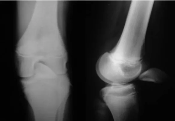
Radiography on the knee showed the low patella and dislocation of the patella, and confirmed the diagnosis of total rupture of the quadriceps muscle of the left knee (Fig. 1). The patient’s clinical history was investigated and blood tests were performed, for all markers for rheumatic and renal diseases. From this, relationships with any systemic disease or steroid use were rejected.
Surgery was performed one day after hospital admission, using a tourniquet and a straight anterior incision in the knee (Fig. 2). The surgical technique used consisted of suturing of the quadriceps muscle by means of transosseous holes, together with repairs to the retinaculum. The suture repair was tested by means of careful flexing of the knee joint.
After the operation, the knee was immobilized for six weeks, using a long splint, which was removed for active rehabilitation exercises, in order to avoid atrophy of the quadriceps. The program consisted of isometric exercises for the quadriceps during the immobilization period, and active exercises for the quadriceps with progressive increases of the range of motion. The complete range of motion and full functioning of the knee were achieved within six months.
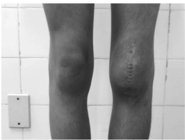
Our patient was evaluated after one week, 15 days, one month, 45 days, two months and monthly thereafter until the sixth month, when the consultations became every three months. We have now been following up this patient for two years, and he had gone back to his habitual activities. The follow-ups consist of a radiological control examination, in which we evaluate the height of the patella and the functional knee score (modified Lysholm classification)1 (Fig. 3). The mean
score is now 90 points, which is considered to be a good result according to this evaluation system (Figs. 4 and 5). The range of motion of the left knee is now from zero to 120 degrees,
Fig. 2 - Intraoperative evaluation of the left knee. Fig. 1 - Preoperative radiograph of the left knee.
Rev Bras Ortop. 2013;48(1):111-113
113
Diagnosing lesions due to quadriceps rupture is basically clinical, and is performed by means of palpation of the gap and observing whether the extensor mechanism has failed. With regard to complementary examinations, radiography on the knee (trauma series) enables good accuracy for the diagnostic confirmation, as well as being inexpensive. We did not use ultrasonography because this is a dependent examination. Magnetic resonance is the gold-standard complementary examination for diagnosing this type of injury. Unfortunately, because of its high cost, it is still not available in all Brazilian hospitals. In the future, as this examination becomes more popular, it will start to contribute greatly towards analysis on the condition of the tendon and the structures around the knee. Diagnosing the lesion and early repair are the secret to success. We did not use reinforcement from the semitendinosus, or any V-Y flap techniques. We prefer to use these techniques when dealing with chronic lesions of the quadriceps tendon. Use of anchors for repairing the quadriceps tendon is another option, and this generates less surgical aggression with this technique, compared with the traditional method.10 In the
literature, in comparatively analyzing biomechanical studies between these two surgical techniques, we did not observe any diference in repair failure, with up to 1,000 cycles.10
Conflicts of interest
The authors declare that there was no conflict of interests in conducting this study.
R E F E R E N C E S
1. Tegner Y, Lysholm J. Rating systems in the evaluation of knee ligament injuries. Clin Orthop Relat Res. 1985;(198):43-9. 2. Aydemir G, Cakmak S, Aydinoz S. Partial rupture of the
quadriceps muscle in a child. BMC Musculoskelet Disord. 2010;11:214.
3. Siwek CW, Rao JP. Ruptures of the extensor mechanism of the knee joint. J Bone Joint Surg Am. 1981;63(6):932-7.
4. Adolphson P. Traumatic rupture of the quadriceps tendon in a 16-year-old girl. A case report. Arch Orthop Trauma Surg. 1992;112(1):45-6.
5. Omololu B, Ogunlade SO, Alonge TO. Quadriceps tendon rupture in an adolescent. West Afr J Med. 2001;20(3):272-3. 6. Naver L, Aalberg JR. Rupture of the quadriceps tendon
following dislocation of the patella. Case report. J Bone Joint Surg Am. 1985;67(2):324-5.
7. Alexander VA, Keilin S, Cohn BT. Adolescent quadriceps mechanism disruption. Orthopedics. 2001;24(6):591-3. 8. West JL, Keene JS, Kaplan LD. Early motion after quadriceps
and patellar tendon repairs: outcomes with single-suture augmentation. Am J Sports Med. 2008;36(2):316-23. 9. Matsumoto K, Hukuda S, Ishizawa M, Kawasaki T, Okabe
H.Partial rupture of the quadriceps tendon (jumper’s knee) in a ten-year-old boy. A case report. Am J Sports Med. 1999;27(4):521-5.
10. Lighthart WA, Cohen DA, Levine RG, Parks BG, Boucher HR Suture anchor versus suture through tunnel fixation for quadriceps tendon rupture: a biomechanical study. Orthopedics. 2008;31(5):441.
and is equal to that of the contralateral knee. The thigh circumference is now 32 cm on the left side and 36 cm on the non-operated side.
Discussion
In the immature skeleton, the muscles, ligaments and tendons are generally stronger than the growth plates.2 In our patient,
a lesion was observed in the upper center of the patella, with hemarthrosis of the knee, thus demonstrating the traumatic and acute nature of the injury.
From reviewing the literature for papers published in English on total rupture of the quadriceps muscle in individuals with an immature skeleton, only five articles were found, thus showing the rarity of this lesion and the importance of case reports.3-7 Siwek and Rao reviewed the literature on ruptures
in the quadriceps between 1880 and 1978. In their study, only two patients under the age of 20 years were seen, out of a total of 69 cases.3 Our patient was only 13 years of age, thus showing
the infrequent nature of such lesions and relevance of our case. There is some controversy regarding whether patients should be kept immobilized during the postoperative period, and how long this immobilization should last.8 It needs to be
borne in mind that this injury occurred in an adolescent, given that in this population group, medical orders are at greater risk of being disrespected. On the other hand, in the immature skeleton, there is less risk of joint stiffness that in the adult population. For this reason, brace use was maintained for six weeks, with daily removal for exercises aimed at achieving gains in range of motion and muscle development.
Research has been conducted on structural alterations to the tendon resulting from microtraumas or degeneration, which give rise to traumatic ruptures.9 On the other hand, other
researchers have maintained that direct trauma to the knee is the cause of injury to the knee extensor mechanism in healthy patients.4 In our study, because this was an adolescent without
previous complaints or systemic diseases, we believe that direct trauma was the mechanism for the injury. Nonetheless, we agree that structural abnormalities increase the risk of injury to the knee extensor apparatus.