Oral Radiology
Jefferson Xavier de Oliveira Jucely Aparecida da Rosa Marcelo Eduardo Pereira Dutra Karina Cecilia Panelli Santos Cibelle Gil
Discipline of Dental Radiology, Department of Stomatology, School of Dentistry, Univ de São Paulo - USP, São Paulo, SP, Brazil.
Corresponding Author: Jucely Aparecida da Rosa E-mail: [email protected]
Assessing joint effusion and bone
changes of the head of the mandible in
MR images of symptomatic patients
Abstract: The aim of the present study was to investigate the relation-ship between degenerative bone changes of the head of the mandible and the presence of joint effusion (JE). This study was based on sagittal mag-netic resonance imaging (MRI) reports of 148 temporomandibular joints (TMJs) of 74 patients complaining of pain and/or dysfunction in the TMJ area. The mandible heads were surveyed for osteoarthritis charac-teristics, which were classiied as osteophytosis, sclerosis or erosion. The presence of JE was checked whenever high signal intensity was observed in the articular space. The results evidenced the presence of bone changes in 30% of the sample. Osteophytes and erosions were the changes most commonly observed. JE was reported in 10% of TMJs. The results from the statistical tests revealed that bone changes in the head of the man-dible are associated with the presence of JE.
Descriptors: Magnetic Resonance Imaging; Temporomandibular Joint; Osteoarthritis.
Introduction
Magnetic resonance imaging (MRI) provides an excellent representa-tion of soft tissues in anatomical and semi-funcrepresenta-tional relarepresenta-tionships. The multi-section images it generates allow a tridimensional analysis of the temporomandibular joint (TMJ), providing the most complete assess-ment of the relationship between TMJ components.1
Examination by MRI may be used to analyze the articular disc posi-tion in the coronal and parasagittal planes, the translaposi-tion movement of the head of the mandible, and the disc movement during mouth opening and closing. MRI may also be used to detect the presence of effusion, bone erosions and degenerative diseases.2,3
Joint effusion (JE) is generally deined as a pathological collection of luid in articular spaces. It appears in MR images as high signal intensity in the TMJ space, observed on T2-weighted images. It often appears ac-companied by disc displacement (DD) and by pathological changes, such as inlammatory processes or synovitis, and is considered a very useful indicator in assessing the progression and severity of temporomandibular dysfunction (TMD).4
Degenerative bone changes of the TMJ are signiicantly more frequent in the head of the mandible than in the articular eminence, and are char-acterized by the development of osteophytes, erosions, avascular necro-Declaration of Interests: The authors
certify that they have no commercial or associative interest that represents a conflict of interest in connection with the manuscript.
Submitted: Jul 17, 2012
ses, subchondrial cysts and intra-articular foreign bodies.4,5 These changes are considered radiological signs of osteoarthritis (OA), and have been observed in symptomatic TMJs.5,6
If a strong association between the presence of JE and OA were to be established, this could be the basis for valuable additional diagnostic information. The aim of this study was thus to investigate the re-lationships between OA in the head of the mandible and the presence of JE, by analyzing MR images of the TMJs of symptomatic patients.
Methodology
MR images of 148 TMJs from 74 adult patients submitted to this examination after indication by their dentists or physicians were used. The images were retrieved from the archives of a private institu-tion. The inclusion criterion was the presence of at least one sign or symptom of TMD reported dur-ing clinical examination. Patients who reported any systemic involvement or who were using medication that could entail bone changes were disregarded.
The exams were performed with a Signa appara-tus (GE Medical Systems, Madison, USA) with 1.5 Tesla ield strength, using T2 and PD weighted se-quences, with the aid of a double surface coil 20 cm in diameter (GE Medical Systems, Madison, USA). The images were obtained in the sagittal plane with 3-mm-thick cuts, saved in DICOM format and eval-uated by two experienced radiologists (a physician and a dentist). Each of them evaluated the images only once and the resulting reports were obtained by consensus.
The mandible heads were surveyed for OA char-acteristics, which were classiied as osteophytosis, sclerosis or erosion.
The presence of JE was checked in the T2-weighted images whenever high signal intensity was observed in the articular space (Figure 1).
The study was approved by the Research Ethics Committee, University of São Paulo, under protocol 119/2009.
The conditions assessed were recorded as present or absent, and then submitted to descriptive statis-tics. The data regarding gender, OA of the mandible head and presence of JE were analyzed through
ab-solute and relative frequency calculation, and then plotted in charts. Following the descriptive analy-sis, Fisher’s exact test was performed to ascertain whether the OA characteristics of the mandible heads were associated with the presence of JE, at a signiicance level of 5% (P < 0.05). The statistical analyses were conducted with Statistical Package for Social Sciences software (SPSS for Windows, ver-sion 11.0, Chicago, USA).
Results
From the 74 patients evaluated (148 TMJs), 51 (68.9%) were female, and 23 (31.1%) were male. The mean age of female patients was 40.4 ± 14.5 years (range: 13–69 years). The mean age of male patients was 35.9 ± 11.2 years (range: 17–58 years).
The TMJs were assessed bilaterally for the oc-currence of OA and JE. OA characteristics were ob-served in 30% of the samples (Figure 2), the most frequent of which were osteophytosis (n = 13, 9%) and sclerosis (n = 8, 5%); erosion was the least fre-quent change observed (n = 1, 1%). The OA charac-teristics observed were associated, as follows:
• erosion and osteophytosis (n = 8, 6%);
• erosion, sclerosis and osteophytosis (n = 6, 4%); • erosion and sclerosis (n = 5, 3%); and
• sclerosis and osteophytosis (n = 3, 2%).
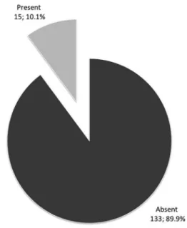
JE was observed in 10% of the samples, as shown in Figure 3.
Table 1 illustrates the descriptive analysis of the association between OA characteristics and the presence of JE.
The results of the statistical tests revealed that the OA characteristics of the head of the mandible were associated with the presence of JE (p = 0.006). Subjects with JE on at least one side presented the same OA proile (p = 0.835). Subjects with no JE on either side presented a higher rate of TMJs with no OA, on both sides, when compared to patients with JE on at least one side (p = 0.048).
Discussion
MRI is considered a choice method to assess the soft components of joints, as well as changes in the quantity of articular luid.5,7
The present study assessed the relationship be-tween mandibular condyle OA and the presence of JE. MRI is a reliable method to detect JE.8 Bone changes may be assessed by viewing the TMJ in the position used in this examination with an accuracy Figure 2 - Distribution of TMJs according to bone changes
of the head of the mandible. Figure 3 - Distribution of TMJs according to JE presence.
of 93%.3
The inclusion criterion was the presence of pain in the TMJ or in the neighboring temporal, frontal or auricular regions. Studies found in the related literature show a strong correlation between TMJ pain, disc displacement and JE.6,9 Pain persisting af-ter TMJ arthrocentesis is generally related to a great amount of JE and erosive condylar OA.9 There are, however, several studies showing a weak correlation between JE and TMJ pain.10,11 Furthermore, JE has also been detected in asymptomatic TMJs, with no observable pathology.12
We also observed a predominance of female over male subjects, an observation consistent with a ten-dency reported in the related literature.5,6,13,14
Osteoarthritis, on the other hand, was observed in 30% of the samples, which is in agreement with other studies stating that bone abnormalities are not always related to symptom reports by patients.5,9 We also observed that the condylar OA characteristics were associated, predominated by the erosion/osteo-phyte combination. This inding is corroborated by a study reporting a higher frequency of osteophytes and erosions combined.5
JE was found in 10.1% of subjects, which agrees with the indings of another study reporting this same percentage in symptomatic patients.12 The
Er: erosion
ErS: erosion and sclerosis ErSO: erosion, sclerosis and
osteophytosis ErO: erosion and osteophytosis S: sclerosis SO: sclerosis and
Bone change status
Intra–articular effusion status
Total
A A A P P A P P
ErS ErS 1 (1.6%) – – – 1 (1.4%)
ErS N 3 (4.8%) – – – 3 (4.1%)
ErSO S – 1 (20.0%) – – 1 (1.4%)
ErSO N 2 (3.2%) – 1 (50.0%) 1 (25.0%) 4 (5.4%)
ErSO O – – 1 (50.0%) – 1 (1.4%)
ErO ErO 1 (1.6%) – – – 1 (1.4%)
ErO S – – – 1 (25.0%) 1 (1.4%)
ErO N 4 (6.3%) – – – 4 (5.4%)
ErO O 1 (1.6%) – – – 1 (1.4%)
S S 2 (3.2%) – – – 2 (2.7%)
SO S – 1 (20.0%) – – 1 (1.4%)
SO N 2 (3.2%) – – – 2 (2.7%)
N Er – – – 1 (25.0%) 1 (1.4%)
N S 1 (1.6%) – – – 1 (1.4%)
N N 39 (61.9%) 2 (40.0%) – 1 (25.0%) 42 (56.8%)
N O 4 (6.3%) 1 (20.0%) – – 5 (6.8%)
O O 3 (4.8%) – – – 3 (4.1%)
Total 63 (100.0%) 5 (100.0%) 2 (100.0%) 4 (100.0%) 74 (100.0%)
Table 1 - Distribution of subjects according to bone changes of the head of the mandible and the presence of intra- articular effusion. AA: absent on both sides; AP: absent on the
right side and present on the left side; PA: present on the right side and absent on the left side; PP: present on both sides; ErS ErS: erosion and sclerosis on both sides; ErS N: erosion and sclerosis on one side and normal on the other side; ErSO S: erosion, sclerosis and osteophytosis on one side and sclerosis on the other side; ErSO N: erosion, sclerosis and osteophytosis on one side and normal on the other side; ErSO O: erosion, sclerosis and osteophytosis on one side and osteophytosis on the other side; ErO ErO: erosion and os-teophytosis on both sides; ErO S: erosion and osteophytosis on one side and sclerosis on the other side; ErO N: erosion and osteophytosis on one side and normal on the other side; ErO O: erosion and osteophytosis on one side and osteophytosis on the other side; S S: sclerosis on both sides; SO S: sclerosis and osteophytosis on one side and sclerosis on the other side; SO N: sclerosis and osteophytosis on one side and normal on the other side; N Er: normal on one side and erosion on the other side; N S: normal on one side and sclerosis on the other side; N N: normal on both sides; N O: normal on one side and osteophytosis on the other side; O O: osteophytosis on both sides.
minute size of the intra-articular space and the simi-larity between the synovial liquid and the JE signals on MR images make it dificult to distinguish these signals. Furthermore, there is no consensus on the amount of luid that would be involved in safely characterizing a JE.2 The criterion used in our study to make the distinction between effusion and sy-novial liquid was high signal intensity in the intra-articular space in T2-weighted images. No quan-titative analysis of luids was performed; only the presence or absence of JE was detected.
The statistical results showed that condylar OA characteristics were associated with the presence of TMJ effusion (p = 0.006). However, JE was also
observed in normal TMJs. Hence, additional data may be necessary to conirm this relationship and to ascertain whether OA characteristics are initial or contributing factors in the onset of JE.
Conclusion
According to the results obtained in this study, the incidence of JE was correlated to OA charac-teristics of the head of the mandible. Erosions and osteophytes were the most frequently observed man-dibular condyle OA characteristics. However, most symptomatic patients evaluated showed neither bone alterations nor signal intensity consistent with joint effusion.
References
1. Hirata FH, Guimarães AS, Oliveira JX, Moreira CR, Ferreira ET, Cavalcanti MG. Evaluation of TMJ articular eminence morphology and disc patterns in patients with disc displace-ment in MRI. Braz Oral Res. 2007 Jul–Sep;21(3):265–71.
3. Tasaki MM, Westesson PL. Temporomandibular joint: di-agnostic accuracy with sagittal and coronal MR imaging. Radiology. 1993 Mar;186(3):723–9.
4. Zarb GA, Carlsson GE. Temporomandibular disorders: os-teoarthritis. J Orofac Pain. 1999;13(4):295–306.
5. Campos MI, Campos PS, Cangussu MC, Guimarães RC, Line SR. Analysis of magnetic resonance imaging characteristics and pain in temporomandibular joints with and without de-generative changes of the condyle. Int J Oral Maxillofac Surg. 2008 Jun;37(6):529–34.
6. Emshoff R, Brandimaier I, Bertram S, Rudisch A. Magnetic resonance imaging findings of osteoarthrosis and effusion in patients with unilateral temporomandibular joint pain. Int J Oral Maxillofac Surg. 2002 Dec;31(6):598–602.
7. Payne M, Nakielny RA. Temporomandibular joint imaging. Clin Radiol. 1996 Jan;51(1):1–10.
8. Ahmad M, Hollender L, Anderson Q, Kartha K, Ohrbach R, Truelove EL, et al. Research diagnostic criteria for tem-poromandibular disorders (RDC/TMD): development of image analysis criteria and examiner reliability for image analysis. Oral Surg Oral Med Oral Pathol Oral Radiol Endod. 2009 Jun;107(6):844–60.
9. Honda K, Yasukawa Y, Fujiwara M, Abe T, Urade M. Causes of persistent joint pain after arthrocentesis of temporoman-dibular joint. J Oral Maxillofac Surg. 2011 Sep;69(9):2311–5.
10. Adame CG, Monje F, Offnoz M, Martin–Granizo R. Effusion in magnetic resonance imaging of the temporomandibular joint: a study of 123 joints. J Oral Maxillofac Surg. 1998 Mar;56(3):314–8.
11. Murakami K, Nishida M, Bessho K, Iizuka T, Tsuda Y, Konishi J. MRI evidence of high signal intensity and tem-poromandibular arthralgia and relating pain. Does the high signal correlate to the pain?. Br J Oral Maxillofac Surg. 1996 Jun;34(3):220–4.
12. Larheim TA, Katzberg RW, Westesson PL, Tallents RH, Moss ME. MR evidence of temporomandibular joint fluid and con-dyle marrow alterations: occurrence in asymptomatic volun-teers and symptomatic patients. Int J Oral Maxillofac Surg. 2001 Apr;30(2):113–7.
13. Emshoff R, Brandlmaier I, Bertram S, Rudisch A. Risk factors for temporomanibular joint pain in patients with disc displace-ment without reduction – a magnetic resonance imaging study. J Oral Rehabil. 2003 May;30(5):537–43.