52 Radiol Bras. 2018 Jan/Fev;51(1):52–57
Fat-containing liver lesions: a pictorial review
Lesões hepáticas que contêm gordura: ensaio iconográicoDaniella Braz Parente1, Jaime Araújo Oliveira Neto2, Antonio Luis Eiras de Araújo3, Rosana Souza Rodrigues1, Renata Mello Perez1, Edson Marchiori4
Parente DB, Oliveira Neto JA, Eiras-Araújo AL, Rodrigues RS, Perez RM, Marchiori E. Fat-containing liver lesions: a pictorial essay. Radiol Bras. 2018 Jan/Fev;51(1):52–57.
Abstract
Resumo
The aim of this pictorial essay is to review the spectrum of fat-containing liver lesions and their characterisation on magnetic reso-nance imaging with focus on the radiological features that aid in the differential diagnoses. Fat-containing liver lesions comprise a heterogeneous group of tumours with variable imaging indings. Magnetic resonance imaging clearly displays the micro- and mac -roscopic fat components of the lesions and other characteristic features that are helpful tools to make the differential diagnosis.
Keywords: Fatty liver/diagnostic imaging; Liver neoplasms/diagnostic imaging; Magnetic resonance imaging.
O objetivo deste ensaio é rever o espectro de lesões hepáticas que contêm gordura e caracterizar seus aspectos de imagem na ressonância magnética, com foco nas características radiológicas que auxiliam no diagnóstico diferencial. As lesões hepáticas que contêm gordura compreendem um grupo heterogêneo de tumores com aspectos de imagem variáveis. A ressonância magnética exibe claramente os componentes de gordura microscópica e macroscópica das lesões e outras características que são úteis para fazer diagnósticos diferenciais.
Unitermos: Fígado gorduroso/diagnóstico por imagem; Neoplasias hepáticas/diagnóstico por imagem; Ressonância magnética.
Study conducted at Universidade Federal do Rio de Janeiro (UFRJ) and at D’Or Institute for Research and Education, Rio de Janeiro RJ, Brazil.
1. MD, PhD, Universidade Federal do Rio de Janeiro (UFRJ) and D’Or Institute for Research and Education, Rio de Janeiro RJ, Brazil.
2. MD, D’Or Institute for Research and Education, Rio de Janeiro, RJ, Brazil. 3. MD, Universidade Federal do Rio de Janeiro (UFRJ) and D’Or Institute for Re-search and Education, Rio de Janeiro RJ, Brazil.
4. MD, PhD, Universidade Federal do Rio de Janeiro (UFRJ), Rio de Janeiro, RJ, Brazil.
Mailing address: Dra. Daniella Braz Parente. Rua General Garzon, 100, ap. 1002, Lagoa. Rio de Janeiro, RJ, Brazil, 22470-010. E-mail: [email protected].
Received August 8, 2016. Accepted after revision August 19, 2016.
smooth muscle and blood vessels. Characteristic features include the presence of fat and prominent central vessels. Fat-rich angiomyolipomas show high signal intensity on
T1-weighted images and a signiicant signal drop on
fat-saturated images. Their enhancement occurs later than does that of hepatocellular carcinomas. Unlike the fatty components of hepatocellular carcinomas, those of angio-myolipomas are well vascularised and enhance early(12–14),
as depicted in Figure 1. The differential diagnosis of an angiomyolipoma typically includes lipomas, hepatocellu-lar adenomas, hepatocelluhepatocellu-lar carcinomas, sarcomas and metastatic neoplasias(13).
Lipoma
Hepatic lipomas are extremely rare. They consist of mature adipose tissue and appear as homogenous fatty le-sions on MRI. Hepatic lipomas show high signal
inten-sity on T1-weighted images and a signiicant signal drop
on fat-saturated images, without enhancement(12,14), as shown in Figure 1.
Pericaval fat
The localised collection of fat posterior to the inferior vena cava is a normal variant that mimics a fat-containing lesion on cross-sectional images. Pericaval fat collections
are rare incidental indings that are frequently associated INTRODUCTION
The increasing use of imaging examinations for ab-dominal evaluation and recent technical advances in radi-ology have led to an increase in the number of liver lesions detected. The evaluation of the liver by imaging methods has been the subject of a series of recent publications in the radiology literature of Brazil(1–10). Most liver lesions
are benign and can be diagnosed on the basis of their im-aging characteristics(11).
This pictorial essay reviews the characteristics of fat-containing liver lesions on magnetic resonance imaging (MRI), with or without gradient-recalled echo (GRE)
se-quences. We highlight the patterns of fat components that
aid in the various differential diagnoses.
LIVER LESIONS CONTAINING MACROSCOPIC FAT
Angiomyolipoma
with chronic liver disease. Their differential diagnosis in-cludes inferior vena cava thrombi and tumours(14,15). Pseudolipoma of Glisson’s capsule
Pseudolipomas of Glisson’s capsule are encapsulated lesions that contain degenerated fat. Serosal metastases
and ibrosing subcapsular necrotic nodules are considered
in the differential diagnosis. On MRI, these pseudolipo-mas appear as well-circumscribed nodules on the liver capsule, with fatty or soft-tissue centres(14,16), as can be
seen in Figure 2.
Liposarcoma
Liposarcomas are rare mesenchymal malignant tu-mours that account for 15% of all sarcomas. Primary or metastatic liver liposarcomas are extremely rare. They
ap-pear as fatty heterogeneous, lobulated, iniltrating masses
with areas of haemorrhage and necrosis(12,14). Metastases
Metastases have the same histology as primary neo-plasms. Fat-containing primary tumours such as teratomas,
liposarcomas, Wilms’ tumours and renal cell carcinomas
can metastasise fat-containing lesions to the liver(12,14), as
depicted in Figure 3.
Hepatic teratoma
Hepatic teratomas are benign, heterogeneous, en-capsulated tumours formed by parts of all three germ cell
layers. These lesions are frequently cystic and contain fat, hair, protein-rich debris and calciications. On MRI,
hepatic teratomas are well-circumscribed and
heteroge-neous, and they can be recognised by the identiication of fat, fluid and calciications. Most hepatic teratomas repre -sent intraperitoneal or retroperitoneal teratomas that have spread to the liver(14).
LIVER LESIONS CONTAINING MICROSCOPIC FAT
Focal hepatic steatosis
Focal hepatic steatosis can appear as a nodule, which leads to the consideration of other focal lesions in the dif-ferential diagnosis. Focal fat deposition occurs preferen-tially in the posterior aspect of segment IV, adjacent to the
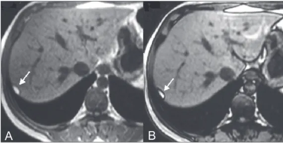
Figure 1. Hepatic lipomatous lesion. A: Axial in-phase T1-weighted GRE image shows a small hyperintense lesion (arrow). B: Axial out-of-phase T1-weighted GRE sequence shows peripheral signal loss (arrow). This is a lipomatous lesion and can rep-resent either a lipoma or an angio-myolipoma.
falciform ligament and along the gallbladder fossa. The lesions are characterised by geographic borders and lack a mass effect; vessels and biliary ducts traverse the area without deviation. On in-phase images, they are iso- to hyperintense with a signal drop on out-of-phase images
and enhancement equal to that of the surrounding liver.
As illustrated in Figures 4 and 5, multifocal steatoses must be differentiated from metastatic diseases(12,14).
Hepatic adenoma
Hepatic adenomas are benign, encapsulated lesions that occur in healthy young women and are strongly related
Figure 3. Teratocarcinoma metas-tasis from an ovarian source with peritoneal dissemination. A: Axial in-phase T1-weighted GRE image shows a small hyperintense lesion on the hepatic dome (arrow). B: Axial out-of-phase T1-weighted GRE se-quence shows peripheral signal loss (arrow). Mild diffuse decrease in the signal intensity of the liver, due to steatosis is also observed.
typically slightly hyperintense on T2-weighted images and iso- to hyperintense on T1-weighted images, with arterial enhancement and washout. Out-of-phase images show signal loss in the fatty component. The main differential diagnosis is focal nodular hyperplasia(11,12,14).
Focal nodular hyperplasia
Focal nodular hyperplasias are the second most com-mon benign liver lesions. They occur most comcom-monly in healthy young women, although their relationship with oral contraceptive use is not as well-established as is that of adenomas. The presence of fat is uncommon in focal nodular hyperplasias and usually associated with hepatic steatosis. Focal nodular hyperplasias appear as iso- to hypointense nodules on T1-weighted images and iso- to hyperintense nodules on T2-weighted images, similar to the surrounding parenchyma. The nodules enhance ho-mogeneously in the arterial phase and show enhance-ment similar to the surrounding liver in the portal phase. A central scar, composed of deformed biliary ducts, blood
vessels and inflammatory cells, is characteristically pres -ent. The scar is hyperintense on T2-weighted images and hypointense on T1-weighted images (Figure 6). In addi-tion, the scar does not enhance in the arterial phase but
does enhance in the equilibrium phase. The central scar
can be absent, especially on small lesions. In such cases, retention of hepatobiliary contrast agents can be diagnos-tic(11,12,14,17).
Steatotic regenerative nodules
Regenerative nodules are the most common nodules in cirrhotic livers. They are usually small, numerous and diffusely distributed throughout the parenchyma. Such nodules can contain fat and show high signal intensity on in-phase images and signal loss on out-of-phase im-ages(18), as illustrated in Figure 7.
Hepatocellular carcinomas
Hepatocellular carcinomas are the most common malignant lesions in cirrhotic livers. They usually show variable signal intensity on T1-weighted images and are hyperintense on T2-weighted images (Figure 8). Hepa-tocellular carcinomas enhance in the arterial phase and have washout in the delayed phases. Pseudocapsule
en-hancement is characteristic in the equilibrium phase.
The fatty components of hepatocellular carcinomas are visible on MRI as a signal drop in fat-suppressed
tech-niques and do not enhance as much as the rest of the
lesion(11,12,14,18).
CONCLUSION
Fat-containing liver lesions constitute a heteroge-neous group of tumours. Careful evaluation of the clinical
history, together with the MRI indings, will facilitate the
differential diagnoses of these lesions.
REFERENCES
1. Siqueira GRS, Guimarães MD, Franco LFS, et al. Exophytic hepa -tocellular carcinoma, simulating a mesenchymal tumor, in a non-cirrhotic liver. Radiol Bras. 2017;50:62–6.
2. Staziaki PV, Teixeira BCA, Pedrazzani BM, et al. Hepatoblastoma with solid and multicystic aspect mimicking a mesenchymal
ham-artoma: imaging and anatomopathologic indings. Radiol Bras.
2017;50:68.
3. Ramalho M, Matos AP, AlObaidy M, et al. Magnetic resonance im-aging of the cirrhotic liver: diagnosis of hepatocellular carcinoma and evaluation of response to treatment – Part 1. Radiol Bras. 2017; 50:38–47.
4. Ramalho M, Matos AP, AlObaidy M, et al. Magnetic resonance im-aging of the cirrhotic liver: diagnosis of hepatocellular carcinoma
Figure 8. Hepatocellular carci-noma with focal fat deposition. A: Axial in-phase T1-weighted GRE se-quence shows a heterogeneously hyperintense lesion (arrow). B: Axial out-of-phase T1-weighted GRE se-quence shows focal areas of signal loss within the lesion (arrow). C,D: On axial T1-weighted image with fat saturation arterial phase image (C), the lesion demonstrates strong homogeneous enhancement with washout (D) and pseudocapsule en-hancement (arrow).
diagnosis of hepatocellular carcinoma without the need of
histo-pathological conirmation – fact! Radiol Bras. 2017;50(1):vii–viii.
7. Cardarelli-Leite L, Fornazari VAV, Peres RR, et al. The value of per-cutaneous transhepatic treatment of biliary strictures following pe-diatric liver transplantation. Radiol Bras. 2017;50:308–13. 8. Candido PCM, Pereira IMF, Matos BA, et al. Giant pedunculated
hemangioma of the liver. Radiol Bras. 2016;49:57–8.
9. Giardino A, Miller FH, Kalb B, et al. Hepatic epithelioid hemangio-endothelioma: a report from three university centers. Radiol Bras. 2016;49:288–94.
10. Cruz JF, Cruz MAF, Machado Neto J, et al. Prevalence and sono-graphic changes compatible with fatty liver disease in patients re-ferred for abdominal ultrasound examination in Aracaju, SE. Radiol Bras. 2016;49:1–5.
14. Prasad SR, Wang H, Rosas H, et al. Fat-containing lesions of the liver:
radiologic-pathologic correlation. Radiographics. 2005;25:321–31. 15. Gibo M, Murata S, Kuroki S. Pericaval fat collection mimicking
an intracaval lesion on CT in patients with chronic liver disease. Abdom Imaging. 2001;26:492–5.
16. Quinn AM, Guzman-Hartman G. Pseudolipoma of Glisson capsule. Arch Pathol Lab Med. 2003;127:503–4.
17. Seale MK, Catalano OA, Saini S, et al. Hepatobiliary-speciic MR
contrast agents: role in imaging the liver and biliary tree. Radio-graphics. 2009;29:1725–48.
18. Parente DB, Perez RM, Eiras-Araujo A, et al. MR imaging of hyper -vascular lesions in the cirrhotic liver: a diagnostic dilemma. Radio-graphics. 2012;32:767–87.