Letters to the Editor
Radiol Bras. 2017 Mai/Jun;50(3):199–208
199
0100-3984 © Colégio Brasileiro de Radiologia e Diagnóstico por Imagem
Letters to the Editor
Dear Editor,
A 44-year-old female presented with a 7-year history of par-oxysmal episodes of dyspnea, headache, palpitation, tremors, and hypertension. In each episode, there had been a sudden onset of symptoms, with no triggering factors, and spontaneous improve-ment after approximately 15 min. On physical examination, she presented no relevant findings or comorbidities. At hospital ad-mission, she reported having had episodes of palpitation, tachy-cardia, and profuse post-micturition sweating, remaining asymp-tomatic between episodes. She was submitted to computed to-mography (CT) and magnetic resonance imaging (MRI), as shown in Figures 1A and 1B, respectively. The CT scan, with intravenous administration of contrast medium, revealed a nodu-lar lesion, measuring 3.5 × 3.0 cm, with lobulated contours and increased density in its soft parts, showing intense, heterogeneous enhancement, in the anteroinferior wall of the bladder. On MRI, the lesion presented a lobular pattern, with a heterogeneous nal on T2-weighted sequences, a predominance of isointense sig-nals, and foci of hyperintense signals in its center. Surgical resec-tion of the lesion (partial cystectomy) was performed. Examina-tion of the surgical specimen, retrieved from the right anterior wall of the bladder, showed a yellowish tumor measuring 3.0 × 3.0 cm, with a macroscopic appearance similar to that of adrenal tissue (Figure 1C). The pathological examination of the speci-men revealed extra-adrenal paraganglioma and tumor-free mar-gins (Figure 1D). In the postoperative period and during the re-mainder of the hospital stay, the patient did not present any of the adrenergic symptoms previously reported.
Pheochromocytomas are tumors of the sympathetic nervous system and can be functioning or nonfunctioning, sometimes
secreting catecholamines, thus causing paroxysmal hypertension, palpitations, headache, and syncope(1). They are most common
between the fourth and sixth decades of life. Approximately 10% are bilateral, 10% are malignant, 10% occur in children, and 10% are extra-adrenal. More than 90% are located in the adrenal gland, and 98% are intra-abdominal. Pheochromocytomas can occur anywhere from the base of the skull to the bladder; when located outside the adrenal gland, they are known as paragangliomas(2).
Pheochromocytoma of the urinary bladder is a rare tumor, origi-nating from chromaffin cells of the sympathetic nervous system and located within the bladder wall, accounting for 0.06% of all bladder tumors and 6% of all paragangliomas(3). In the bladder, it
can produce symptoms typical of pheochromocytoma, including hematuria and micturition syncope resulting from the release of catecholamines by bladder contraction. In 10–15% of cases, paragangliomas of the bladder are nonfunctioning; another 10% show hormonal activity without clinical expression(4).
Recent studies have discussed the role of imaging examina-tions in the investigation of pelvic lesions(5–10). The diagnostic
imaging methods used in the investigation of pheochromocyto-mas include ultrasound, CT, MRI, and scintigraphy. For the de-tection of adrenal pheochromocytomas > 1.0 cm in diameter, CT and MRI have a sensitivity of nearly 95% and 100%, respectively, and MRI has greater specificity than does CT(11). On MRI,
pheo-chromocytoma typically manifests as an expansive lesion with low signal intensity on T1-weighted sequences and high signal in-tensity on T2-weighted sequences, with intense impregnation after contrast administration. However, in rare cases, pheochromocy-toma can present low signal intensity on T2-weighted se-quences(2). The treatment of choice for paraganglioma is
surgi-cal resection, because most are benign and can be completely resected(12).
Pheochromocytoma of the urinary bladder
Figure 1. A: Intravenous contrast-enhanced axial CT scan showing a hypervascular nodule in the antero-inferior wall of the bladder (arrow).
B: T2-weighted MRI sequence show-ing a lesion with an isointense sig-nal at the same site, with a hetero-geneous signal and foci of hyper intense signals in its center (arrow).
C: Surgical specimen (resection of the lesion). D: Hematoxylin and eosin-stained histological section, showing a lesion with a standard zellballen (nested) pattern (solid ar-row), tumor capsule (dashed arar-row), and the bladder wall (arrowhead).
A
B
Letters to the Editor
Radiol Bras. 2017 Mai/Jun;50(3):199–208
200
André Martins Fernandes1, Bernardo Vieira Paim1, Ana
Paula Aguiar Vidal1, Edson Marchiori1, Daniella Braz
Parente2
1. Universidade Federal do Rio de Janeiro (UFRJ), Rio de Janeiro, RJ, Brazil. 2. Instituto D’Or de Pesquisa e Ensino, Rio de Janeiro, RJ, Brazil. Mailin address: Dr. André Martins Fernandes. Hospital Universitário Cle-mentino Fraga Filho. Rua Rodolpho Paulo Rocco, 255, Cidade Universitá-ria. Rio de Janeiro, RJ, Brazil, 21941-913. E-mail: [email protected].
REFERENCES
1. Beilan J, Lawton A, Hajdenberg J, et al. Pheochromocytoma of the uri-nary bladder: a systematic review of the contemporary literature. BMC Urol. 2013;13:22.
2. Martins DL, Baroni RH, Blasbalg R, et al. Evaluation of adrenal tumors by magnetic resonance imaging with histological correlation. Radiol Bras. 2008;41:55–62.
3. Wong EMH, Lai TCT, Tsu JHL, et al. Primary paraganglioma of uri-nary bladder: case series and review of the literature. Surgical Practice. 2015;19:82–5.
4. Peng C, Bu S, Xiong S, et al. Non-functioning paraganglioma occur-ring in the urinary bladder: a case report and review of the literature. Oncol Lett. 2015;10:321–4.
5. Montón CS, Esparza JFO, Ventura AB, et al. Mesothelioma of the tu-nica vaginalis in a patient with giant hydrocele. Radiol Bras. 2016;49: 63–4.
6. Rondina RG, Volpato R, Guerra LFA, et al. Differential diagnosis of anterior sacral meningocele during the evaluation of post-hysterectomy pelvic collections. Radiol Bras. 2016;49:203–4.
7. Queiroz RM, Costa PP, Oliveira NYF, et al. Female urethral diverticu-lum containing a urothelial carcinoma. Radiol Bras. 2016;49:406–7. 8. Lopes PM, Sepúlveda L, Ramos R, et al. The role of transrectal
ultra-sound in the diagnosis of prostate cancer: new contributions. Radiol Bras. 2015;48:7–11.
9. Ferreira DM, Bezerra ROF, Ortega CD, et al. Magnetic resonance imaging of the vagina: an overview for radiologists with emphasis on clinical decision making. Radiol Bras. 2015;48:249–59.
10. Salvadori PS, Bomfim LN, von Atzingen AC, et al. Spontaneous rupture of ovarian cystadenocarcinoma: pre- and post-rupture computed tomog-raphy evaluation. Radiol Bras. 2015:330–2.
11. Qiao HS, Feng XL, Yong L, et al. The MRI of extraadrenal pheochro-mocytoma in the abdominal cavity. Eur J Radiol. 2007;62:335–41. 12. Young WF Jr. Paragangliomas: clinical overview. Ann N Y Acad Sci.
2006;1073:21–9.
http://dx.doi.org/10.1590/0100-3984.2015.0204
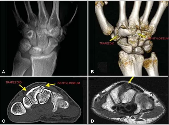
Carpal boss syndrome: os styloideum fused to the trapezoid
Dear Editor,
A 29-year-old White female presented with chronic pain on dorsiflexion of the right hand and a hard prominence, which was painful on palpation, at the base of the second and third metacar-pal muscles. An X-ray of the hand (Figure 1A) revealed a bony prominence in the region identified as palpable in the physical examination, as well as showing that there was lack of definition of the joint space between the trapezoid and the capitate. In multiplanar and three-dimensional computed tomography recon-structions, which provided greater detail (Figures 1B and 1C), an os styloideum was seen to be fused to the trapezoid bone and in neoarticulation with the base of the third metacarpal. Magnetic
resonance imaging showed a hypointense signal on a T1-weighted image (Figure 1D) and increased intensity in a T2-weighted short-tau inversion-recovery sequence, with bone edema adjacent to the neoarticulation, which is indicative of apophysitis.
Os styloideum is an anatomical variation characterized by an accessory ossicle on the dorsum of the wrist, between the trap-ezoid and capitate, at the base of the second and third metacarpal bones(1). When it produces symptoms, mainly local pain, it is known
as a carpal boss(2,3). The true incidence of carpal boss syndrome
is unknown; it is probably underestimated and often confused, clini-cally, with other causes of tumor in the dorsum of the carpus(4).
Although a carpal boss can be classified as acquired (osteo-phytic), congenital (secondary to os styloideum), or of mixed etiol-ogy, the clinical presentations appear to be similar across the
Figure 1. A: Digital X-ray showing a lack of definition of the joint space between the trapezoid and the capitate. B,C: Three-di-mensional computed tomography recon-struction and axial computed tomography slice showing an os styloideum fused to the trapezoid and in neoarticulation with the capitate. D: Magnetic resonance imaging in a T1-weighted sequence, showing os styloideum with bone edema adjacent to the neoarticulation (apophysitis).