A simple method to measure cell viability
in proliferation and cytotoxicity assays
Abstract: Resazurin dye has been broadly used as indicator of cell vi-ability in several types of assays for evaluation of the biocompatibility of medical and dental materials. Mitochondrial enzymes, as carriers of diaphorase activities, are probably responsible for the transference of electrons from NADPH + H+ to resazurin, which is reduced to resoruin.
The level of reduction can be quantiied by spectrophotometers since re-sazurin exhibits an absorption peak at 600 ηm and resoruin at 570 ηm wavelengths. However, the requirement of a spectrophotometer and spe-ciic ilters for the quantiication could be a barrier to many laboratories. Digital cameras containing red, green and blue ilters, which allow the capture of red (600 to 700 ηm) and green (500 to 600 ηm) light wave-lengths in ranges bordering on resazurin and resoruin absorption bands, could be used as an alternative method for the assessment of resazurin and resoruin concentrations. Thus, our aim was to develop a simple, cheap and precise method based on a digital CCD camera to measure the reduction of resazurin. We compared the capability of the CCD-based method to distinguish different concentrations of L929 and normal Hu-man buccal ibroblast cell lines with that of a conventional microplate reader. The correlation was analyzed through the Pearson coeficient. The results showed a strong association between the measurements of the method developed here and those made with the microplate reader (r2 = 0.996; p < 0.01) and with the cellular concentrations (r2 = 0.965;
p < 0.01). We concluded that the developed Colorimetric Quantiication System based on CCD Images allowed rapid assessment of the cultured cell concentrations withsimple equipment at a reduced cost.
Descriptors: Colorimetry / methods; Cells, cultured; Mitochondria / metabolism.
Ricardo Carneiro Borra(a) Mônica Andrade Lotufo(a) Sonia Maria Gagioti(a)
Fabiana de Mesquita Barros(b) Priscila Maria Andrade(c)
(a) PhD, Professor; (b)MSc, Graduate Student
– Graduate Program in Biodentistry, Ibirapuera University (UNIB), São Paulo, SP, Brazil.
(c) Fellow Researcher, Department of Urology
(LIM 55), School of Medicine, University of São Paulo (FMUSP), São Paulo, SP, Brazil.
Corresponding author:
Ricardo Carneiro Borra
Universidade Ibirapuera, Curso de Odontologia
Av. Interlagos, 1329 São Paulo - SP - Brazil CEP: 04661-100 E-mail: [email protected]
Introduction
The biocompatibility of medical and dental ma-terials is routinely evaluated utilizing in vitro meth-odologies. Cell lines are often cultivated in contact with the target materials, and after a variable period of time, the proliferation (estrogenicity) and death (toxicity) rates are measured.1,2,3
Resazurin dye (7-hydroxy-3H-phenoxazin-3-one 10-oxide) has been broadly used as an indicator of cell viability in several types of proliferation and cy-totoxicity assays.4-8 The reduction of resazurin
cor-relates with the number of live organisms, such as bacterial, fungi and mammalian cells. Mitochondri-al enzymes, as carriers of diaphorase activities, like NADPH dehydrogenase, are probably responsible for the transference of electrons from NADPH + H+
to resazurin, which is reduced to resoruin.9
Cellular viability quantiication based on resa-zurin has advantages, including rapidity, reliabil-ity, sensitivreliabil-ity, safety and cost. In addition, it keeps cells intact, which permits other parallel analyses, such as mRNA, cytogenetic, apoptosis, and im-munophenotyping.6 The level of reduction can be
quantiied by spectrophotometers using appropriate ilters, since resazurin exhibits an absorption peak at 600 ηm and resoruin at 570 ηm wavelengths.10
However, the requirement of a spectrophotometer and speciic ilters for the quantiication of resazurin reduction levels could be a barrier to many laborato-ries. Digital cameras, containing red, green and blue (RGB) ilters, which allow the capture of red (600 to 700 ηm) and green (500 to 600 ηm) light wave-lengths in ranges bordering on resazurin and resoru-in absorption bands, could be used as an alternative method for the assessment of resazurin reduction levels. Therefore, using a color digitalized image of microplate load with resazurin and common image-processing software it might be possible to isolate red and green intensity values to calculate the level of reduction of resazurin of different samples.
Thus, our aim was to develop a simple, cheap, precise and reliable method based on RGB signals generated by a digital CCD camera to measure the reduction of resazurin dye into resoruin. To test this method, reduction levels of resazurin measured by the digital camera-based system developed were
com-pared with those measured by the conventional ELI-SA microplate reader. Then the viability of the sys-tem in quantifying different concentrations of L929 and buccal ibroblast cells in culture was tested.
Material and Methods
Reagents
AlamarBlue™ was purchased from Biosource
(Camarillo, CA, USA, cat.# DAL1025); resazur-in sodium salt (cat.#R7017) and L-ascorbic acid (cat.#A5960) were acquired from Sigma (St. Louis, MO, USA). DMEM and RPMI-1640 media supple-mented with 2 mM of L-glutamine, 100 U/mL of penicillin and 100 µg/mL of streptomycin; fetal bo-vine serum, a solution of Trypsin-EDTA and TPP 96-well tissue culture microplates were purchased from CultiLab (Campinas, SP, Brazil). The resazur-in solutions were prepared usresazur-ing phosphate buffered saline from Sigma. L929 cells were purchased from ATCC (Manassas, VA, USA) and a normal Human buccal ibroblast cell line (FLM1) was obtained from the Ibirapuera University cell bank.
Colorimetric Quantification System based on CCD images (CQS)
The image from the microplate loaded with sam-ples was used to calculate the density of the red (Wr) and green (Wg) spectra of each well, and an image of an empty microplate was used to calculate the red (Br) and green (Bg) background from the cor-related wells. Afterwards, the highest red (MAXBr) and green (MAXBg) background values were deter-mined and the CQS transmittance index (CQSTI) was calculated according to the following formula:
ied using the CQS methodology. This experiment was repeated at two different times.
Comparison between AlamarBlueTM and
resazurin solutions
Cell proliferation and cytotoxicity assays are of-ten carried out using a commercial resazurin-based product known as AlamarBlue™ (Biosource,
Ca-marillo, CA, USA), but a cheaper laboratory-pre-pared resazurin solution could also be used with-out compromising rapidity, reliability, sensitivity or safety.7 In order to evaluate the sensitivity of both
solutions, a colorimetric assay using CQS methodol-ogy was performed comparing the reduction curve of AlamarBlue™ with curves of different
concentra-tions of resazurin soluconcentra-tions. In this case, wells con-taining 20 µL of AlamarBlue™ or different
concen-trations of resazurin (175, 350, 525 and 700 µM) diluted in 180 µL of RPMI-1640 complete medium were incubated in triplicate for thirty minutes with different amounts of vitamin C (0.5 × 10³, 1.0 × 10³,
2.0 × 10³, 3.0 × 10³ and 4.0 × 10³µg/mL), which
were used as a reduction agent capable of simulating diaphorase activity. This experiment was repeated at two different times.
Comparison between the CQS method and microplate reader quantification
With the aim of comparing the reliability and sen-sitivity of the CQS methodology with data obtained from a spectrophotometer, a curve of reduction con-structed using a laboratory-prepared resazurin solu-tion was submitted to a reading by the CQS method and by an ELISA microplate reader. Briely, wells containing solutions of 180 µL of RPMI-1640 and 20 µL of the combination of ten different propor-tions of 700 µM oxidized resazurin (20, 18, 16, 14, 12, 10, 8, 6, 4 and 0 µL) and of 700 µM reduced re-sazurin (0, 2, 4, 6, 8, 10, 12, 14, 16 and 20 µL) were analyzed at wavelengths of 595 and 490 nm using the microplate reader (Bio Tek ELX – 800 – BioTek Instruments, Inc., VT, USA) and compared with measurements performed using the CQS method. Since the autoclaving procedure was not eficient in reducing the resazurin in PBS, the resoruin stock (700 µM) was reduced with 500 µg/mL of vitamin where:
m = microplate column n = microplate row
Wrmn = red spectrum density of the well located on the mth colum, nth row of a sample
illed microplate image
Wgmn = green spectrum density of the well located on the mth colum, nth row of a
sample illed microplate image
Brmn = red spectrum density of the well located on the mth colum, nth row of an empty
microplate image
Bgmn = green spectrum density of the well located on the mth colum, nth row of an
empty microplate image MAXBred = the highest value of Br MAXBgreen = the highest value of Bg
The inal CQSTI is adjusted by discounting the CQSTI mean of the wells used as negative controls (oxidized resazurin).
Evaluation of the CQS method
After reducing an aliquot of AlamarBlue™
(Bio-source, Camarillo, CA, USA), by autoclaving for 15 minutes and allowing it to cool to room temperature, a curve of reduction was constructed mixing 20 µL of eleven different proportions of the oxidized (re-sazurin: 20, 18, 16, 14, 12, 10, 8, 6, 4, 2 and 0 µL) and reduced (resoruin: 0, 2, 4, 6, 8, 10, 12, 14, 16, 18 and 20 µL) form of AlamarBlue™ in 180 µL of
C. Thus, the correlation of pairs of optical density (OD) and CQSTI values was veriied using the Pear-son Correlation Statistic Test.
Reliability of the CQS method to quantify different cell densities
Conluent monolayer adherent L929 and FLM1 cells, grown in RPMI-1640 and DMEM medium respectively, supplemented with 10% fetal bovine serum, 2 mM of L-glutamine + 100 U/mL of peni-cillin / 100 µg/mL of streptomycin, and maintained at 37°C in a 5% CO2 atmosphere, were trypsinized and resuspended at a concentration of 106 cells/mL.
Different cell concentrations of L929 and FLM1 cells diluted in 180 µL of RPMI-1640 and DMEM complete medium respectively were seeded in a 96-well tissue culture microplate and incubated in triplicate (FLM1) and quadruplicate (L929) with AlamarBlue™ and resazurin (700 µM) for up to 35
hours. Images were taken and analyzed at different time points using the CQS methodology during the
entire period of incubation.
Statistical analysis
To determine the level of correlation between Ala-marBlue™ / resazurin and CQSTI and the correlation
between the optical density (OD) of the microplate reader with the CQSTI, the values were plotted in a scatter plot and the Pearson Correlation was calcu-lated using SPSS 10.0 software (SPSS Inc. Chicago, IL, USA). p <0.05 was considered signiicant.
Results
Evaluation of the CQS method
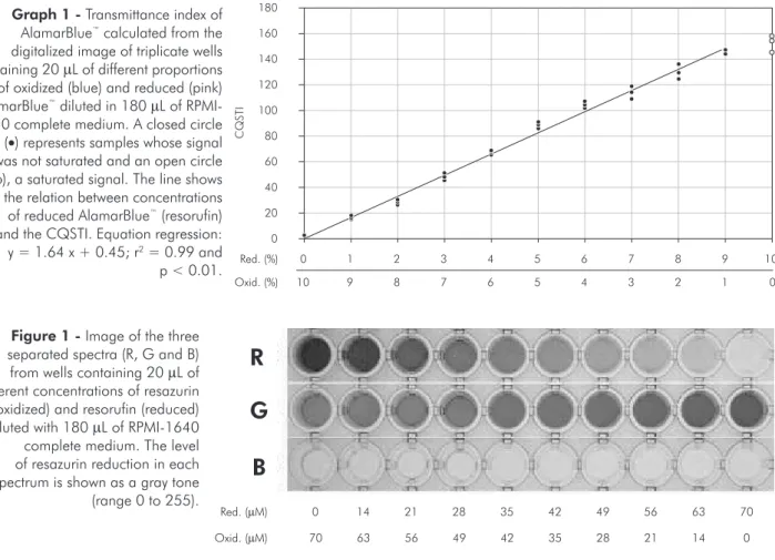
The CQSTI was strongly correlated with the in-tensity of the AlamarBlue™ reduction level. This
cor-relation was linear until the mixture of 9% of reso-ruin + 1% of resazurin (y = 1.64 x + 0.45; r2 = 0.99;
p < 0.01) (Graph 1). The densities of red and green spectra correlated directly with the level of resoruin and resazurin respectively, in a dose dependent man-ner, while the density of the blue did not (Figure 1).
0 1 2 3 4 5 6 7 8 9 10
0
10 9 8 7 6 5 4 3 2 1
C
QSTI
Red. (%)
Oxid. (%) 0 20 40 80
60 100 120 140 160 180 Graph 1 - Transmittance index of
AlamarBlue™ calculated from the
digitalized image of triplicate wells containing 20 µL of different proportions of oxidized (blue) and reduced (pink) AlamarBlue™ diluted in 180 µL of
RPMI-1640 complete medium. A closed circle (•) represents samples whose signal was not saturated and an open circle (Ο), a saturated signal. The line shows the relation between concentrations of reduced AlamarBlue™ (resorufin)
and the CQSTI. Equation regression: y = 1.64 x + 0.45; r2 = 0.99 and
p < 0.01.
R
Figure 1 - Image of the three separated spectra (R, G and B) from wells containing 20 µL of different concentrations of resazurin (oxidized) and resorufin (reduced) diluted with 180 µL of RPMI-1640 complete medium. The level of resazurin reduction in each spectrum is shown as a gray tone (range 0 to 255).
Red. (µM)
Oxid. (µM)
0 14 21 28 35 42 49 56 63 70
0 14 21 28 35 42 49 56 63 70
G
Comparison between AlamarBlueTM and resazurin solutions
Vitamin C reduced both resazurin and Alamar-Blue proportionally to its concentration. Resazurin at a concentration of 525 µM was more closely associated with the AlamarBlue reduction curve (Graph 2). The equivalent concentration of resazur-in related to AlamarBlue was calculated as 560 µM (data not shown).
Comparison between the CQS method and microplate reader quantification
The concentration of resazurin measured by the CQS was strongly correlated with the values mea-sured by the ELISA microplate reader (y = 206.8 x; r2 = 1.0 and p < 0.01) (Graph 3).
Reliability of the CQS method to quantify different cell densities
Graph 4 shows that the numbers of L929 and FLM1 cells were strongly correlated with values cal-culated using the CQS method (CQSTI). The time of incubation and the number of cells inluenced direct-ly the level of reduction of both resazurin 700 µM and AlamarBlue. The results showed that the best incubation time range for quantifying FLM1 was
11 to 19 hours; for quantifying L929, a murine tu-mor cell line, the best incubation time range was 6 to 10 hours. The sensitivity of the method was su-perior when 700 µM of the resazurin solution were employed for quantiication of the concentrations of both cell types. The 700 µM solution of resazurin reached the saturation point around a CQSTI of 160, contrasting with AlamarBlue, which reached that point around a CQSTI of 120 (Graph 4).
Discussion
Resazurin dye (7-hydroxy-3H-phenoxazin-3-one 10-oxide) has been broadly used as a reliable indi-cator of cell viability in proliferation and cytotox-icity assays.4-13 Analogous to resazurin, the dye
3-[4,5-dimethylthiazol-2-yl]-2,5-diphenyl tetrazolium bromide (MTT) has been also employed to quantify cell viability; however, the resazurin assay seemed to be slightly more sensitive than the MTT assay.9,12
Resazurin is probably a better redox indicator for cell viability because it does not interfere with the reactions of the electron transport chain since the midpoint redox potential of resazurin is greater than that of any of the components of the electron trans-port chain while indicators with an inferior redox potential such as MTT cannot detect the reduction
4x10³
3x10³
2x10³
1x10³
RZ - 175 µM RZ - 350 µM AlamarBlue
RZ - 700 µM RZ - 525 µM
0
C
QSTI
Vitamin C (µg/mL) 0
20 40 80
60 100
Graph 2 - Relation between AlamarBlue and resazurin (RZ) curves as a function of vitamin C concentration. Transmit-tance index calculated from the digitalized image of trip-licate wells containing 20 µL of AlamarBlue and different concentrations of resazurin diluted in 180 µL of RPMI-1640 complete medium with different amounts of vitamin C.
Graph 3 - Comparison between the transmittance indexes obtained by the CQS and microplate reader methods. Trip-licate wells containing 20 µL of ten different concentrations of resazurin and resorufin diluted with 180 µL of RPMI-1640 complete medium, read using the CQS and the microplate reader. Equation regression: y = 206.8 x; r2 = 1.0 and
p < 0.01. MRI: Microplate reader index.
0 0.1 0.2 0.3 0.4 0.5 0.6
C
QSTI
(R
ed
-
G
reen)
MRI (595 - 490 nm wavelength) 0
20 40 80
mediated by cytochromes.13 After comparing the
sensitivity of six assays for quantiication of the bio-ilms formed by a broad range of microorganisms in 96-well microtiter plates, the resazurin-based assay has proven to be one of the best alternatives.14
However, in order to quantify resazurin reduc-tion, a spectrophotometer and speciic ilters at 600 and 570 ηm are required. The use of an RGB CCD digital camera could be an alternative method, since
it contains ilters that work between 600 to 700 ηm (red) and 500 to 600 ηm (green). In this case, a sim-ple inexpensive methodology capable of capturing and analyzing images from a microplate, denomi-nated Colorimetric Quantiication based on CCD Images (CQS), was developed and tested regarding its ability to monitor and quantify cell viability.
Quantiication was based on assessment of the amount of red and green light emanating from the Graph 4 - Comparison of profiles of reduction of different concentrations of L929 and FLM1 cells assessed by AlamarBlue and 700 µM resazurin solution dyes. The CQSTI was represented by mean ± standard deviation.
5 .0 x1 0 4 3 .1 x1 0 3 7 .8 x1 0 2 C QSTI
Number of buccal fibroblasts Resazurin assay 1 .3 x1 0 4 1 .6 x1 0 3 6 .3 x1 0 3 1 0 5 2 .5 x1 0 4 -20 0 20 40 80 60 100 120 140 160 180 11 hours 27 hours 1 hour 19 hours 35 hours 7 hours 5 .0 x1 0 4 6 .3 x1 0 3 7 .8 x1 0 2 1 .6 x1 0 3 C QSTI
Number of buccal fibroblasts Alamar blue assay
1 .3 x1 0 4 3 .1 x1 0 3 1 0 5 2 .5 x1 0 4 -20 0 20 40 80 60 100 120 140 160 180 2 .5 x1 0 5 3 .1 x1 0 4 2 .0 x1 0 3 3 .9 x1 0 3 7 .8 x1 0 3 C QSTI
Number of L929 cells Resazurin assay 6 .3 x1 0 4 1 .6 x1 0 4 5 .0 x1 0 5 1 .3 x1 0 5 -20 0 20 40 80 60 100 120 140 160 180 6 hours 10 hours 2 hours 8 hours 12 hours 4 hours 2 .5 x1 0 5 3 .1 x1 0 4 2 .0 x1 0 3 3 .9 x1 0 3 7 .8 x1 0 3 C QSTI
Number of L929 cells Alamar blue assay
resazurin samples (transmittance). In order to obtain the intensity of red and green light, the microplate was digitalized, and each component R and G from the color image was separated using the ImageLab software. Next, the density of each component cor-responding to the bottom well area was determined and used to create the transmittance index.
The normalization procedures based on back-ground reference and the use of the difference be-tween the red and green spectra as a mean param-eter in the formula was able to neutralize systematic errors related to the parallax effect and non-perfect homogeneity of illumination. This was possible because the difference between the red and green components remains constant regardless of the in-tensity of light that passes through the wells (data not shown). The use of a scanner with a transpar-ency adaptor could be another alternative. In 2002, Gabrielson et al.11 evaluated the use of AlamarBlue
indicators for measuring relative growth of micro-organisms in microplate using a scanner-digitalized image. Nevertheless, since they did not separate the color components, the difference of reduction level was not detectable, although visible to the naked eye.
The relationship between the CQSTI and the re-sazurin / resoruin standard curve showed a strong linear association, indicating that the index could be useful for measuring the reduction level of re-sazurin and, indirectly, cell viability. Furthermore, the results obtained using a microplate reader were strongly correlated with the CQS method.
Although cell viability is often assessed using
a commercial product known as AlamarBlue™, a
laboratory-prepared resazurin solution could be employed as a cheaper alternative. Analysis of the performance of AlamarBlue™ and different
concen-trations of the resazurin solution demonstrated that a resazurin concentration of 560 µM produced an identical curve in a vitamin C reduction assay. More-over, the assay based on a 700 µM resazurin solution was more sensitive than that based on AlamarBlue™.
In order to test the usefulness of the method, buc-cal ibroblast FLM1 and L929 cells were incubated in triplicate and quadruplicate, respectively, with 700 µM of resazurin solution and AlamarBlue™ for
35 hours. The results showed a strong correlation between the CQSTI and the number of cells regard-less of the type of solution employed. The CQSTI increased in a time- and cell density-dependent man-ner until a saturation point. The correlation between cell number and time of incubation was more stable and had a higher amplitude when the 700 µM resa-zurin solution was used. The method was capable of distinguishing narrow variations in the number of cells between 7 × 10² up to 5 × 10⁵ cells, principally
after 11 hours of incubation for FLM1 and 6 hours for L929 cells.
Conclusions
It was concluded that the Colorimetric Quantii-cation System based on CCD Images allowed rapid assessment of many cultured cell samples with sim-ple equipment at a reduced cost, thus offering many laboratories a cost-effective alternative to the cur-rently used methods.
References
1. Oh SH, Choi SY, Choi SH, Lee YK, Kim KN. The influence of lithium fluoride on in vitro biocompatibility and bioactivity of calcium aluminate-pMMA composite cement. J Mater Sci Mater Med. 2004 Jan;15(1):25-33.
2. Taira M, Toguchi MS, Hamada Y, Takahashi J, Itou R, Toyosawa S et al. Studies on cytotoxic effect of nickel ions on three cultured fibroblasts. J Mater Sci Mater Med. 2001 May;12(5):373-6.
3. Moretti Neto RT, Mello I, Moretti AB, Robazza CR, Pereira AA. In vivo qualitative analysis of the biocompatibility of
different cyanoacrylate-based adhesives. Braz Oral Res. 2008 Jan-Mar;22(1):43-7.
4. Ahmed SA, Gogal RM Jr, Walsh JE. A new rapid and simple non-radioactive assay to monitor and determine the prolifera-tion of lymphocytes: an alternative to [3H]thymidine incorpo-ration assay. J Immunol Methods. 1994 Apr 15;170(2):211-24.
of bladder cancer. Urol Oncol. 2009 Mar 7. [in press] [Epub ahead of print]
6. Zhi-Jun Y, Sriranganathan N, Vaught T, Arastu SK, Ahmed SA. A dye-based lymphocyte proliferation assay that permits multiple immunological analyses: mRNA, cytogenetic, apop-tosis, and immunophenotyping studies. J Immunol Methods. 1997 Dec 15;210(1):25-39.
7. Nakayama GR, Caton MC, Nova MP, Parandoosh Z. Assess-ment of the Alamar Blue assay for cellular growth and viability
in vitro. J Immunol Methods. 1997 May 26;204(2):205-8. 8. Nociari MM, Shalev A, Benias P, Russo C. A novel one-step,
highly sensitive fluorometric assay to evaluate cell-mediated cytotoxicity. J Immunol Methods. 1998 Apr 15;213(2):157-67.
9. O’Brien J, Wilson I, Orton T, Pognan F. Investigation of the Alamar Blue (resazurin) fluorescent dye for the assess-ment of mammalian cell cytotoxicity. Eur J Biochem. 2000 Sep;267(17):5421-6.
10. Zalata AA, Lammertijn N, Christophe A, Comhaire FH. The correlates and alleged biochemical background of the resazur-in reduction test resazur-in semen. Int J Androl. 1998 Oct;21(5):289-94.
11. Gabrielson J, Hart M, Jarelöv A, Kühn I, McKenzie D, Möllby R. Evaluation of redox indicators and the use of digital scanners and spectrophotometer for quantification of microbial growth in microplates. J Microbiol Methods. 2002 Jun;50(1):63-73.
12. Hamid R, Rotshteyn Y, Rabadi L, Parikh R, Bullock P. Com-parison of alamar blue and MTT assays for high through-put screening. Toxicol In Vitro. 2004 Oct;18(5):703-10. 13. Petrenko YA, Gorokhova NA, Tkachova EN, Petrenko AY.
The reduction of Alamar Blue by peripheral blood lympho-cytes and isolated mitochondria. Ukr Biokhim Zh. 2005 Sep-Oct;77(5):100-5.