A comparative study of ascending urethrogram and
sono-urethrogram in the evaluation of stricture urethra
_______________________________________________
B.R. Ravikumar
1, Chiranjeevi Tejus
1, K.M. Madappa
1, Dharakh Prashant
1, G.S. Dhayanand
11 JSS Medical college and hospital, Mysore, Karnataka, India
_______________________________________________________________________________________
ABSTRACT
To compare the efficacy of sono-urethro-gram and ascending urethrosono-urethro-gram in the evalua-tion of stricture urethra.
Materials and Methods: In this prospective study 40 patients with obstructive lower urinary tract symptoms and suspected to be having stric-ture urethra were subjected to ascending urethro-gram and sonourethrourethro-gram. The radiologist was blinded to the findings of ascending urethrogram. All the sonourethrograms were done by the same radiologist. The findings of sonourethrogram & ascending urethrogram were compared with the findings of cystoscopy and intra-operative findin-gs. The specificity, sensitivity,positive predictive value and negative predictive value of each moda-lity in the diagnosis of various urethral anomalies were estimated.
Results: The sonourethrogram identified stricture disease in all the patients who had ab-normal ascending urethrogram. In addition, other abnormalities like spongiofibrosis, diverticula and stones which were not picked up in ascending urethrogram were diagnosed by sonourethrogram. The cystoscopic and intra-operative findings with respect to stricture length, diameter and spongio-fibrosis correlated well with sono-urethrogram findings. 5 patients who had stricture in the as-cending urethrogram were found to be having the normal urethra in sonourethrogram and confir-med by cystoscopy.
Conclusion: sonourethrogram is an effec-tive alternaeffec-tive to ascending urethrogram in the evaluation of stricture urethra. It is more sensitive in the diagnosis of anterior urethral strictures than posterior urethral strictures. It is superior to as-cending urethrogram in the identification of spon-giofibrosis, diameter and length of the stricture. The complications were lower in sonourethrogram group compared to ascending urethrogram.
INTRODUCTION
Stricture urethra in Males is a common problem encountered by the urologists.
Besides history and physical examination, ascending urethrogram remained the Gold Stan-dard for evaluating Male Urethral Stricture (Cun-ningham et al. 1910) (1-3). It has a Sensitivity of 91% and specificity of 72% for diagnosing ante-rior urethral strictures (4).
Ascending urethrogram leads to radiation exposure of 1-2 msv, equivalent to 6 months of background radiation and 20 chest X-rays (5, 6). Procedure related infection- Contributes to 0.6% to 1.6% of all hospital acquired infections (7, 8).
The initial experiences with ultrasound evaluation of the urethra were described separa-tely in the late 1980s by McAninch et al. (2) and Merkle and Wagner (5). Early studies identified not only the ability of ultrasound to demonstrate the exact length of strictures but also the added abili-ty to define the periurethral tissues, as opposed to contrast urethrography, which only demonstrates the lumen. In particular, the presence and degree of periurethral fibrosis can be shown with a view to guiding surgery (9, 10).
The aim of the study was to compare the efficacy of Sono-urethrogram with Ascending urethrogram in the evaluation of Male urethral strictures.
MATERIALS AND METHODS
This study was conducted in the depart-ment of urology, JSS hospital, Mysore, done be-tween march 2011 to March 2012. Male Patients with age group between 25-75 years (mean 43 years) presenting with obstructing voiding symp-toms suggestive of stricture urethra were subjec-ted to ascending urethrogram (AUG) under asep-tic precautions and antibioasep-tic coverage. A written consent was taken from all the patients before subjecting to the study.
40 patients with evidence of stricture in AUG further underwent sono-urethrogram. Among 40 patients with evidence of stricture in AUG; 13 were secondary to traumatic, 12 inflam-matory, 9 BXO and 1 post TURP respectively (Fi-gure-1). Patients with recurrent stricture were
ex-TECHNIQUE
AUG: 20 mL of contrast medium (urogra-ffin 76%) was injected into the urethra using 20 ml syringe and spot films were taken. The proce-dure time was 15-20 min.
The following data were recorded
• Stricture location, length and diameter were measured using a scale. 20% de-duction was made to correct for mag-nification.
• Complications.
Sono-Urethrogram: Was done using 10 MHz fre-quency linear array transducer. Xylocaine jelly or sterile water (20-30 ml) injected using 20 ml syringe taking care not to inject air bubble. The urethra was screened up to BMJ-using trans-scro-tal and trans-perineal approach. Longitudinal and transverse images were obtained. Procedure time was 10 to 15 min.
The following data were recorded
• The stricture location, diameter, leng-th, spongiofibrosis.
All the statistical calculations were done through SPSS 16.0 (2007) for windows.
RESULTS
Among 40 patients who were diagnosed as having stricture urethra by Ascending ure-throgram, 5 were found to be normal in Sono--urethrogram. The findings were confirmed later by cystoscopy. One patient who had evidence of posterior urethral stricture in SUG had normal urethra in cystoscopy. The assessment of stricture was incomplete in 2 patients due to posterior lo-cation. These pts subsequently required MCUG for complete stricture evaluation (Table-1).
Average stricture length in AUG group was 9.3 mm (Figure-2). Stricture length could not be assessed correctly in 5 patients in AUG group due to complete cut- off. Average stricture leng-th in SUG group was 14.1 mm (Figure-3). Mean difference between 2 groups was 4.8 mm (P va-lue- <0.01). The cystoscopic and intra-operative findings correlated well with the findings of SUG.
Average diameter of stricture in AUG group was 0.9 mm. Average diameter of stricture in SUG group was 1.1 mm. Mean difference be-tween 2 modalities was 0.2 mm (P-value- <0.01).
Spongiofibrosis was demonstrated in 12 patients with traumatic strictures, 4 patients with inflammatory strictures, 7 patients with BXO and in 1 patient with Post- operative (TURP) stricture. The findings of SUG correlated well with intra-op findings who underwent open surgery. Ascending urethrogram did not identify spongiofibrosis in any patient.
In Ascending urethrogram group, 3 pa-tients had False tracts, 2 papa-tients had urethral calculi and 4 patients had urethral diverticulum. In Sono-urethrogram group, 3 patients had False tracts, 3 patients had urethral calculi, 5 patients had urethral diverticulum and 2 patients had Pe-riurethral abscess (Figures 4 and 5).
Minor bleeding was seen in 5 patients, In-travasation of contrast in 6 patients and Dysuria in 6 patients in ascending urethrogram group. In SUG group, Minor bleeding was seen in 2 patients and Dysuria in 4 patients. None of the patients developed procedure related infection.
Table 1 – Results of Sono-urethrogram.
Sensitivity Specificity Positive predictive
value
Negative predictive
value
Anterior 100% 100% 100% 100%
Posterior 75% 50% 75% 50%
Figure 2 - Stricture as seen in Ascending urethrogram.
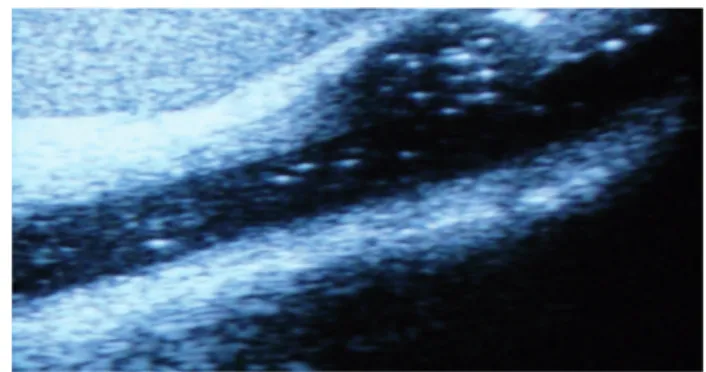
Figure 3
-
Same stricture as seen in sono-urethrogram.DISCUSSION
Ascending urethrogram has been the gold standard investigation in the evaluation of stric-ture urethra. But it is associated with radiation exposure and underestimates the length of the stricture. Hence to overcome these shortcomings of ascending urethrogram, ultrasound evaluation of stricture urethra called sono-urethrogram has been tried.
Sono-urethrogram was more sensitive and specific in diagnosing urethral stricture disease in our study as compared to Ascending urethro-gram. The false positive rate was less with sono--urethrogram as compared to ascending urethro-gram. Sono-urethrogram was 100% specific, 100% sensitive in identifying anterior urethral strictu-res, with positive and negative predictive values being 100% (Table-1). But the accuracy of sono--urethrogram decreased dramatically in evalua-ting posterior urethral strictures. It was only 75% sensitive and 50% specific in diagnosing posterior urethral strictures. Voiding cysto-urethrogram is the gold standard investigation in the evaluation
limited in its ability to define the posterior ure-thral strictures at present. The accuracy of sono--urethrogram may be improved by the addition of antegrade dynamic study using perineal USG or TRUS. In our study we have not directly compared VCUG and sono-urethrogram in the evaluation of posterior urethral strictures. Till the time advances in sono-urethrogram happens, VCUG along with antegrade cystoscopy is considered as standard for the evaluation of posterior urethral strictures.
Ascending urethrogram underestimates the length of anterior urethral stricture due to end on view. It results in wrong decision making re-garding the type of intervention. This is may be overcome by the addition of sono-urethrogram which accurately estimates the length of the ure-thral stricture in real time. In our study the ave-rage length of the stricture in Ascending urethro-gram group was 9.3 mm, whereas the length in sono-urthrogram group was 14.1 mm. the diffe-rence between 2 groups was 4.8 mm, which was statistically significant (P value-0.01). The findin-gs of sono-urethrogram correlated well with the cystoscopic and intra-operative findings. But the sono-urethrogram is not the ideal study for esti-mating the length of posterior urethral strictures. Combining voiding cysto-urethrogram with the retrograde study still remains the gold standard in evaluating posterior urethral stricture. Sono-ure-throgram is better than Ascending ureSono-ure-throgram in estimating the length of anterior urethral stricture.
Sono-urethrogram accurately estimates the thickness and length of spongiofibrosis in all the patients which helps in better planning of surgery. Hence sono-urethrogram scores over Ascending urethrogram in estimating the spongiofibrosis.
Stricture urethra is complicated in some cases. It may be associated with diverticulum, sto-nes, false tracts and abscesses which may com-plicate the surgery (False tracts in our study are because of attempted catheterization or urethral dilatation before AUG or Sono-urethrogram). Hence identifying these complicating factors be-fore surgery is very important.
Sono-urethrogram identifies all these fac-tors more accurately than Ascending urethrogram. Periurethral abscess cannot be identified by As-cending uretrhrogram, whereas sono-urethrogram identifies the abscess more precisely. Hence sono--urethrogram helps in identifying the complica-ting factors and better planning of surgery.
Ascending urethrogram is associated with complications like urinary tract infection, Bleeding and intravasation. Eventhough sono-urethrogram is also associated with similar complications, the incidence is less compared to Ascending urethro-gram. Hence sono-urethrogram is more accurate in assessing the diameter of the stricture as com-pared to sono-urethrogram.
CONCLUSIONS
Sono-urethrogram is a highly sensitive and specific investigation in the diagnosis of an-terior urethral strictures.
Sono-urethrogram is not ideal for the eva-luation of posterior strictures.
Stricture length can be estimated more precisely with Sono-urethrogram compared to As-cending urethrogram.
Spongiofibrosis-The thickness and length can be appreciated by Sono-urethrogram, which is not possible with Ascending urethrogram.
Associated findings such as diverticulum and peri-urethral abscess can be detected with hi-gher sensitivity by Sono-urethrogram.
In future Sono-urethrogram might repla-ce Asrepla-cending urethrogram as the investigation of
ARTICLE INFO
Int Braz J Urol. 2015; 41: 388-92
_____________________
Submitted for publication: June 16, 2014
_____________________
REFERENCES
1. McCallum RW. The adult male urethra: normal anatomy, pathology, and method of urethrography. Radiol Clin North Am. 1979;17:227-44.
2. McAninch JW, Laing FC, Jeffrey RB Jr. Sonourethrography in the evaluation of urethral strictures: a preliminary report. J Urol. 1988;139:294-7.
3. Cunningham JH. The diagnosis of stricture of the urethra by Roentgen rays.Trans Am Assoc Genitourinary Surg 1910; 369. 4. Mahmud SM, El KS, Rana AM, Zaidi Z. Is ascending
urethrogram mandatory for all urethral strictures? J Pak Med Assoc. 2008;58:429-31.
5. Merkle W, Wagner W. Sonography of the distal male urethra--a new diagnostic procedure for urethral strictures: results of a retrospective study. J Urol. 1988;140:1409-11.
6. Hart D, Wall BF. Radiation exposure of the UK population from medical and dental x-ray examinations, NRPB-W4 (2002). 7. Klosterman PW, Laing FC, McAninch JW. Sonourethrography
in the evaluation of urethral stricture disease. Urol Clin North Am. 1989;16:791-7.
8. Gallentine ML, Morey AF. Imaging of the male urethra for stricture disease. Urol Clin North Am. 2002;29:361-72. 9. Chiou RK, Anderson JC, Tran T, Patterson RH, Wobig R,
Taylor RJ. Evaluation of urethral strictures and associated abnormalities using high-resolution and color Doppler ultrasound. Urology. 1996;47:102-7.
10. Das S. Ultrasonographic evaluation of urethral stricture disease. Urology.1992;40:237-42.
_______________________ Correspondence address: