S
ho
rt
R
e
p
o
rt
J. Braz. Chem. Soc., Vol. 19, No. 1, 199-202, 2008. Printed in Brazil - ©2008 Sociedade Brasileira de Química 0103 - 5053 $6.00+0.00
*e-mail: [email protected]
Oxidation of 5,6-Diamino-1,3-dimethyl-2,4-dioxopyrimidine by Perrhenate.
The Crystal Structure of 1,3,6,8-Tetramethylpyrimidopteridine-2,4,5,7-tetrone
Irvin Booysen,
aThomas I.A. Gerber
*
,aand Peter Mayer
baDepartment of Chemistry, Nelson Mandela Metropolitan University, 6031 Port Elizabeth, South Africa
bDepartment of Chemistry, Ludwig-Maximilians University, D-81377 München, Germany
A oxidação de 5,6-diamino-1,3-dimetil-2,4-dioxopirimidina (H2ddd) por perrenato (ReO4)
leva à formação de 1,3-dimetilaloxano, que condensa com H2ddd não-oxidado para produzir 1,3,6,8-tetrametilpirimidopteridino-2,4,5,7-tetrona (tppt). A estrutura do tppt consiste de um anel pirazínico central e dois anéis pirimidínicos terminais em posições cis. Os ângulos diedros entre os anéis pirazina e pirimidina são de 1,08° e 1,20°.
The oxidation of 5,6-diamino-1,3-dimethyl-2,4-dioxopyrimidine (H2ddd) by perrhenate (ReO4)
led to the formation of 1,3-dimethylalloxan, which condenses with unoxidized H2ddd to yield the product 1,3,6,8-tetramethylpyrimidopteridine-2,4,5,7-tetrone (tppt). The structure of tppt consists of a central pyrazine ring and two terminal pyrimidine rings in cis positions. The dihedral angles between the pyrazine and pyrimidine rings are 1.08° and 1.20°.
Keywords: oxidation, diaminouracil, perrhenate, crystal structure
Introduction
The pyrimidine ring forms a constituent of nucleic acids, antibiotics, coenzymes and vitamins, and its coor-dination properties are important in understanding the role
of metal ions in biological systems.1 The interest in uracil
(2,4-dioxopyrimidine) derivatives arises from their poten-tial biological activity. For example, some fluoro derivatives
have shown antitumour activity2 and anti-inflammatory
ac-tion.3 Uracil and its derivatives are known to bind to metal
centres by using various combinations of their donor atoms,
which make them versatile ligands.4-6 For example, we have
recently synthesized the complex salt [Re(ddd)(Hddd)
I(PPh3)2](ReO4) (H2
ddd=5,6-diamino-1,3-dimethyl-2,4-dioxopyrimidine), where ddd is coordinated monodentately through the doubly deprotonated amino nitrogen, and Hddd
is coordinated bidentately via a neutral amino nitrogen and
a singly deprotonated amido nitrogen.7
In this account we report on the oxidation of H2ddd to
1,3,6,8-tetramethylpyrimidopteridine-2,4,5,7-tetrone (tppt)
by perrhenate (ReO4).
Experimental
Ammonium perrhenate and H2ddd were obtained
com-mercially from Aldrich. Infrared spectra were obtained us-ing KBr pellets on a Nicolet 20 DXC FTIR
spectrophotom-eter in the 4000-200 cm-1 range, and 1H NMR spectra were
recorded at 300.13 MHz (7.05 T) on a Bruker AMX-300
Oxidation of 5,6-Diamino-1,3-dimethyl-2,4-dioxopyrimidine by Perrhenate J. Braz. Chem. Soc.
200
tetramethylsilane (tms) as reference. Elemental analysis was carried out by the Department of Chemistry at the University of the Western Cape in Cape Town.
Synthesis of tppt
Equimolar quantities of (NH4)[ReO4] (100 mg,
115 µmol) and H2ddd (20 mg, 117 µmol) were added to
methanol (20 cm3), and the mixture was heated under reflux
for 3 h under nitrogen. After cooling to room temperature, the solution was filtered (no precipitate formed) and left to evaporate slowly at room temperature. After four days, pale yellow crystals were collected by filtration, washed with acetone and dried under vacuum. Yield 85%, mp
255 °C. Anal. Calc. (%) for C12H12N6O4: C, 47.37; H, 3.98;
N, 27.62. Found: C, 47.58; H, 3.89; N, 27.93. IR Nmax/cm-1:
N(C=O) 1696, 1635; N(C=N) 1542; D(CH3) 1428. 1H NMR
(295 K, D, ppm): 2.89 (s, 6H, C(9)H3, C(11)H3); 2.73 (s,
6H, C(10)H3, C(12)H3).
X-ray crystallography
Intensity data for tppt were collected at 200(2) K on a Nonius Kappa CCD single-crystal diffractometer, using
Mo-KA radiation. Unit cell and space group determinations
were carried out in the usual manner.8For the structure
factors, corrections for Lorentz and polarization effects and absorption were made. The structure was solved by direct methods and refined by full matrix least-squares
procedures using SHELXL-97.9 All non-hydrogen atoms
were geometrically constrained.
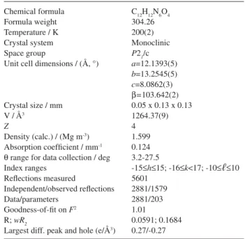
Crystal data and structure refinement details for tppt are given in Table 1, with selected bond distances and angles shown in Table 2.
Results and Discussion
The reaction of equimolar quantities of (NH4)[ReO4]
with H2ddd in methanol under reflux led to the isolation
of tppt as the only product. The heating of H2ddd in the
absence of perrhenate led to no reaction. The [ReO4]
anion is therefore instrumental in the formation of tppt. It has been shown previously that perrhenate reacts with
1,2-diaminobenzene (H2dab) to form the rhenium(VII)
cationic complex [Re(dab)3]+, where dab is coordinated as
the diamide.10 The dab dianion can be readily oxidized by
two electrons to the neutral benzoquinonediimine.11
The product tppt contains the terminal pyrimidine rings
incis positions. This phenomenon has also been observed
by the reaction of H2ddd with Fe3+, which also gives tppt
as product.12 However, the metal ions Au3+, Hg
22+, Ag+,
and Tl3+ also promote the oxidation of H
2ddd, but then the
product tppt contains a trans disposition of the terminal
pyrimidine rings.13
In the infrared spectrum of tppt there are two very strong
absorption bands at 1696 and 1653 cm-1, which correspond
to the C=O stretching vibrations. Another strong peak at
1542 cm-1 is assigned to N(C=N). The CH
3 deformation
vibrations are assigned to the medium-intensity band at Table 1. Crystal data and structure refinement data for tppt
Chemical formula Formula weight Temperature / K Crystal system Space group
Unit cell dimensions / (Å, °)
Crystal size / mm V / Å3
Z
Density (calc.) / (Mg m-3) Absorption coefficient / mm-1 Q range for data collection / deg Index ranges
Reflections measured
Independent/observed reflections Data/parameters
Goodness-of-fit on F2 R;wR2
Largest diff. peak and hole (e/Å3)
C12H12N6O4 304.26 200(2) Monoclinic
P21/c
a=12.1393(5)
b=13.2545(5)
c=8.0862(3)
B=103.642(2) 0.05 x 0.13 x 0.13 1264.37(9) 4 1.599 0.124 3.2-27.5
-15aha15; -16ak<17; -10aрa10 5601 2881/1579 2881/203 1.01 0.0591; 0.1684 0.27/-0.27
Table 2. Selected bond lengths [Å] and bond angles [°] for tppt
Booysen et al. 201
Vol. 19, No. 1, 2008
atoms. The angular strain around the nitrogen atoms of the two pyrimidine rings is reflected in the C-N-C bond angles, which vary from 122.1(2)° to 125.4(2)°. In the pyrazine ring, the bond angles C(4)-N(3)-C(5)=115.9(2)°
and C(3)-N(4)-C(6)=117.1(2)° indicate sp2 hybridization
of the nitrogens.
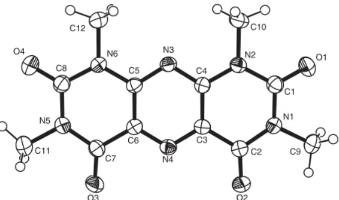
The packing of the molecule in the unit cell is com-plemented by the strong inter-molecular hydrogen bonds
C(10)H(10C)···N(4) and C(12)H(12C)···O(3) (see Figure
2), in addition to four intramolecular hydrogen bonds (see Table 3).
Conclusions
The oxidative deamination of H2ddd can be achieved
by its reaction with perrhenate in methanol, to yield 1,3,6,8-tetramethylpyrimidopteridine-2,4,5,7-tetrone (tppt) as product. This compound is probably formed by conden-sation of the oxidation product alloxan with unoxidized
H2ddd. The crystal structure of tppt consists of a central
pyrazine ring and two condensed terminal pyrimidine rings
incis positions.
Acknowledgments
I.B. is grateful to the NMMU and NRF for financial support.
1428 cm-1, as was suggested in the literature.14Two singlet
peaks of equal intensity at 2.89 and 2.73 ppm in the 1H
NMR spectrum are ascribed to the C(9)H3/C(11)H3 and
C(10)H3/C(12)H3 hydrogens respectively. The compound
is soluble in a wide variety of solvents, including water, chloroform, acetonitrile, dimethylsulfoxide, dimethylfor-mamide and dichloromethane.
The X-ray crystal structure of tppt is shown in Figure 1, together with the atom labelling scheme. The structure consists of tricyclic rings, comprising a central pyrazine ring and two terminal pyrimidine rings. The pyrimidine and pyrazine rings are essentially planar, with maximum deviations from the calculated mean plane of 0.008 Å and 0.004 Å respectively. In the pyrimidine rings there are some steric interactions involving the exocyclic groups, as shown by the short C(9)···O(1), O(1)···C(10), O(4)···C(11) and O(4)···C(12) contacts, which are 0.6 Å less than the sum of the van der Waals radii. Because of these steric interactions, the exocyclic groups deviate considerably from the pyri-midine mean plane. Deviations from the plane range from -0.166(1) Å for C(9) to 0.096(1) Å for O(2). The dihedral angles between the pyrazine and pyrimidine rings are 1.08° and 1.20°, indicating that the whole molecule is almost planar. The shortest intermolecular contact distance of 2.788(3) Å [O(2)···C(1)(x, ½ - y, ½ + z)] indicates that only van der Waals forces are present between molecules.
All C-N bonds (Table 2) in the two pyrimidine rings are single, varying in the range 1.365(3) Å [N(2)-C(4)] to 1.397(3) Å [N(1)-C(1)]. In the pyrazine, there is electron delocalization over the C-N-C parts of the ring, with the C-N bond lengths varying in the narrow range 1.325(3) to 1.336(3) Å.
The bond angles around the ketonic carbons, e.g. O(1)-C(1)-N(1)=121.1(2)°, O(2)-C(2)-N(1)=121.7(2)°, O(3)-C(7)-N(5)=120.9(2)° and O(4)-C(8)-N(6)=121.3(2)°,
are indicative of the sp2 hybridization of these carbon
Figure 1. ORTEP view of tppt, showing the atom labelling and 40%
probability displacement ellipsoids.
Figure 2. ORTEP drawing of the intermolecular hydrogen bonds in tppt.
Table 3. Hydrogen-bonding geometry for tppt (Å, deg)
D H A DH H···A D···A DH···A C9
C10 C10 C11 C12 C12
H9A H10B H10C H11B H12B H12C
O2 O1 N4 O4 O4 O3
0.98 0.98 0.98 0.98 0.98 0.98
2.30 2.33 2.56 2.26 2.27 2.46
2.742(3) 2.719(3) 3.386(3) 2.713(3) 2.714(3) 3.252(3)
Oxidation of 5,6-Diamino-1,3-dimethyl-2,4-dioxopyrimidine by Perrhenate J. Braz. Chem. Soc.
202
Supplementary Information
Crystallographic data have been deposited with the Cambridge Crystallographic Data Centre as supplementary material (deposition number CCDC 643784). Copies of the data can be obtained, free of charge, via www.ccdc. cam.ac.uk/conts/retrieving.html (or from the Cambridge Crystallographic Data Centre, CCDC, 12 Union Road, Cambridge CB2 1EZ, UK; fax: +44 1223 336033; or e-mail: [email protected]).
References
1. Sorenson, J. R. J. In Metal Ions in Biological Systems; Marcel
Dekker: New York, 1976, ch. 14.
2. Hibino, S.; Cancer Chemother. Rep.1961,13, 141.
3. Inoue, Y.; Tsobe, M.; Shiohara, T.; Hayashi, H.; Int. Arch. Al-lergy Immunol.2003,131, 143.
4. Kivekas, R.; Colacio, E.; Ruiz, J.; Lopez-Gonzalez, J. D.; Leon, P.; Inorg. Chim. Acta1989,159, 103.
5. Romero, M. A.; Moreno, M. N.; Ruiz, J.; Sanchez, M. P.; Nieto, F.; Inorg. Chem.1986,25, 1498.
6. Arriotua, M. I.; Pitarra, J. L.; Ruiz, J.; Moreno, J. M.; Colacio, E.;Inorg. Chim. Acta1995,231, 103.
7. Booysen, I.; Gerber, T. I. A.; Hosten, E.; Mayer, P.; J. Coord. Chem.2007,60, 1755.
8. Sheldrick, G. M.; Acta Crystallogr.1990,A46, 467.
9. Sheldrick, G. M.; SHELXL-97; Program for Crystal Structure Refinement,University of Göttingen: Germany, 1997.
10. Danopoulos, A. A.; Wong, A. C. C.; Wilkinson, G.; Hursthouse, M. B.; Hussain, B.; J. Chem. Soc., Dalton Trans.1990, 315. 11. Mederos, A.; Domínguez, S.; Hernandez-Molina, R.; Sanchiz,
J.; Brito, F.; Coord. Chem. Rev.1999,193, 913.
12. Okamoto, Y.; Ogura, K.; Kurasawa, Y.; Kinoshita, T.; Hete-rocycles1984,22, 1231.
13. Romeresa, A.; Colacio, E.; Suarez-Varela, J.; Avila-Roson, J. C.; Hidalgo, M. A.; Romero-Garzon, J.; Acta Crystallogr. 1995,
C51, 1005.
14. Al-Arab, M. M.; Hamilton, G. A.; J. Am. Chem. Soc.1986,108,
5972.
Received: April 18, 2007