ABSTRACT
Masculinisation of internal and external genitalia during foetal development depends on the existence of two discrete testicular hormones: Leydig cell-secreted testosterone drives the differentiation of the Wolffian ducts, the uro-genital sinus and the external uro-genitalia, whereas Sertoli cell-produced anti-Müllerian hormone (AMH) provokes the regression of anti-Müllerian ducts. The absence of AMH action in early foetal life results in the formation of the Fal-lopian tubes, the uterus and the upper third of the vagina. In 46,XY foetuses, lack of AMH may result from testicular dysgenesis affecting both Leydig and Sertoli cell populations: in this case persistence of Müllerian remnants is asso-ciated with ambiguous or female external genitalia. Alternatively, defective AMH action may result from mutations of the genes encoding for AMH or its receptor: in this condition known as Persistent Müllerian Duct Syndrome, testosterone production is normal and external genitalia are normally vir-ilised. Finally, AMH may be normally secreted in intersex patients with defects restricted to androgen synthesis or action, resulting in patients with female or ambiguous external genitalia with no Müllerian derivatives. (Arq Bras Endocrinol Metab 2005;49/1:26-36)
Keywords:Androgens; Cryptorchidism; Germ cells; Intersex states; Persistent Müllerian Duct Syndrome; Sertoli cells
RESUMO
Hormônio Anti-Mülleriano nos Distúrbios da Determinação e Diferenciação do Sexo.
A masculinização dos genitais internos e externos durante o desenvolvi-mento fetal depende da existência de dois hormônios testiculares distintos: a testosterona secretada pelas células de Leydig conduz à diferenciação dos dutos de Wolff, do seio urogenital e dos genitais externos, enquanto que o hormônio anti-Mülleriano (HAM) produzido pelas células de Sertoli provo-ca a regressão dos dutos de Müller. A falta de ação do HAM no início da vida fetal resulta na formação de tubas uterinas, útero e terço superior da vagina. Em fetos 46,XY, a falta do HAM pode resultar de disgenesia testicu-lar, afetando tanto as células de Leydig quanto as de Sertoli: nesse caso, a presença de remanescentes Müllerianos está associada a genitais externos femininos ou ambíguos. Por outro lado, distúrbios na ação do HAM podem resultar de mutações em genes que codificam o HAM ou seu receptor: nessa afecção, conhecida como síndrome da Persistência dos Dutos de Müller, a produção de testosterona é normal e os genitais externos são vir-ilizados normalmente. Finalmente, o HAM costuma ser secretado normal-mente em pacientes com intersexo decorrente de defeitos restritos à síntese ou à ação de andrógenos, resultando em indivíduos com genitais externos femininos ou ambíguos com ausência de derivados de Müller. (Arq Bras Endocrinol Metab 2005;49/1:26-36)
Descritores: Andrógenos; Criptorquidismo; Células germinativas; Estados intersexuais; Síndrome da persistência dos dutos de Müller; Células de Ser-toli
Rodolfo Rey
Centro de Investigaciones Endocrinológicas (CONICET), División de Endocrinología, Hospital de Niños Ricardo Gutiérrez, and Departamento de Histología, Embriología, Biología Celular y Genética, Facultad de Medicina, Universidad de Buenos Aires, Argentina.
T
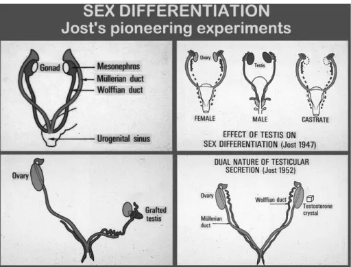
HE EXISTENCE OF ANTI-MÜLLERIANhormone wasfirst suggested by the pioneering experiments per-formed by the French scientist Alfred Jost in the 1940’s. In early stages of development in mammals, foetuses of both sexes have two pairs of unipotential internal genital ducts: the mesonephric (Wolffian) ducts and the paramesonephric (Müllerian) ducts, and bipotential primordial of external genitalia. Testicular differentiation from the gonadal ridge, occurring in humans during the 7th week of foetal life, drives the
fate of internal and external genitalia. First Jost grafted testicular tissue to castrated foetuses and observed that Wolffian ducts gave rise to epididymes, vasa deferentia and seminal vesicles while Müllerian ducts regressed. In subsequent experiments, he noticed that a crystal of testosterone propionate was capable of masculinising Wolffian ducts in the castrated foetus but did not affect Müllerian ducts, which formed the Fallopian tubes, the uterus and the upper third of the vagina. This proved that a testicular product different from testosterone, that he named “hormone inhibitrice” or “Müllerian inhibitor”, was responsible for the regression of Mül-lerian ducts in the male foetus (1) (figure 1).
However, the identification of the “Müllerian inhibitor” did not prove easy. A test for the detection of testicular anti-Müllerian activity was developed in
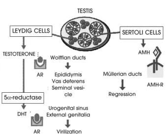
1969 (2) but it was only until 1978 that the first evi-dence was produced indicating the glycoprotein dimeric nature of this testicular factor (3). Finally, AMH could be purified to homogeneity in 1984 (4), and in 1986 the human and bovine genes were isolat-ed and sequencisolat-ed (5,6). Along its history, the Müller-ian inhibitor received several names, of which anti-Müllerian hormone (AMH) and anti-Müllerian inhibiting substance (MIS) are the two most widely used nowa-days. In the last 10 years, the use of molecular tech-niques has allowed the identification of AMH recep-tors and its signalling pathways, as well as the under-standing of the mechanisms involved in the regulation of AMH expression, which will be discussed hereafter. All this knowledge has helped understand the respon-sibility of AMH in disorders of sex differentiation. As already shown by Jost’s pioneering experiments almost 60 years ago, the testis has a determinant importance in foetal sex differentiation, via two independent path-ways. Leydig cells secrete androgens, necessary for the masculinization of Wolffian ducts, the urogenital sinus and external genitalia, whereas Sertoli cells secrete AMH, required for the regression of Müllerian ducts (figure 2). With this simple scheme in mind, the com-prehension of intersex states can be more easily achieved.
AMH: The Protein And The Gene
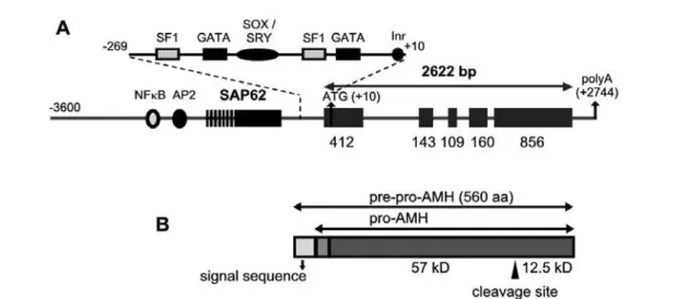
AMH is a 140kD homodimeric glycoprotein (3). Human AMH is synthesised as a 560 amino acid pre-cursor with a 24-25 amino acid leader containing a 16-18 amino acid signal sequence and a putative 7-8 residue pro-sequence (5) (figure 3). The carboxyl-ter-minal region of AMH shares homology with that of members of the transforming growth factor-β (TGF-β
superfamily). Most members of this family require pro-teolytic cleavage at a site 110 amino acids from the car-boxyl terminus to be active. While the full-length AMH molecule is active in organ culture, a cleavage site 109 amino acids from the C-terminus releases a more active fragment (7). However, the cleaved N-terminal domain interacts and enhances the activity of the C-terminus (8). While AMH can be experimentally cleaved using plasmin (8), PC5 and furin, two members of the pro-protein convertase family, have been reported to be able to process AMH in embryonic rat testes (9).
AMH is encoded by a 2.75kb gene divided into 5 exons (figure 3), characterised by a high GC content. The human AMH gene maps on chromosome 19 p13.3 (10). While the mouse and rat promoters con-tain an almost perfect TATA box, the human AMH promoter lacks consensus TATA or CCAAT box ele-ments (5). AMH transcription of the human gene has been shown to contain a functional initiator (Inr) ele-ment that is specifically recognised by transcription
fac-tor TFII-I (11). Cloning of 3.6kb of the 5’-flanking sequences of the human AMH gene allowed to identi-fy a major transcription initiation site and three minor sites, a putative oestrogen response element at –1772 and an Sp1 binding site at –303 (12). The more recent finding of the existence of the SAP62/SF3A2 house-keeping gene, encoding for a spliceosome protein, at –789 of the human AMH ATG codon (and –434 in the mouse), has generated doubts about the real length of the AMH promoter (13) and abridged research studies to the proximal promoter sequences (less than 400bp), where binding elements for SOX/SRY pro-teins (14,15), SF-1 (16,17) and GATA factors (17,18) have been identified (figure 3). However, recent stud-ies have shown that more distant sequences are required for the normal chronology of expression of AMH in the testis (19,20).
The Ontogeny of AMH Expression
In the mammalian foetus
AMH is one of the earliest Sertoli cell-specific proteins expressed by the gonad (21). As soon as testicular cords begin to assemble in the foetal gonad, AMH expression is triggered. AMH is secreted by the human testis from the 8th week of amenorrhea and provokes
irreversible Müllerian duct regression, which is com-pleted by the end of week 9 (22). After that time, Mül-lerian ducts become insensitive to AMH, which high-lights the importance of a tightly regulated chronology of its secretion by the foetal testis. Although AMH is no longer active on Müllerian ducts, its expression by Sertoli cells remains at high levels until puberty, which indicates that the end of the critical window of Müller-ian duct regression is dependent on the expression pat-tern of AMH type II receptor (reviewed in ref. 23).
In the postnatal testis
Except for a transient decline in the peri-natal period, testicular AMH secretion is maintained at high levels until puberty, when Sertoli cell maturation is charac-terised by a decreasing AMH activity (24,25). There-fore, determination of serum AMH levels is useful to assess Sertoli cell maturation (26-28). During pubertal development, AMH expression faints in coincidence with the increase of androgen production by Leydig cells and the onset of germ cell meiosis (24,25,29,30).
In the ovary
Ovarian granulosa cells, the homologous to testicular Sertoli cells, also produce AMH but with several dif-ferences: AMH expression only begins at the peri-natal
Figure 2.The hormonal control of male foetal sex differen-tiation. Leydig cells secrete testosterone, which drives Wolf-fian duct differentiation into the epididymes, vasa deferen-tia and seminal vesicles acting through the androgen receptor. In the anlagen of the external genitalia, testos-terone is transformed by 5α-reductase into dihydrotestos-terone (DHT), a more potent androgen that binds the androgen receptor to induce external virilisation. Sertoli cells secrete anti-Müllerian hormone (AMH), which binds to a membrane receptor in Müllerian ducts and provokes their regression.
TESTIS
TESTOTERONE
Wolffian ducts Epididymis Vas deferens Seminal
vesi-cle Urogenital sinus External genitalia
Virilization
Regression
AMH-R AMH AR
AR DHT
Müllerian ducts
LEYDIG CELLS
5α-reductase
period (25,31), remains low throughout reproductive life and becomes undetectable after menopause (32). Gonadal AMH secretion shows a clear-cut sexual dimorphism in prepubertal ages, when serum AMH is significantly lower in females; in adults, serum AMH is similarly low in both sexes (26,32).
AMH Action on Target Organs: Receptors and Signalling Pathways
The AMH signalling pathway began to be unravelled in 1994, when the specific AMH receptor type II was cloned (33,34). This specific AMH receptor is present on the cell membrane of target organs and is responsi-ble for ligand binding. It subsequently recruits a non-specific type I receptor in order to transduce its signal. Three different type I receptors are considered to mediate AMH response in target cells: ALK6, named BMPRI-B (35), ALK2, named ActRI (36,37), and ALK3, also known as BMPRI-A (38). The intracellu-lar transduction pathways involved after type I recep-tor recruitment by the specific type II AMH receprecep-tor seem to vary according to the target cell (reviewed in ref. 39). AMH receptor type II is a single transmem-brane protein with serine/threonine kinase activity, that is encoded by a 8kb gene composed of 11 exons and mapping to chromosome 12q13 (40).
AMH receptor type II has been identified in the mesenchymal cells surrounding the epithelium of Mül-lerian ducts in both sexes. Its expression follows a cra-nial-to-caudal chronology that explains why the cranial-most end of the ducts, that are near the testes, regress first and the more distant portions of the ducts regress later (41). After AMH binding to its receptor in mes-enchymal cells, paracrine-mediated mechanisms trigger
epithelial cell apoptosis and epithelial-mesenchymal transformation finally resulting in complete Müllerian duct regression in the male. The absence of AMH (e.g. in the normal female foetus) or of its signalling mecha-nisms (e.g. mutations of the AMH receptor) results in the stabilization of Müllerian ducts. AMH receptors are also present in granulosa cells of the ovary and in Sertoli and Leydig cells of the testes. In different experimental conditions, AMH has been shown to inhibit Leydig cell differentiation and steroidogenesis as well as granulosa cell response to LH and FSH, but whether it plays any essential role in gonadal physiology still needs to be determined (42,43).
The Regulation of AMH Expression
Owing to its sex-specific and time-restricted require-ment during foetal developrequire-ment, AMH expression needs to be tightly regulated (reviewed in ref. 44). Uncontroversial proof exists indicating that SOX9 binding to the proximal AMH promoter is essential for the initiation of AMH expression in early foetal development (45). SF-1 (16), GATA-4 (18) and WT-1 (46) enhance, while DAX-WT-1 impairs (46), AMH transcription.
As already mentioned, testicular AMH produc-tion remains high during foetal life and childhood and is downregulated at puberty. The decline of AMH pro-duction by Sertoli cells is related to the stage of puber-tal development: the most significant decrease in serum AMH is observed between stages II and III of pubertal development (29), in coincidence with the increase of intratesticular testosterone concentration (figure 4): the decline in AMH production is a marker of the elevation of intratesticular androgen concentration, which
inhibits AMH production at puberty. Independently from androgen action, meiotic germ cells seem to play a role in downregulation of AMH expression at puber-ty (30,47). Interestingly, AMH is not down-regulated by testosterone during foetal life and in the first months after birth owing to the lack of expression of the andro-gen receptor in Sertoli cells (30). The physiological androgen insensitivity of foetal and neonatal Sertoli cells explains, thus, the transient coexistence of high concen-trations of androgens and AMH (figure 4). In the absence of the negative effect of androgens and meiotic germ cells (e.g. in foetal and neonatal periods), FSH upregulates testicular AMH production. On one hand, FSH induces Sertoli cell proliferation and, on the other, it enhances AMH transcription in each Sertoli cell through a cAMP-PKA mediated signalling pathway involving transcription factors AP2 and NFB, which bind to specific response elements in distant sequences of the AMH promoter (20).
Abnormal AMH Secretion or Action
When AMH is not produced between the 8thand the
9thweeks of foetal life, or if the AMH receptor
path-way is not capable of transducing AMH signalling, Müllerian ducts differentiate to form the Fallopian tubes, the uterus and the upper vagina. Absence of AMH action is the normal situation in the XX foetus: although the AMH receptor and its signalling path-ways are present in Müllerian ducts, the foetal ovaries do not express AMH. In the XY foetus, the absence of AMH action results in an abnormal persistence of Müllerian derivatives. This can be an isolated situa-tion, known as the persistent Müllerian duct syn-drome, or a disorder in which the existence of female internal genitalia is associated with defective virilisa-tion of the Wolffian ducts, urogenital sinus and external genitalia.
The Persistent Müllerian Duct Syndrome (PMDS)
The uncontroversial and unique role of AMH in foetal sex differentiation can be deduced from the phenotype of patients with AMH or AMH receptor mutations (48) or of transgenic mice with manipulations of AMH expression or signalling (49-51). It seems clear that the absence of AMH activity in early foetal life results in one conspicuous phenotype, the persistence of Müllerian ducts. No other growth factor or hor-mone is capable of replacing AMH in this function.
For PMDS to be considered as a possible diag-nosis in an XY patient, a sine qua noncondition is that the external genitalia, urogenital sinus and Wolffian ducts have normally differentiated, accounting for nor-mal androgen activity. Therefore, PMDS patients are genotypic and externally phenotypic males with cryp-torchidism, sometimes associated with inguinal hernia. The presence of Müllerian derivatives is usually not sus-pected and discovered at surgery. Two anatomical
forms of PMDS have been described. The most com-mon one is known as transverse testicular ectopia: one testis descends into the scrotum dragging the ipsilateral Fallopian tube into the inguinal canal (a condition known as hernia uteri inguinalis) and pulling the uterus and the contralateral Fallopian tube together with the testis, which becomes located in the abdomen. More rarely, PMDS presents as bilateral cryptorchidism, the uterus is fixed in the pelvis and both testes are embed-ded in the broad ligament in ovarian position. These clinical variants are not genetically determined and may occur within the same sibship (52). In PMDS, the testes are abnormally mobile because they are not anchored to the scrotum (53), which favours testicular torsion (54). Testes have a normal histological appearance. It is usu-ally difficult to bring the testes down to a normal posi-tion. The spermatic cord is usually very short because the vasa deferentia are embedded in the mesosalpynx,
lateral uterine wall and cervix. Lack of proper commu-nication between the testis and excretory ducts and dif-ficulties at orchidopexy probably explain why fertility is rare in PMDS patients (55).
Two biological variants of PMDS can be distin-guished: those with normal AMH production (AMH-positive) and those with impaired or null AMH secre-tion (AMH-negative). The AMH-negative variant is due to mutations in the AMH gene, resulting in com-plete lack of AMH production or secretion. These patients have very low or undetectable serum AMH concentrations because (56), in rare cases with AMH mutations, serum AMH concentration may be normal for age; these mutations impair bioactivity but not secretion (57). The AMH-positive variant is generally due to mutations in the gene coding for AMH recep-tor type II (AMHR-II); as expected, these patients have a normal AMH serum concentration for their age (58) (figure 5). Serum testosterone and response to hCG are normal in all cases. Female relatives of AMH-resistant patients who share their genetic background are phenotypically normal and fertile.
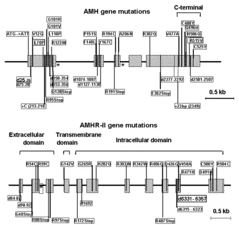
As could be expected the inheritance of PMDS is according to an autosomal recessive pattern: affect-ed patients are either homozygotes or compound he-terozygotes. AMH gene mutations have been report-ed in 47% of PMDS families: 35 different mutations, for the most part missense type, have been detected along the gene, with no hot spots (figure 6). AMHR-II mutations have been found in 38% of PMDS fami-lies: 25 different mutations of AMHR-II, mostly mis-sense, were spread out over the whole length of the gene (figure 6). Five mutations were recurrent, with a
deletion of 27bp in exon 10 present in 45% of families of this group (48). In 15% of the families studied to date, no mutation of either the AMH or the AMHR-II genes was detectable and no large DNA rearrange-ments were seen by Southern-blotting. Associated dis-eases, such as jejunal atresia, lipoatrophic diabetes and vitamin D resistant rickets, lymphangectasia and other malformations were present in approximately half the cases. Unexplained PMDS may reflect mutations in the proteolytic enzyme involved in AMH processing or in components downstream of the type II receptor in the AMH transduction cascade; alternatively, in view of the high incidence of associated defects, unex-plained PMDS may be part of complex malformative syndromes with no relationship to the AMH pathway.
Persistence of Müllerian derivatives associated with ambiguous external genitalia
Based on the endocrinology of foetal sex differentia-tion (figure 2), it can be easily deduced that a com-bined defect of androgen and AMH secretion should be suspected if Müllerian derivatives are associated in an XY patient with ambiguous or female external gen-italia. This phenotype excludes the diagnosis of PMDS and genetic analysis of the AMH or AMHR-II genes is unnecessary. Since both Leydig and Sertoli cells are affected, gonadal dysgenesis is the most probable aeti-ology. Depending on the degree of gonadal dysgene-sis, testosterone and AMH levels might be from low to undetectable (27). This usually correlates to the anatomic phenotype: low testicular hormones are observed in 46,XY patients with ambiguous external genitalia and rudimentary Wolffian and Müllerian
derivatives (a condition commonly referred to as dys-genetic male pseudohermaphroditism). Undetectable AMH and no response of testosterone to hCG is usu-ally observed in 46,XY females (pure gonadal dysgen-esis, also known as Swyer syndrome).
In 46,XX patients bearing a uterus and with very low or undetectable AMH levels, the most prob-able diagnosis is congenital adrenal hyperplasia (59). However, most often the diagnosis is made early in the neonate owing to adrenal insufficiency.
In patients with rudimentary Müllerian deriva-tives with XX karyotype or with mosaicisms, the degree of Müllerian structure development is indicative of the amount of AMH produced by the gonads. AMH acts as a local factor on the homolateral Müllerian duct. There-fore, an ovotestis with a scarce testicular component is usually associated with a homolateral hemi-uterus and Fallopian tube, but these structures are very hypoplastic or even absent if the ovotestis is composed of abundant testicular parenchyma. Similarly, in patients with asym-metric gonadal differentiation (mixed gonadal dysgene-sis), a Fallopian tube and hemi-uterus are present on the side of the streak gonad; contralaterally the degree of tes-ticular dysgenesis will determine the development of Müllerian derivatives.
Absence of Müllerian derivatives in XY patients with ambiguous external genitalia
Once again, based on the endocrinology of foetal sex differentiation (figure 2), the absence of Müllerian ducts in a 46,XY patient indicates that the gonads are not dysgenetic, since Sertoli cells have correctly differ-entiated and secreted sufficient amounts of AMH (27). In these cases, referred to as non-dysgenetic male pseudohermaphroditism, only the androgen pathway is affected, and the probable diagnoses are defects of the LH receptor, of steroidogenic proteins or of the androgens receptor or its coactivators. When the impairment of androgen secretion or action is severe, a female external phenotype results together with a smaller vagina and the absence of uterus and tubes. These patients may only present at puberty owing to primary amenorrhea.
Clinical Utility of AMH Measurement in Inter-sex Patients
Except for the first 7-14 days of postnatal life, when endocrine testicular activity seems to be low, serum AMH levels reflect the amount of functional Sertoli cells (i.e. testicular parenchyma) in prepubertal boys, before they decline owing to the inhibitory effect
exerted by androgens and meiotic germ cells (24,28). Therefore, in most cases serum AMH can be used to estimate what might have happened in foetal life (one should be aware that this is an extrapolation which could overlook, for instance, a late onset of AMH secretion after the 9thweek, in which case high AMH
would coexist with persistent Müllerian ducts). Normally high serum AMH indicates that Ser-toli cells are quantitatively and qualitatively normal: the predictive value of serum AMH for the existence of normal testicular tissue is higher than that of testos-terone response to hCG (60). In 46,XY patients with female or ambiguous external genitalia, the androgen pathway is clearly impaired. The possibilities are numerous and androgen response to hCG may not always elucidate the diagnosis. For example, a low response to hCG can be observed in gonadal dysgene-sis but also in patients with LH receptor or steroido-genic enzyme defects. Serum AMH determination can be very helpful, since levels are low in testicular dysge-nesis but normal to extremely high in patients with mutations in the LH receptor or steroidogenic proteins (27) (table 1). Another difficult differential diagnosis is that between androgen insensitivity owing to a muta-tion in the androgen receptor and a defect in DHT owing to a mutation in 5α-reductase, serum AMH may also be helpful here. AMH is downregulated by intrat-esticular testosterone in normal conditions. This inhi-bition is absent, resulting in high AMH, in patients
with androgen insensitivity (27) (table 1). Conversely, in patients with 5α-reductase deficiency, AMH down-regulation by testosterone occurs normally (61).
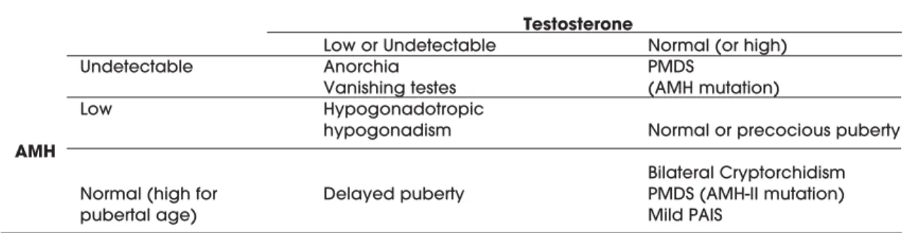
In 46,XY normally virilised boys with nonpal-pable gonads, normal serum AMH clearly indicates the existence of testes in ectopic position (62,63). Undetectable AMH would indicate anorchia (or van-ishing testes, if previously present), with the sole and rare exception of cryptorchidism owing to AMH-neg-ative PMDS. Normal AMH also suggests that FSH activity has been adequate during foetal and neonatal periods, whereas low AMH may suggest congenital hypogonadotropic hypogonadism (64) (table 2). Conversely, high AMH has been reported in a patient with Sertoli cell hyperplasia due to an activating muta-tion of the Gsαsubunit involved in the FSH receptor signalling pathway (65). Declining serum AMH is indicative of pubertal development of the testes. An early decrease of AMH is observed in patients with central precocious puberty or with testotoxicosis (table 2); serum AMH recovers normal values after treatment (29). Finally, persistently high levels of AMH in boys of pubertal age might indicate delayed puberty if androgens are low or mild androgen insen-sitivity if androgen levels have risen. Hypopgo-nadotropic hypogonadism seems to be characterised by low levels of both androgens and AMH in patients with micropenis or cryptorchidism but no ambiguous genitalia (table 2).
Table 1.Serum AMH and testosterone in 46,XY intersex patients.
Testosterone
Low or Undetectable Normal or High Low or Undetectable Gonadal dysgenesis 5α-reductase defect
AMH
Normal or High LH receptor defects Androgen insensitivity Steroidogenic protein defects
Table 2.Serum AMH and testosterone in 46,XY boys with normally virilised external genitalia.
Testosterone
Low or Undetectable Normal (or high)
Undetectable Anorchia PMDS
Vanishing testes (AMH mutation)
Low Hypogonadotropic
hypogonadism Normal or precocious puberty
AMH
Bilateral Cryptorchidism Normal (high for Delayed puberty PMDS (AMH-II mutation)
REFERENCES
1. Jost A. Problems of fetal endocrinology: the gonadal and hypophyseal hormones. Recent Prog Horm Res 1953;8:379-418.
2. Picon R. Action of the fetal testis on the development in vitro of the Müllerian ducts in the rat. Arch Anat Microsc Morphol Exp 1969;58:1-19.
3. Picard JY, Tran D, Josso N. Biosynthesis of labelled anti-Müllerian hormone by fetal testes: evidence for the gly-coprotein nature of the hormone and for its disulfide-bonded structure. Mol Cell Endocrinol 1978;12:17-30.
4. Picard JY, Josso N. Purification of testicular anti-Müller-ian hormone allowing direct visualization of the pure glycoprotein and determination of yield and purifica-tion factor. Mol Cell Endocrinol 1984;34:23-9.
5. Cate RL, Mattaliano RJ, Hession C, Tizard R, Farber NM, Cheung A, et al. Isolation of the bovine and human genes for Müllerian inhibiting substance and expression of the human gene in animal cells. Cell 1986;45:685-98.
6. Picard JY, Benarous R, Guerrier D, Josso N, Kahn A. Cloning and expression of cDNA for anti-Müllerian hor-mone. Proc Natl Acad Sci USA 1986;83:5464-8.
7. Pepinsky RB, Sinclair LK, Chow EP, Mattaliano RJ, Man-ganaro TF, Donahoe PK, et al. Proteolytic processing of Müllerian inhibiting substance produces a transforming growth factor-?-like fragment. J Biol Chem 1988;263:18961-5.
8. Wilson CA, di Clemente N, Ehrenfels C, Pepinsky RB, Josso N, Vigier B, et al. Müllerian inhibiting substance requires its N-terminal domain for maintenance of bio-logical activity, a novel finding within the transforming growth factor-beta superfamily. Mol Endocrinol 1993;7:247-57.
9. Nachtigal MW, Ingraham HA. Bioactivation of Mullerian inhibiting substance during gonadal development by a kex2/subtilisin-like endoprotease. Proc Natl Acad Sci USA 1996;93:7711-6.
10. Cohen-Haguenauer O, Picard JY, Mattei MG, Serero S, Nguyen VC, de Tand MF, et al. Mapping of the gene for anti-Müllerian hormone to the short arm of human chro-mosome 19. Cytogenet Cell Genet 1987;44:2-6.
11. Morikawa N, Clarke TR, Novina CD, Watanabe K, Haqq C, Weiss M, et al. Human Müllerian inhibiting substance promoter contains a functional TFII-I-binding initiator.
Biol Reprod 2000;63:1075-83.
12. Guerrier D, Boussin L, Mader S, Josso N, Kahn A, Picard JY. Expression of the gene for anti-Müllerian hormone. J Reprod Fertil 1990;88:695-706.
13. Dresser DW, Hacker A, Lovell-Badge R, Guerrier D. The genes for a spliceosome protein (SAP62) and the anti-Müllerian hormone (AMH) are contiguous. Hum Mol Genet 1995;4:1613-8.
14. Haqq CM, King CY, Ukiyama E, Falsafi S, Haqq TN, Dona-hoe PK, et al. Molecular basis of mammalian sexual determination: activation of Müllerian inhibiting sub-stance gene expression by SRY. Science 1994 ;266:1494-500.
15. De Santa Barbara P, Bonneaud N, Boizet B, Desclozeaux M, Moniot B, Sudbeck P, et al. Direct interaction of
SRY-related protein SOX9 and steroidogenic factor 1 regu-lates transcription of the human anti-Müllerian hormone gene. Mol Cell Biol 1998;18:6653-65.
16. Shen WH, Moore CC, Ikeda Y, Parker KL, Ingraham HA. Nuclear receptor steroidogenic factor 1 regulates the Müllerian inhibiting substance gene: a link to the sex determination cascade.Cell 1994;77:651-61.
17. Watanabe K, Clarke TR, Lane AH, Wang X, Donahoe PK. Endogenous expression of Müllerian inhibiting sub-stance in early postnatal rat Sertoli cells requires multi-ple steroidogenic factor-1 and GATA-4-binding sites.
Proc Natl Acad Sci USA 2000;97:1624-9.
18. Tremblay JJ, Viger RS. Transcription factor GATA-4 enhances Mullerian inhibiting substance gene tran-scription through a direct interaction with the nuclear receptor SF-1. Mol Endocrinol 1999;13:1388-401.
19. Beau C, Vivian N, Münsterberg A, Dresser DW, Lovell-Badge R, Guerrier D. In vivo analysis of the regulation of the anti-Müllerian hormone, as a marker of Sertoli cell differentiation during testicular development, reveals a multi-step process. Mol Reprod Dev 2001;59:256-64.
20. Lukas-Croisier C, Lasala C, Nicaud J, Bedecarras P, Kumar TR, Dutertre M, et al. Follicle-stimulating hormone increases testicular anti-Müllerian hormone (AMH) pro-duction through Sertoli cell proliferation and a nonclas-sical cyclic adenosine 5’-monophosphate-mediated activation of the AMH gene. Mol Endocrinol 2003;17:550-61.
21. Tran D, Meusy-Dessolle N, Josso N. Anti-Müllerian hor-mone is a functional marker of foetal Sertoli cells.
Nature 1977;269:411-2.
22. Taguchi O, Cunha GR, Lawrence WD, Robboy SJ. Timing and irreversibility of Müllerian duct inhibition in the embryonic reproductive tract of the human male. Dev Biol 1984;106:394-8.
23. Josso N, di Clemente N, Gouédard L. Anti-Müllerian hormone and its receptors. Mol Cell Endocrinol 2001;179:25-32.
24. Rey R, Al-Attar L, Louis F, Jaubert F, Barbet P, Nihoul-Fekete C, et al. Testicular dysgenesis does not affect expression of anti-müllerian hormone by Sertoli cells in premeiotic seminiferous tubules. Am J Pathol 1996;148:1689-98.
25. Rajpert-De Meyts E, Jørgensen N, Græm N, Müller J, Cate RL, Skakkebæk NE. Expression of anti-Müllerian hormone during normal and pathological gonadal development: association with differentiation of Sertoli and granulosa cells. J Clin Endocrinol Metab 1999;84:3836-44.
26. Lee MM, Donahoe PK, Hasegawa T, Silverman B, Crist GB, Best S, et al. Mullerian inhibiting substance in humans: normal levels from infancy to adulthood. J Clin Endocrinol Metab 1996;81:571-6.
27. Rey RA, Belville C, Nihoul-Fekete C, Michel-Calemard L, Forest MG, Lahlou N, et al. Evaluation of gonadal func-tion in 107 intersex patients by means of serum antimül-lerian hormone measurement. J Clin Endocrinol Metab 1999;84:627-31.
in serum from human foetuses and children: pattern and clinical interest. Mol Cell Endocrinol 2003 ;211:55-63.
29. Rey R, Lordereau-Richard I, Carel JC, Barbet P, Cate RL, Roger M, et al. Anti-müllerian hormone and testosterone serum levels are inversely related during normal and precocious pubertal development. J Clin Endocrinol Metab 1993;77:1220-6.
30. Al-Attar L, Noel K, Dutertre M, Belville C, Forest MG, Bur-goyne PS, et al. Hormonal and cellular regulation of Ser-toli cell anti-Müllerian hormone production in the post-natal mouse. J Clin Invest 1997;100:1335-43.
31. Bézard J, Vigier B, Tran D, Mauleon P, Josso N. Immuno-cytochemical study of anti-Müllerian hormone in sheep ovarian follicles during fetal and post-natal develop-ment. J Reprod Fertil 1987;80:509-16.
32. Rey RA, Lhomme C, Marcillac I, Lahlou N, Duvillard P, Josso N, et al. Anti-Müllerian hormone as a serum mark-er of granulosa-cell tumors of the ovary: comparative study with serum alpha-inhibin and estradiol. Am J Obstet Gynecol 1996;174:958-65.
33. di Clemente N, Wilson C, Faure E, Boussin L, Carmillo P, Tizard R, et al. Cloning, expression, and alternative splic-ing of the receptor for anti-Müllerian hormone. Mol Endocrinol 1994;8:1006-20.
34. Baarends WM, van Helmond MJ, Post M, van der Schoot PJ, Hoogerbrugge JW, de Winter JP, et al. A novel mem-ber of the transmembrane serine/threonine kinase receptor family is specifically expressed in the gonads and in mesenchymal cells adjacent to the müllerian duct. Development 1994;120:189-97.
35. Gouédard L, Chen YG, Thevenet L, Racine C, Borie S, Lamarre I, et al. Engagement of bone morphogenetic protein type IB receptor and Smad1 signaling by anti-Müllerian hormone and its type II receptor. J Biol Chem 2000;275:27973-8.
36. Visser JA, Olaso R, Verhoef P, Kramer P, Themmen AP, Ingraham HA. The serine/threonine transmembrane receptor ALK2 mediates Müllerian inhibiting substance signaling. Mol Endocrinol 2001;15:936-45.
37. Clarke TR, Hoshiya Y, Yi SE, Liu X, Lyons KM, Donahoe PK. Müllerian Inhibiting Substance signaling uses a Bone Morphogenetic Protein (BMP)-like pathway mediated by ALK2 and induces Smad6 expression. Mol Endocrinol 2001;15:946-59.
38. Jamin SP, Arango NA, Mishina Y, Hanks MC, Behringer RR. Requirement of Bmpr1a for Müllerian duct regres-sion during male sexual development. Nat Genet 2002;32:408-10.
39. Josso N, di Clemente N. Transduction pathway of anti-Müllerian hormone, a sex-specific member of the TGF-beta family. Trends Endocrinol Metab 2003;14:91-7.
40. Imbeaud S, Faure E, Lamarre I, Mattei MG, di Clemente N, Tizard R, et al. Insensitivity to anti-mullerian hormone due to a mutation in the human anti-mullerian hormone receptor. Nat Genet 1995;11:382-8.
41. Xavier F, Allard S. Anti-Mullerian hormone, [beta]-catenin and Mullerian duct regression. Mol Cell Endocrinol 2003;211:115-21.
42. Visser JA. AMH signaling: from receptor to target gene.
Mol Cell Endocrinol 2003;211:65-73.
43. di Clemente N, Josso N, Gouedard L, Belville C. Com-ponents of the anti-Mullerian hormone signaling path-way in gonads. Mol Cell Endocrinol 2003;211:9-14.
44. Rey R, Lukas-Croisier C, Lasala C, Bedecarrás P. AMH/MIS: what we know already about the gene, the protein and its regulation. Mol Cell Endocrinol 2003;211:21-31.
45. Arango NA, Lovell-Badge R, Behringer RR. Targeted mutagenesis of the endogenous mouse Mis gene pro-moter: in vivo definition of genetic pathways of verte-brate sexual development. Cell 1999;99:409-19.
46. Nachtigal MW, Hirokawa Y, Enyeart-VanHouten DL, Flanagan JN, Hammer GD, Ingraham HA. Wilms’ tumor 1 and Dax-1 modulate the orphan nuclear receptor SF-1 in sex-specific gene expression. Cell 1998;93:445-54.
47. Hong CY, Park JH, Seo KH, Kim JM, Im SY, Lee JW, et al. Expression of MIS in the Testis Is Downregulated by Tumor Necrosis Factor Alpha through the Negative Reg-ulation of SF-1 Transactivation by NF-{kappa}B. Mol Cell Biol 2003;23:6000-12.
48. Belville C, Josso N, Picard JY. Persistence of Müllerian derivatives in males. Am J Med Genet 1999;89:218-23.
49. Behringer RR, Cate RL, Froelick GJ, Palmiter RD, Brinster RL. Abnormal sexual development in transgenic mice chronically expressing müllerian inhibiting substance.
Nature 1990;345:167-70.
50. Behringer RR, Finegold MJ, Cate RL. Müllerian-inhibiting substance function during mammalian sexual develop-ment. Cell 1994;79:415-25.
51. Mishina Y, Rey R, Finegold MJ, Matzuk MM, Josso N, Cate RL, et al. Genetic analysis of the Müllerian-inhibit-ing substance signal transduction pathway in mam-malian sexual differentiation. Genes Dev 1996 ;10:2577-87.
52. Guerrier D, Tran D, Vanderwinden JM, Hideux S, Van Outryve L, Legeai L, et al. The persistent Müllerian duct syndrome: a molecular approach. J Clin Endocrinol Metab 1989;68:46-52.
53. Hutson JM, Hasthorpe S, Heyns CF. Anatomical and functional aspects of testicular descent and cryp-torchidism. Endocr Rev 1997;18:259-80.
54. Imbeaud S, Rey R, Berta P, Chaussain JL, Wit JM, Lustig RH, et al. Testicular degeneration in three patients with the persistent müllerian duct syndrome. Eur J Pediatr 1995;154:187-90.
55. Farag TI. Familial persistent müllerian duct syndrome in Kuwait and neighboring populations. Am J Med Genet 1993;47:432-4.
56. mbeaud S, Carré-Eusèbe D, Rey R, Belville C, Josso N, Picard JY. Molecular genetics of the persistent müllerian duct syndrome: a study of 19 families. Hum Mol Genet 1994;3:125-31.
58. Imbeaud S, Belville C, Messika-Zeitoun L, Rey R, di Clemente N, Josso N, et al. A 27 base-pair deletion of the anti-müllerian type II receptor gene is the most com-mon cause of the persistent müllerian duct syndrome.
Hum Mol Genet 1996;5:1269-77.
59. Misra M, MacLaughlin DT, Donahoe PK, Lee MM. The role of Müllerian inhibiting substance in the evaluation of phenotypic female patients with mild degrees of vir-ilization. J Clin Endocrinol Metab 2003;88:787-92.
60. Misra M, MacLaughlin DT, Donahoe PK, Lee MM. Mea-surement of Müllerian inhibiting substance facilitates management of boys with microphallus and cryp-torchidism. J Clin Endocrinol Metab 2002;87:3598-602.
61. Stuchi-Perez EG, Lukas-Croisier C, De Castro M, Baptista MT, Ribeiro Scolfaro M, Marques-De-Faria AP, et al. Eval-uation of the tubular and interstitial functions of the testis in 46,XY patients with ambiguous genitalia. J Pediatr Endocrinol Metab 2000;13:605-12.
62. Josso N. Paediatric applications of anti-Müllerian hor-mone research. 1992 Andrea Prader Lecture. Horm Res 1995;43:243-8.
63. Lee MM, Donahoe PK, Silverman BL, Hasegawa T, Hasegawa Y, Gustafson ML, et al. Measurements of serum Müllerian inhibiting substance in the evaluation of children with nonpalpable gonads. N Engl J Med 1997;336:1480-6.
64. Young J, Rey R, Couzinet B, Chanson P, Josso N, Schai-son G. Antimüllerian hormone in patients with hypogo-nadotropic hypogonadism. J Clin Endocrinol Metab 1999;84:2696-9.
65. Coutant R, Lumbroso S, Rey R, Lahlou N, Venara M, Rouleau S, et al. Macroorchidism due to autonomous hyperfunction of Sertoli cells and Gsa gene mutation: an unusual expression of McCune-Albright syndrome in a prepubertal boy. J Clin Endocrinol Metab 2001 ;86:1778-81.
Endereço para correspondência:
Rodolfo Rey