Structure Refinement of (Sr,Ba)Nb
2O
6Ceramic Powder
from Neutron and X-Rays Diffraction Data
J.G. Carrioa, Y.P. Mascarenhasb, W. Yelonc#, I.A. Santosd,
D. Garciad*, J.A. Eirasd
aUniversidade Estadual Norte Fluminense, Campos dos Goytacazes,
28015-620 Campos dos Goytacazes - RJ, Brasil
bInstituto de Física de São Carlos, Universidade de São Paulo,
13560-250 São Carlos - SP, Brazil
cMissouri University Research Reactor, University of Missouri-Columbia,
Research Park, 65211-3400 Columbia - MO, USA
dDepartamento de Física,Universidade Federal de São Carlos,
13565-905 São Carlos - SP, Brazil
Received: January 31, 2001; September 25, 2001
The structure of polycrystalline strontium barium niobate at room temperature was refined by the Rietveld method. Sintered ceramic samples were used to collect powder neutron and X-ray diffraction data. The ratio Sr/Ba ≈ 64/36 was found from the initial batch composition
Sr0.61Ba0.39Nb2O6, corroborating with the quantitative X-ray dispersive spectroscopy (EDS)
meas-urements. The structure is tetragonal with cell parameters a, b = 12.4504(3) Å and c = 3.9325(1) Å and space group P4bm. It was not necessary to introduce any positional disorder for the oxygen atoms. Cation Nb+5 displacements not parallel to the c direction are presented, which can influence the behavior of the ferroelectric properties.
Keywords:strontium barium niobate, tungsten bronze structure, X-ray diffraction, neu-tron diffraction
1. Introduction
Ferroelectric compounds with tungsten bronze struc-ture have the general chemical formula (A1,A2)(B1,B2)O6,
where the A sites are usually filled by divalent metals and the B sites, by Nb+5 or Ta+5 atoms. This structure consists of an arrangement of corner sharing BO6 octahedrons
form-ing three interstitial sites. The primitive unit cell contains five molecules and six sites that can be occupied by the A atoms1. SrxBa1-xNb2O6 (SBN) ceramics for 0.25 ≤ x ≤ 0.75
form solid solutions that crystallize with the tungsten bronze structure2. These materials can show high pyroelec-tric coefficients, strong electro-optic effects and photore-fractive behavior2,3.
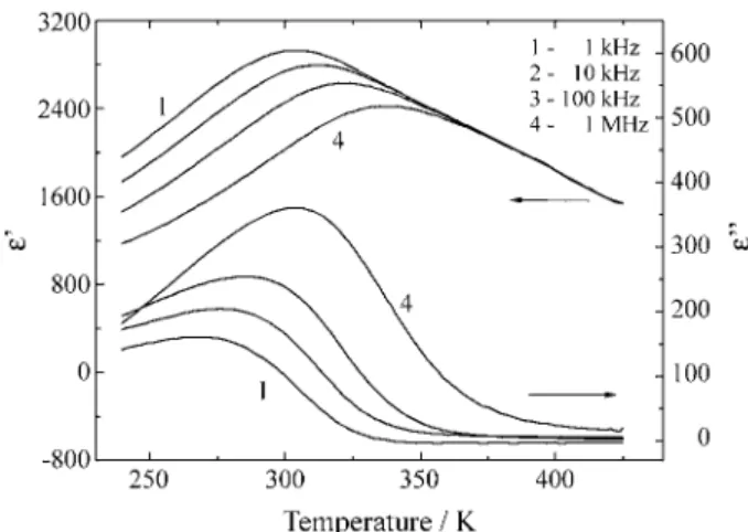
Previous works4,5 have shown that the basic features of SBN dielectric behavior are: (a) a strong dielectric disper-sion in the radio frequency region around and below the temperature of maximum dielectric constant (Tm), which
does not obey the Debye theory; (b) a broadening of the peak of the dielectric constant as a function of temperature; (c) a Tm shift towards high temperatures with a frequency
increase; and (d) strong frequency dependence of the di-electric absorption curves, i.e., the dielectric absorption increases with increasing frequency. These characteristics are typical of SBN materials with high concentrations of strontium (Sr/Ba > 1), as it can be seen in the Fig. 1. However, reported SBN dielectric parameters, such as Tm,
show large differences from one study to another, espe-cially between ceramic and single crystals samples of the same composition6-8.
Generally, the properties of this kind of displacive ferroelectric material can be correlated to the distortion and the relative orientation of the oxygen octahedron and to the shift of some of the atoms from their high temperature symmetry positions6. Moreover, they are very sensitive to the stoichiometric ratios. Because of this, a precise
struc-*e-mail: [email protected]
#
ture determination of these materials is required to under-stand some of their fundamental properties.
Jamiesonet al.1 first determined the crystalline struc-ture of Sr0.76Ba0.24Nb2O5.78. Using X-ray single crystal
data, these authors have shown the relationship between structure and some ferroelectric properties. Later the struc-tures of Sr0.72Ba0.28Nb2O6, Sr0.6Ba0.4Nb2O6 a nd
Sr0.5Ba0.5Nb2O6 were refined by Trubelja et al.7 and of
Sr0.61Ba0.39Nb2O6doped with Ce by Chernaya et al.8, using
X-ray data from powdered and sphere shaped single crys-tals, respectively, assuming the same crystal structure. Al-though many of the SBN ferroelectric and dielectric properties have been analyzed in ceramic samples, there is no report on structural studies of its polycrystalline form.
In this work the structure of SBN ceramic was deter-mined from sintered polycrystalline samples, combining X-ray and neutron diffraction powder data, allowing a good refinement of the positional and vibrational parameters of the heavy metals and oxygen atoms.
2. Experimental
2.1. Sample preparation
The SBN ceramics with nominal composition Sr0.61Ba0.39Nb2O6 were prepared by the conventional
mixed oxides method as described elsewhere9. The precur-sors (Ba(NO3)2, Nb2O5 and SrCO3) were ball mill/mixer
using ZrO2 balls and isopropyl alcohol. The mixture was
calcined in air at 1200 °C for 3 h and ball milled again for 10 h. Discs with 15 mm in diameter were prepared by uniaxial and isostactic cold pressing and fired for 3 h at 1350 °C and then crushed for the measurements. Scanning electronic microscopy (SEM) on the ceramic bodies, using a Philips FEG XL microscope equipped with quantitative X-rays dispersive spectroscopy (EDS), showed a Sr/Ba ratio = 1.71-1.78 which deviates from the batch ratio (1.56). It was found that Nb2O5- rich and Ba-deficient liquid
phases could be formed at the initial sintering stage of SBN ceramics10. These authors reported that, during a conven-tional sintering procedure, the grains rapidly absorb the phase, contributing to the abnormal grain growth behavior of the SBN-like materials. In our case, it can be believed that the diffusion of such phase into the grains generated a continuous distribution of Ba/Sr ratios from the boundary (where the composition would be more Ba-deficient) to the grain core (where the Ba/Sr would reach the batch ratio). This fact could explain the EDS data, which can be inter-preted as a result of an average of this grain Ba/Sr ratio distribution.
2.2. Conventional X-ray powder diffraction
X-ray powder diffraction data were collected with CuKα radiation on a rotating anode Rigaku Denki powder automatic diffractometer. The data were collected in the 2θ = 20°-60° and 60°-120° ranges with counting times of 5 and 10 s, respectively, in order to increase the collected intensities, which are much smaller in the second range. For both ranges, the step was 0.02°.
2.3. Neutron powder diffraction
The data were collected at the position sensitive detec-tor diffractometer of the Missouri University of Research Reactor (MURR), USA. About 1 g of sample was contained in a 3 mm diameter vanadium thin walled sample holder. The wavelength used was 1.4875 Å and the data was acquired in five 2θ spans, from 5° to 105° range, in approxi-mately 36 h of measurement.
3. Results and Discussions
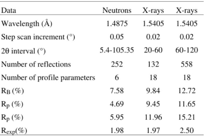
The structure of the polycrystalline SBN was refined by the Rietveld Method using the programs GSAS11 and DBWS12. With GSAS the refinement was performed com-bining the X-ray and the neutron powder diffraction data. The structural parameters related to the heavy atoms could be well refined with preliminary X-ray diffraction data and the neutron diffraction data allowed better determination of the positional and vibrational parameters of the oxygen atoms. Simultaneous use of both data resulted in the best structural model. The effective barium and strontium con-centrations were 0.636 and 0.364, resulting in a Ba/Sr≈1.75, in agreement with the results found by EDS analysis. The best fitting was achieved for the neutron data, with Rp = 4.69% and Rwp = 5.95% and the total residuals
were Rp = 7.95% and Rwp = 9.66%. Figure 2 shows the
fitting of the three powder patterns and the details of the Rietveld refinement are presented in Table 1. The obtained structural parameters and the thermal parameters for the SBN are summarized in Tables 2 and 3, respectively. Sr
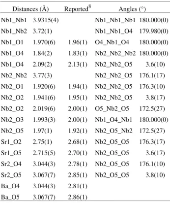
atoms are located in the A1 (2a) sites and in part of the A2 (4c) sites. Their site occupation factors were refined with constraints in order to maintain the compound stoichiometry. Ba atoms are located only in the A2 (4c) sites and Nb atoms are occupying the B1 (2b) and B2 (8d) sites. Selected interatomic distances and angles are listed in Table 4, in which other reported values are included for comparison.
Jamiesonet al.1 reported a O5 positional disorder in the results for the SBN single crystal samples of composition Sr0.76Ba0.24Nb2O5.78. For the structural refinement of
pow-dered (crushed) SBN-based single crystals, Chernaya et al.8 and Trubelja et al.7 considered such positional disorder for both O4 and O5 atoms. In this work, the values of the final anisotropic temperature coefficients (Table 1), ob-tained from the refinement of X-ray and neutron data, do not present anomalies that could suggest the need for assuming a positional disorder site occupancy for O4 and O5 in the structure of the sintered powder.
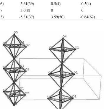
Figure 3 shows the octahedral representation obtained with the program ATOMS for Windows13 using the refined atomic coordinates. Further representations of details of the structure are presented to better illustrate the mechanism of formation of the ferroelectric dipoles. The displacement of Nb cations from the planes formed by oxygen is shown in Fig. 4. Parallel to the c direction displacement occurs only for the Nb1 atoms, as shown in Fig. 5a. In the case of Nb2, there are components in a and b directions, as shown in Fig. 5b. From the distance and angle calculations, a quantitative measurement of these displacements was obtained. Nb1 atoms are 0.12 Å displaced from the middle of the distance O4-O4 bond in the c direction. The displacement of Nb1 atoms from the center of the octahedra is 0.14 Å. Nb2 atoms are 0.05 Å displaced from the middle of the distance O5-O5 bond, which yields an angle of 3.8° with the c direction (see Table 2). This small inclination can be observed in the disposition of the Nb2-O5 bonds shown in Fig. 5b, where it is remarkable that the Nb1 centered octahedra do not present them, as a consequence of the position of O4. The displacement of Nb2 atoms from the center of the octahedra is 0.17 Å.
Table 1. Rietveld refinement details for SBN using neutron and X-rays powder diffraction data simultaneously.
Data Neutrons X-rays X-rays
Wavelength (Å) 1.4875 1.5405 1.5405
Step scan increment (°) 0.05 0.02 0.02
2θ interval (°) 5.4-105.35 20-60 60-120
Number of reflections 252 132 558
Number of profile parameters 6 18 18
RB(%) 7.58 9.84 12.72
Rp (%) 4.69 9.45 11.65
Rp (%) 5.95 11.96 15.21
Rexp(%) 1.98 1.97 2.50
Number of structural parameters: 96. Totals: Rp = 7.25% Rwp = 9.66%.
Table 3. Thermal parameters (multiplied by 100) for SBN ceramic refinement.
Atom U11 U22 U33 U12 U13 U23
Nb1 0.10(18) 0.10(18) 4.91(42) -0.46(19) 0 0
Nb2 0.61(20) 0.62(16) 0.97(16) -0.83(16) 1.9(5) 1.1(4)
Sr1 0.71(33) 0.71(33) 9.7(9) 0 0 0
Sr2 2.16(29) 2.16(29) 4.9(6) 4.52(90) -1.7(12) -1.7(12)
Ba 6.10(29) 6.10(29) 0.7(4) -7.7(4) 1.3(10) 1.3(10)
O1 2.21(38) 1.22(38) 3.8(6) 0.80(34) -0.4(7) -2.7(5)
O2 0.34(24) 0.25(27) 6.77(41) 0.61(20) -1.93(49) 1.77(59)
O3 0.17(32) 0.17(32) 0.18(36) 3.61(39) -0.5(4) -0.5(4)
O4 7.2(7) 7.2(7) 0.2(10) 3.0(8) 0 0
O5 4.72(39) 6.05(39) 0.16(23) -5.31(37) 3.59(50) -0.64(67)
Table 2. Refinement results for SBN ceramic.
Atom X Y Z Ue*100 Fractn Mult Site sym
Nb1 0 0.5 -0.0176(52) 1.70 1 2 MM2d001
Nb2 0.07529(16) 0.21097(17) -0.0187(19) 0.73 1 8 1
Sr1 0 0 0.4507(57) 3.69 0.7253 2 4(001)
Sr2 0.17097(18) 0.67097(18) 0.4760(33) 3.07 0.3999 4 M(+-0)
Ba 0.17097(18) 0.67097(18) 0.4760(33) 4.31 0.4875 4 M(+-0)
O1 0.3413(5) 0.0055(4) -0.054(4) 2.41 1 8 1
O2 0.1413(5) 0.0647(5) -0.059(4) 2.45 1 8 1
O3 0.2817(5) 0.7817(5) -0.054(4) 0.17 1 4 M(+-0)
O4 0 0.5 0.513(5) 4.87 1 2 MM2d001
O5 0.2965(9) 0.4175(8) 0.468(4) 3.65 1 8 1
Space group: P 4 b m; The lattice is acentric; primitive cell: tetragonal. Lattice parameters: a = b = 12.4603(3) Å, c = 3.9315(2) Å.
Cell volume = 610.41(3) Å3.
Figure 3. Octahedral representation obtained with the program Atoms for Windows using the refinement results.
4. Conclusions
Previous structural works about this compound were performed using single crystalline samples or polycrystal-line materials obtained by grinding single crystals1,7,8, but this is the first time that structural analysis is performed using sintered ceramic material. The sintered compound presents the crystalline tungsten bronze structure with space group P4bm and the structure could be well described without including disorder in oxygen positions. The Nb1 centered octahedra exhibit orthorhombic point group mm2 symmetry (distorted tetragonal). These octahedra corre-spond to only two of the ten that are contained in the unit cell. As a consequence, their influence in the resultant direction of the ferroelectric properties should be smaller than that of the Nb2 centered octahedra. The general posi-tion of the Nb2 atoms produces a triclinic point group symmetry (orthorhombic distorted), which permits a dis-torted orthorhombic symmetry for their octahedra and a cation displacement that is non-parallel to c direction. This fact could cause a ferroelectric contribution at a direction non-parallel to the c direction. Nevertheless, for an unpoled material, the resultant of these contributions (non-parallel to c) is zero because of the tetragonal crystal symmetry.
Acknowledgements
The authors would like to thank FAPESP, contract no. 95.00791-8, and PADCT/CNPq, contract no.620007/98-2 for financial support. J.G.C. and Y.P.M. are respectively also grateful to CAPES for a doctoral and CNPq for a research fellowship.
References
1. Jamieson, P.B.; Abrahams, S.C.; Bernstein, J.L. J. Chem. Phys., v. 48, p. 5048-5057, 1968.
2. Fang, T.; Wu, N.; Shiau, F.; J. Mat. Sc. Letters, v. 13, p. 1746-1748, 1994.
3. Xu, Y.; Li, Z.; Wang, H.; Chen, H. Phys. Rev. B, v. 40, p. 11902-11908, 1989.
4. Spínola, D.U.; Santos, I.A.; Bássora, L.A.; Garcia, D.; Eiras, J.A. J. Eur. Ceram. Soc., v. 19, p. 1111-1114, 1999.
5. Fan, H.; Zhang, L.; Yao, X. J. Mater. Science, v. 33, p. 895-900, 1998.
Figure 5. (a) The displacement occurs parallel to the c direction only for the Nb1 atoms; (b) in the Nb2 also there are components in a and b directions (small inclination of 3.8° observed in the disposition of the Nb2-O5 bonds).
Table 4. Selected interatomic distances and angles. Other reported values are included for comparison.
Distances (Å) Reported8 Angles (°)
Nb1_Nb1 3.9315(4) Nb1_Nb1_Nb1 180.000(0)
Nb1_Nb2 3.72(1) Nb1_Nb1_O4 179.980(0)
Nb1_O1 1.970(6) 1.96(1) O4_Nb1_O4 180.000(0)
Nb1_O4 1.84(2) 1.83(1) Nb2_Nb2_Nb2 180.000(0)
Nb1_O4 2.09(2) 2.13(1) Nb2_Nb2_O5 3.6(10)
Nb2_Nb2 3.77(3) Nb2_Nb2_O5 176.1(17)
Nb2_O1 1.920(6) 1.94(1) Nb2_Nb2_O5 176.3(10)
Nb2_O2 1.941(6) 1.95(1) Nb2_Nb2_O5 3.8(17)
Nb2_O2 2.019(6) 2.00(1) O5_Nb2_O5 172.5(27)
Nb2_O3 1.993(3) 2.00(1) Nb1_O4_Nb1 180.000(0)
Nb2_O5 1.97(1) 1.92(1) Nb2_O5_Nb2 172.5(27)
Sr1_O2 2.75(1) 2.68(1) Nb2_O5_O5 176.3(17)
Sr1_O5 2.715(5) 2.70(1) Nb2_O5_O5 3.6(17)
Sr2_O4 3.044(3) 2.78(1) Nb2_O5_O5 176.1(10)
Sr2_O5 3.067(7) 2.85(1) Nb2_O5_O5 3.8(10)
Ba_O4 3.044(3) 2.81(1)
6. Abrahams, S.C.; Kurtz, S.K.; Jamieson, P.B. Phys. Rev., v. 172, p. 551-553, 1968.
7. Trubelja, M.P.; Ryba, E.; Smith, D.K. J. Mat. Science, v. 31, p. 1435-1443, 1996.
8. Chernaya,T.S.; Maksimov, B.A.; Verin, I.A.; Ivleva, L.I.; Simonov, V.I. Crys. Rep., v. 43, p. 986-990, 1998.
9. Santos, I.A.; Spínola, D.U.; Garcia, D.; Eiras, J.A.; Manoel, E.R.; Hernandes, A.C.; Carrio, J.A.G.; Mas-carenhas, Y.P. Ferroelectrics, v. 238, p. 711-718, 2000.
10. Lee, H.; Freer, R.J. Mater. Science, v. 33, p. 1703-1708, 1998.
11. Larson, A.C.; Von Dreele, R.B. GSAS, Generalized Structure Analysis System, Document LAUR 86-748 (Los Alamos National Laboratory, Los Alamos), 1993.
12. Young, R.A.; Larson, C.A.; Paiva-Santos, C.O. J. Appl. Cryst., v. 28, p. 366-367, 1995.
13. ATOMS for Windows, Version 3.1, Shape Software, 1995.