RESUMO: “Rastreamento histoquímico, itoquímico e biológico de folhas de Plinia caulilora (DC.) Kausel (Myrtaceae)”. No presente trabalho, foram investigadas a composição química e atividades biológicas de extratos brutos obtidos com etanol 50%, etanol 70%, acetona:água (7:3; v/v) e clorofórmio das folhas de Plinia caulilora (DC.) Kausel, Myrtaceae, uma árvore nativa
de várias regiões do Brasil. Os rastreamentos histoquímico e itoquímico foram feitos de acordo com reações de caracterização e cromatograia em camada delgada. Para auxiliar na padronização dos extratos foram realizadas determinações do teor de fenóis totais e de lavonoides totais,
espectrofotometricamente. A atividade antioxidante foi analisada pela porcentagem de sequestro de
radicais livres usando solução de DPPH. A atividade antimicrobiana foi avaliada frente a bactérias patogênicas Gram-positivas, Gram-negativas e espécies de Candida utilizando os métodos de
difusão em ágar e determinação da concentração inibitória mínima (MIC) de acordo com métodos
padronizados. As folhas apresentaram lipídeos nas cavidades secretoras e fenóis, principalmente
taninos, nas nervuras e parênquima paliçádico. Os extratos polares apresentaram lavonoides, taninos, alto teor de fenóis totais e de lavonoides totais. Os extratos mostraram elevada atividade antioxidante e a atividade antimicrobiana foi melhor contra as espécies de Candida do que contra
as bactérias.
Unitermos: Plinia caulilora, Myrtaceae, rastreamento químico, atividade antioxidante, atividade antimicrobiana.
ABSTRACT: In this work, chemical and biological activities of crude extracts obtained with 50% ethanol, 70% ethanol, acetone:water (7:3; v/v) and chloroform of Plinia caulilora (DC.) Kausel,
Myrtaceae, leaves, a native tree from several regions of Brazil, was investigated. Histochemical
and phytochemical screenings were done according to characterization reactions and thin layer
chromatography. To assist in extracts standardization, total phenol and lavonoids content
spectrophotometric was performed. Antioxidant activity was analyzed by percentage of radical
scavenging using DPPH solution. Antimicrobial activity was evaluated against Gram-positive,
Gram-negative pathogenic bacteria and species of Candida using agar diffusion and minimal inhibitory concentration (MIC) determination methods according to standard methods. The leaves presented lipids at secretory cavity and phenols, mainly tannins, in nervures and palisade
parenchyma. Polar extracts showed lavonoids, tannins and high content of phenols and lavonoids.
The extracts showed great antioxidant activity and antimicrobial activity was better against Candida species than against bacteria.
Keywords: Plinia caulilora, chemical screening, antioxidant activity, antimicrobial activity.
Myrciaria caulilora (Mart.) O. Berg, Myrtus caulilora Mart., Eugenia caulilora DC. Myrciaria jaboticaba (Vell.) O. Berg, Myrciaria tenella (DC.) O. Berg and Myrciaria
truncilora O. Berg are scientiic synonims of P. caulilora. In Brazil, it is called popularly as “jabuticabeira” or “jaboticaba” (Lorenzi, 2000).
Morphologically, it is a tree about ten-ifteen 20(1): 48-53, Jan./Mar. 2010
Ar
tigo
Received 15 September 2008; Accepted 17 December 2008
Histochemical, phytochemical and biological screening of
Plinia caulilora
(DC.) Kausel, Myrtaceae, leaves
Tatiana M. Souza-Moreira,
1Raquel R. D. Moreira,
2Luis V. S. Sacramento,
2Rosemeire C. L. R. Pietro
*,11Departamento de Fármacos e Medicamentos, Pós-graduação em Ciências Farmacêuticas, Faculdade de Ciências Farmacêuticas, Universidade Estadual Paulista “Júlio de Mesquita Filho”, Rodovia Araraquara-Jaú, km 1,
14801-902 Araraquara-SP, Brasil
2Departamento de Princípios Ativos Naturais e Toxicologia, Faculdade de Ciências Farmacêuticas, Universidade Estadual Paulista “Júlio de Mesquita Filho”, Rodovia Araraquara-Jaú, km 1, 14801-902 Araraquara-SP, Brasil.
INTRODUCTION
Plinia caulilora (DC.) Kausel is a plant from the family Myrtaceae, which has one hundred twenty nine genders. The several species from this family are source of essential oils, condiments, food and many of them are
meters of height, which bark scales every year, that has
simple leaves, with lowers and fruits together the stem,
feature by what this plant has been named. This tree occurs,
preferentially, at lood plain and open forests being ind at forests from Brazil, Argentine and Paraguay. P. caulilora
produces lots of comestible and delicious fruits twice or more times a year (Lorenzi, 2000; Barros et al., 1996). The bark of P. caulilora is astringent and used by Brazilian people to treat diarrhoea and skin irritations (Lorenzi, 2000).
It has not been scientiically reported the
chemical composition and biological properties of the leaves of P. caulilora so that, the purpose of this study was to characterize histochemically and phytochemically the composition and to investigate antioxidant and antimicrobial activities of different crude extracts of these leaves.
MATERIAL AND METHODS
Plant material and preparation of extracts
The leaves of Plinia caulilora (DC.) Kausel (Myrtaceae), collected from São Carlos, Brazil,
(22º01’16.6”S; 47º53’57.0”W) in 2004, were identiied by Marcos Sobral and deposited at Herbário da Escola Superior de Agricultura “Luiz de Queiroz”, ESALQ, USP-Piracicaba, Brazil (voucher ESA nº 96038). The leaves
were dried at 40 ºC for 4 d, comminuted and percolated
individually with 50% ethanol (50 EtOH), 70% ethanol (70 EtOH), acetone:water (7:3; v/v) (Ac:H2O) and chloroform
(CHCl3). The extracts were concentrated under reduced
pressure and lyophilization. The yield of each extract was 35%, 30%, 20% and 2%, respectively.
Bacterial and fungal strains
Tests were performed on reference strains obtained from American Type Culture Collection (ATCC): Bacillus
subtilis (ATCC 9362); Staphylococcus aureus (ATCC 25923) and Staphylococcus epidermidis (ATCC 12228);
Escherichia coli (ATCC 10536); Candida albicans (ATCC 64548); Candida parapsilosis (ATCC 22019); Candida
tropicalis (ATCC 750). After overnight growth at 37 ºC the inocula were prepared by adjusting the turbidity of the suspension to match the 0.5 McFarland standard in order to achieve the suitable suspension of 1.5 x 10⁸ bacteria/
mL and 1.0 x 10⁶ to 5.0 x 10⁶ yeast/mL (NCCLS, 2002;
NCCLS, 2003a).
Histochemical screening
Thin transverse sections of the midvein were handmade with a stainless steel razor. Some sections were treated with 2% sodium hypochloride solution, submitted to ethanol-cetone-hexane sequence, rehydrated, washed with
distilled water and used as negative control. Anatomical
identiication was done with 0.05% Toluidin Blue in buffer at pH 6.8 (Kraus & Arduin, 1997). Identiication
of hydrophilic substances used: ferric chloride for
phenolic compounds (Johansen, 1940), vanillin in HCl
(9%) solution for tannins (Valette et al., 1998) and picric
acid for alkaloids. Identiication of lipophilic substances
used: Sudan red III for total lipids (Johansen, 1940), Nile Blue sulphate for acid and neutral lipids (Cain, 1947), osmium tetroxide for unsaturated lipids (Ganter
& Jollés, 1969-1970), Nadi staining for essential oil and
oleoresin (David & Carde, 1964), antimony trichloride
for steroids (Hardman & Sofowora, 1972), sulphuric acid for sesquiterpene lactones (Geissman & Grifin, 1971)
and 2,4-dinitrophenylhydrazine for terpenoids (Ganter &
Jollés, 1969-1970).
Preliminary phytochemical screening and thin-layer chromatography (TLC) proile
Powdered leaves were extracted appropriately
to perform characterization reactions of secondary
metabolites classes according to Henriques et al. (2004),
Zuanazzi and Montanha (2004), Schenkel et al. (2004) and Santos and Mello (2004). Dragendorf’s, Bouchardat’s, Mayer’s and Bertrand’s reagents were used to indicate the presence of alkaloids by precipitate formation. Flavonoids
were characterized by luorescence by complexation
with aluminum chloride visualized at 235 and 355 nm. The formation and persistence of foam after 20 min of previous agitation and addition of chloridric acid 2 N was used to characterized saponins. For tannins, it was done precipitation reactions with gelatin, ferric chloride to detect condensed and/or hydrolyzed tannins and lead acetate to detect only hydrolyzed tannin. One-dimensional TLC silica gel analysis of the four extracts was performed in chloroform:methanol:n-propanol:water (5:6:1:4; v/v) and
visualized by spraying anisaldehyde/H2SO4 and heating
with UV irradiation at 235 nm and by spraying 1% ferric
chloride in methanol (Wagner et al., 1984).
Determination of total phenolics and total lavonoids
Total phenolics in the extracts were determined with Folin-Denis reagent according to the method from
AOAC (1984). Briely, 100 μL of aqueous solutions (20
mg/mL) of extracts were diluted 1:200 and samples from 0.2 to 0.5 mL of these solutions were added to 7 mL of water and 0.5 mL of Folin-Denis reagent. After 3 min, it was added 1 mL of 35% Na2CO3 solution completing the volume to 10 mL with water. The absorbance was measured
at 760 nm using Shimadzu UV-1603 spectrophotometer
standard curve. Total lavonoids content was determined
by colorimetrical method described by Djeridane et al. (2006). The extracts (0.2 g) were dissolved in 20 mL of 80% methanol, extracted for 2 h at room temperature and centrifuged at 3000 g for 15 min. The volume of the supernatant was made up to 100 mL with 80% methanol. A portion of 2 mL was taken and added to 2 mL of 2% AlCl3 methanol solution. The absorbance was measured at 430 nm after 15 min at room temperature. A standard curve was plotted using different concentrations of rutin solution
in 80% methanol. The percentage of total lavonoids in the
extracts was calculated using a linear equation based on the standard curve.
Antioxidant activity
Antioxidant potential of the extracts at 250 µg/mL in methanol was evaluated by radical scavenging capacity
with 0.004% 2,2-diphenyl-1-picryl-hydrazyl (DPPH)
in methanol using spectrophotometry at 517 nm. Gallic acid, rutin and vitamin C were used as positive control
(250 µg/mL). It was added 2.50 mL of DPPH solution to
1.00 mL of samples, shaked and kept at dark place for 30 min. Blank solutions were done with 1.00 mL of extract solution added to 2.50 mL of methanol and negative control consisted of 1.00 mL of methanol added to 2.50 mL
of DPPH solution. Anti-radical activity was calculated as discolour percentage of DPPH according to the equation: % = (DA-SA/DA).100; where: DA = DPPH absorbance;
SA = sample absorbance (Falcão et al., 2006).
Antimicrobial assay of crude extracts
Stock solutions of 100 mg/mL of each extract were prepared in dimethylsulfoxide (DMSO) and were conveniently diluted in culture medium. All tests were performed according to standard methods and to Ostrosky et al. (2008) review applied for medicinal plants.
Agar diffusion method (NCCLS, 2003b)
The test was made with 1 x 10⁶ CFU/mL in Petri
dishes. Steel templates were placed on the solid medium
and 50 μL of the extracts (50 mg/mL), 50 μL of DMSO:BHI (BHI) (1:1; v/v) and 50 μL of ampicillin solution (50 μg/
mL) were separately added to each well. After 2 h at 4 °C the plates were incubated at 37 ºC for 24 h. Bacterial growth inhibition was determined by inhibition zones diameter around the wells.
Assay for antimicrobial activity
Minimal inhibitory concentration (MIC) values were determined by microdilution method (NCCLS, 2002; NCCLS, 2003a). Final dilutions ranging from 5 mg/mL to 0.078 mg/mL of crude extract were prepared
in sterile 96-well microplates. The wells were illed with BHI for antibacterial approach; RPMI-1640 medium
with 2% glucose and 0.165 mol/L 3-[N-morpholine]
propanesulfonic acid (MOPS) at pH 7 for antifungal. Final
bacterial inocula were 2.5 x 10⁵ CFU/mL and incubated
aerobically at 35 ºC for 24 h. Final yeast inocula were 2.5 x 10³ CFU/mL incubated at 35 ºC for 48 h at 100 rpm.
Ampicillin and amphotericin B were used as positive control to bacteria and yeast, respectively. Bacterial growth was indicated by addition of 0.01% resazurin
aqueous solution and MIC values were identiied as
the lowest extract concentration in which no growth is visible showed by changing colour of resazurin from blue (abscence of growth) to pink (growth) (Gabrielson et al.,
2002). Yeast growth was indicated by addition of 20 μL
of 2% 2,3,5-triphenil tetrazolium chloride (TTC) aqueous
solution (Eloff, 1998). Yeast MIC values were identiied
as the lowest extract concentration in which there was no growth and therefore TTC changed the colour from yellow (absence of growth) to red (growth). Minimal bactericidal concentration (MBC) and minimal fungal concentration (MFC) were determined by subculturing the microplates
in Petri dishes with Müller-Hinton-agar for bacteria and
Sabouraud-agar for yeast, incubated at 35 ºC for 24 h, and
deined as the lowest extract concentration which there
was no visible growth of colony.
RESULTS AND DISCUSSION
It was noted that the leaves of Plinia caulilora (DC.) Kausel (Myrtaceae) has the same anatomical organization as Myrtaceae family, showing dorsiventral mesophyll emphasing secretory cavities and idioblasts containing calcium oxalate crystals (druseans and
monocristals). It was veriied the presence of phenols,
tannins and alkaloids in midvein and smaller veins;
phenols were also veriied in palisade parenchyma. Lipids were veriied mainly in secretory cavities and reactions
were positive for acid and unsaturated lipids, steroids, sesquiterpene lactones and terpenoids; it was noted weak
positive result for essential oil. Preliminary phytochemical
analysis of the drug showed only the presence of
hydrolysable tannins and lavonoids. TLC conirmed these results since three extracts showed two yellow luorescent spots of lavonoids (Rf = 0.37 and 0.42) with anisaldehyde/
H2SO4, heating and observation under UV light and one
gray spot of tannin (Rf = 0.25). One strong blue spot of tannin (Rf = 0.25) was observed by spraying ferric chloride 1% in methanol. In contrary of histochemical reaction, TLC tests did not indicate the presence of alkaloids, which
must be investigated. CHCl3 extract showed only a weak
blue spot around start point. Terpenoids were detected
with anisaldehyde/H2SO4 with spot around Rf = 1.00.
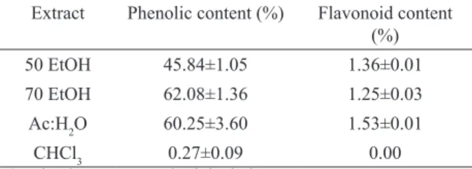
Except to CHCl3 extract, the three extracts showed content
of phenolic compounds higher than 45%. Total lavonoids content were not found in CHCl3 extract and about 1% in
Extract Phenolic content (%) Flavonoid content (%)
50 EtOH 45.84±1.05 1.36±0.01
70 EtOH 62.08±1.36 1.25±0.03
Ac:H2O 60.25±3.60 1.53±0.01
CHCl3 0.27±0.09 0.00
a) value is mean±standard deviation.
Polar extracts showed high antioxidant activity, beyond 90%, but signiicantly smaller than the controls
gallic acid, rutin and vitamin C (Figure 1). Choi et al. (2006) suggested that many biological activities in plants
are due to the content of total phenols like lavonoids and tannins. Plant phenols have potential antioxidant
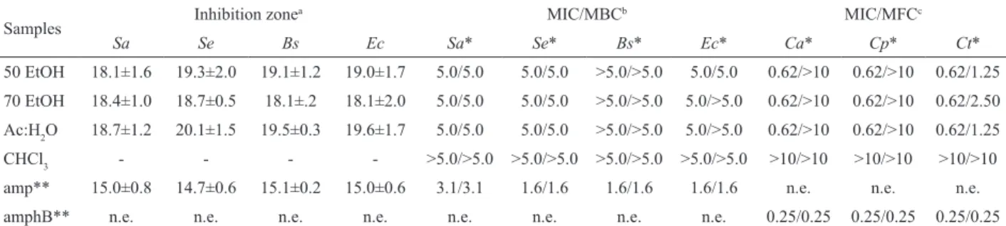
activity, mainly by acting as oxygen radical scavengers (Quettier-Deleu et al., 2000). Other studies (Souza et al., 2007; Tadhani et al., 2007) showed that extracts with high content of phenols have high antioxidant activity and that was seen in this study since chloroformic extract exhibited the smaller phenol content and antioxidant activity. Agar diffusion method showed great inhibition
zones (Table 2) for polar extracts. However, MIC and MBC determination of 50 EtOH, 70 EtOH and Ac:H2O
extracts have showed weak antibacterial activity whereas
CHCl3 had not any activity at the highest concentration
tested (Table 2). The bacteria selected for this study are present in the human being daily as normal microbiota, e.g.,
Staphylococcus aureus and Staphylococcus epidermidis; as food contaminants, e.g., Escherichia coli or used to sterility control, e.g., Bacillus subtilis (Chomnawang et
al., 2005; Beovic, 2006; Abdou et al., 2007; Pesavento et al., 2007). Polar extracts showed great activity against Table 1. Total phenolic and lavonoid content in the extracts of
the leaves of P. caulilora.
Figure 1. Radical scavenging capacity. Percentual ± standard deviation. A) gallic acid. B) rutin. C) vitamin C. D) 50 EtOH extract. E) 70 EtOH extract. F) Ac:H2O extract. G) CHCl3 extract.
Candida species and C. tropicalis was more sensible to
them since MFC values were the smallest. CHCl3 extract
did not showed activity at the highest concentration tested (Table 2). This work had interesting results against
Candida species. Candida albicans is the yeast species
more often isolated from biological samples being an usual skin and mucosa colonizing agent with many hosts in the oral cavity, and higher incidence rates among young
children and patients infected with HIV, but is necessary
to pay attention to the emergence of other Candida species as infecting agents (McCullough et al., 1996; Runyoro et al., 2006; Zhang et al., 2006). Resistance to commonly used agents, toxicity and costs impelled the search for new agents and it can be noted that in recent years the research for new active compounds from natural sources has been increasing (Duarte et al., 2005; Runyoro et al., 2006).
Histochemical and phytochemical results showed
phenolic prevalence in the composition of the leaves and extracts of P. caulilora and can contribute to the standardization of the extracts. Besides, it was possible to indicate that polar extracts of the leaves have biological activities as antioxidant and antimicrobial that can be due to their phenolic content. It should be emphasize
the presence of lavonoids and tannins derivates in polar
extracts and the activity against Candida species showed
by them. It was noticed that CHCl3 extract showed small
composition of phenolic content, therefore it could explain the smallest antioxidant and antimicrobial activities.
This study is important since it is the irst to
indicate the leaves extracts of P. caulilora as new source of antioxidant and antimicrobial molecules and because it was determined the composition of the leaves. These investigations showed a preliminary standardization of the leaves by histochemical and phytochemical methods.
ACKNOWLEDGEMENTS
This work was supported by CAPES, FAPESP and PADC-FCF-UNESP. The authors wish to thank Juliana A. Severi and Prof. Dr. Wagner Vilegas from Departamento de Química Orgânica, Instituto de Química, UNESP, Araraquara-SP, Brazil for phytochemical help. We also thank Luís Eduardo dos Santos and Maria Angélica Lima
Barretto for technical assistance.
REFERENCES
Abdou AM, Higashiguchi S, Aboueleinin AM, Kim M, Ibrahim HR 2007. Antimicrobial peptides derived from hen egg
lysozyme with inhibitory effect against Bacillus species. Food Control 18: 173-178.
AOAC-Association of Oficial Analytical Chemists 1984. Oficial
methods of analysis of the association of chemists. 13.
ed. Washington, D.C.: Association of Oficial Analytical
Chemists.
Barros RS, Finger FL, Magalhães MM 1996. Changes in non-structural carbohydrates in developing fruit of Myrciaria jaboticaba. Sci Hortic-Amsterdam 66: 209-215.
Beovic B 2006. The issue of antimicrobial resistance in human medicine. Int J Food Microbiol 112: 280-287.
Cain AJ 1947. The use of Nile Blue in the examination of lipids. Q J Microsc Sci 88: 383-392.
Choi YM, Noh DO, Cho SY, Suh HJ, Kim KM, Kim JM 2006.
Antioxidant and antimicrobial activities of propolis from several regions of Korea. LWT-Food Sci Technol 39: 756-761.
Chomnawang MT, Surassmo S, Nukoolkarn VS, Gritsanapan W 2005. Antimicrobial effects of Thai medicinal plants against acne-inducing bacteria. J Ethnopharmacol 101: 330-333.
Costa AF 1994. Farmacognosia. Lisboa: Calouste Gulbenkian.
David R, Carde JP 1964. Coloration différentielle des inclusions
lipidique et terpeniques des pseudophylles du Pin maritime au moyen du reactif Nadi. C R Hebd Seances Acad Sci D 258: 1338-1340.
Djeridane A, Yousi M, Nadjemi B, Boutassouna D, Stocker P,
Vidal N 2006. Antioxidant activity of some Algerian medicinal plants extracts containing phenolic compounds. Food Chem 97: 654-660.
Samples Inhibition zone
a MIC/MBCb MIC/MFCc
Sa Se Bs Ec Sa* Se* Bs* Ec* Ca* Cp* Ct*
50 EtOH 18.1±1.6 19.3±2.0 19.1±1.2 19.0±1.7 5.0/5.0 5.0/5.0 >5.0/>5.0 5.0/5.0 0.62/>10 0.62/>10 0.62/1.25
70 EtOH 18.4±1.0 18.7±0.5 18.1±.2 18.1±2.0 5.0/5.0 5.0/5.0 >5.0/>5.0 5.0/>5.0 0.62/>10 0.62/>10 0.62/2.50
Ac:H2O 18.7±1.2 20.1±1.5 19.5±0.3 19.6±1.7 5.0/5.0 5.0/5.0 >5.0/>5.0 5.0/>5.0 0.62/>10 0.62/>10 0.62/1.25
CHCl3 - - - - >5.0/>5.0 >5.0/>5.0 >5.0/>5.0 >5.0/>5.0 >10/>10 >10/>10 >10/>10
amp** 15.0±0.8 14.7±0.6 15.1±0.2 15.0±0.6 3.1/3.1 1.6/1.6 1.6/1.6 1.6/1.6 n.e. n.e. n.e.
amphB** n.e. n.e. n.e. n.e. n.e. n.e. n.e. n.e. 0.25/0.25 0.25/0.25 0.25/0.25 Sa: Staphylococcus aureus; Se: Staphylococcus epidermidis; Bs: Bacillus subtilis; Ec: Escherichia coli; Ca: Candida albicans; Cp: Candida parapsilosis; Ct: Candida tropicalis. amp: ampicillin; amphB: amphotericin B; a) values expressed, in milimeters, as mean of three determinations ± standard deviation; b) MIC and MBC determination; c) MIC and MFC determination; -: no inhibition; n.e.:
not evaluated; * values expressed in mg/mL; ** values expressed in μg/mL.
Duarte MCT, Figueira GM, Sartoratto A, Rehder VLG, Delarmelina C 2005. Anti-Candida activity of Brazilian medicinal plants. J Ethnopharmacol 97: 305-311.
Eloff JN 1998. A sensitive and quick microplate method to
determine the minimal inhibitory concentration of plant extracts for bacteria. Planta Med 64: 711-713.
Falcão DQ, Costa ER, Alviano DS, Alviano CS, Kuster RM,
Menezes FS 2006. Rev Bras Farmacogn 16: 73-76.
Gabrielson J, Hart M, Jarelöv A, Kuhn I, Mckenzie D, Möllby R 2002. Evaluation of redox indicators and the use of digital scanners and spectrophotometer for quantiication
of microbial growth in microplates. J Microbiol Meth 50: 63-73.
Ganter P, Jollés G 1969-1970. Histochimie normale et pathologique. Paris: Gauthier-Villars.
Geissman TA, Grifin TS 1971. Sesquiterpenes lactones:
acid-catalyzed color reactions as an aid structure determination. Phytochemistry 10: 2475-2485.
Hardman R, Sofowora EA 1972. Antimony trichloride as test
reagents for steroids, specially diosgenin and yamogenin, in plant tissues. Stain Technol 47: 205-208.
Henriques AT, Limberger RP, Kerber VA, Moreno PRH 2004.
Alcalóides: generalidades e aspectos básicos. In: Simões
CMO, Schenkel EP, Gosmann G, Mello JCP, Mentz LA, Petrovick PR 2004. Farmacognosia: da planta ao medicamento. 5. ed. Porto Alegre/ Florianópolis: Ed. Universidade/ UFRGS/ Ed. da UFSC. p. 765-792.
Johansen DA 1940. Plant Microtechnique. New York:
McGraw-Hill.
Kraus JE, Arduin M 1997. Manual básico de métodos em morfologia vegetal. Rio de Janeiro: Edur.
Lorenzi H 2000. Árvores brasileiras: manual de identiicação e cultivo de plantas arbóreas nativas do Brasil. São Paulo: Instituto Plantarum.
McCullough MJ, Ross BC, Reade PC 1996. Candida albicans: a review of its history, taxonomy, epidemiology, virulence attributes, and methods of strain differentiation. Int J Oral Max Surg 25: 136-144.
National Committee for Clinical Laboratory Standards 2002. Method broth dilution antifungal susceptibility testing of yeasts. 2. ed. Approved Standard. NCCLS document
M27-A2. Wayne, Pennsylvania: NCCLS.
National Committee for Clinical Laboratory Standards 2003a. Methods for dilution antimicrobial susceptibility tests for bacteria that grow aerobically. 6. ed. Approved Standard.
NCCLS.
National Committee for Clinical Laboratory Standards
2003b. Performance standards for antimicrobial disk
susceptibility tests. 8. ed. Approved Standard. NCCLS
document M2-A8. Wayne, Pennsylvania: NCCLS. Ostrosky EA, Mizumoto MK, Lima MEL, Kaneko TM, Nishikawa
SO, Freitas BR 2008. Métodos para avaliação da
atividade antimicrobiana e determinação da concentração mínima inibitória (CMI) de plantas medicinais. Rev Bras Farmacogn 18: 301-307.
Pesavento G, Ducci B, Comodo N, Lo Nostro A 2007. Antimicrobial resistance proile of Staphylococcus aureus isolated from raw meat: A research for methicillin resistant Staphylococcus aureus (MRSA). Food Control 18: 196-200.
Quettier-Deleu C, Gressier B, Vasseur J, Dine T, Brunet C, Luyckx M, Cazin M, Cazin JC, Bailleul F, Trotin F
2000. Phenolic compounds and antioxidant activities of
buckwheat (Fagopyrum esculentum Moench) hulls and
lour. J Ethnopharmacol 72: 35-42.
Runyoro DKB, Ngassapa OD, Matee MIN, Joseph CC, Moshi MJ 2006. Medicinal plants used by Tanzanian traditional healers in the management of Candida infections. J Ethnopharmacol 106: 158-165.
Santos AC, Mello JCP 2004. Taninos. In: Simões CMO, Schenkel EP, Gosmann G, Mello JCP, Mentz LA, Petrovick PR
2004. Farmacognosia: da planta ao medicamento. 5. ed.
Porto Alegre/ Florianópolis: Ed. Universidade/ UFRGS/ Ed. da UFSC. p. 615-656.
Schenkel EP, Gosmann G, Athayde ML 2004. In: Simões CMO, Schenkel EP, Gosmann G, Mello JCP, Mentz LA, Petrovick PR 2004. Farmacognosia: da planta ao medicamento. 5. ed. Porto Alegre/ Florianópolis: Ed. Universidade/ UFRGS/ Ed. da UFSC. p. 711-740. Souza TM, Severi JA, Silva VYA, Santos E, Pietro RCLR 2007.
Bioprospecção de atividade antioxidante e antimicrobiana da casca de Stryphnodendron adstringens (Mart.) Coville (Leguminosae-Mimosoidae). Rev Cienc Farm Basica Apl 28: 221-226.
Stasi LC, Hiruma-Lima CA 2002. Myrtales medicinais. In: Stasi LC, Hiruma-Lima CA. Plantas Medicinais na Amazônia e na Mata Atlântica. 2. ed. São Paulo: Editora UNESP,
p. 321-330.
Tadhani MB, Patel VH, Subhash R 2007. In vitro antioxidant
activities of Stevia rebaudiana leaves and callus. J Food Compost Anal 20: 323-329.
Valette C, Andary C, Geiger JP, Sarah JL, Nicole M 1998. Histochemical and cytochemical investigations of
phenols in roots of banana infected by the burrowing nematode Radopholus similes. Phytopathology 88: 1141-1148.
Wagner H, Bladt S, Zgainski EM 1984. Plant Drug Analysis. Berlin: Springer-Verlag.
Zhang JD, Xu Z, Caoa YB, Chenb HS, Yan L, An MM, Gao PH,
Wang Y, Jia XM, Jiang YY 2006. Antifungal activities and action mechanisms of compounds from Tribulus terrestris L. J Ethnopharmacol 103: 76-84.
Zuanazzi JAS, Montanha JA 2004. Flavonóides. In: Simões