Article
ISSN 0102-695X
http://dx.doi.org/10.1590/S0102-695X2011005000160 Received 8 Mar 2011 Accepted 17 Jul 2011 Available online 2 Sep 2011
Suzana Guimarães Leitão,
*,1Tereza Cristina dos Santos,
#,2Franco Delle Monache,
4Maria Eline Matheus,
3Patrícia Dias
Fernandes,
3Bruno Guimarães Marinho
31Faculdade de Farmácia, Departamento de Produtos Naturais e Alimentos, Universidade Federal do Rio de Janeiro, Brazil,
2Núcleo de Pesquisas de Produtos Naturais, Universidade Federal do Rio de Janeiro, Rio de Janeiro, Brazil,
3Departamento de Farmacologia, ICB, Universidade Federal do Rio de Janeiro, Brazil,
4Dipartimento di Studi di Chimica e Tecnologia delle Sostanze Biologicamente Attive, Università La Sapienza. Italy.
Abstract: Vitex cymosa Bertero ex Spreng., Lamiaceae, is found in Central and Amazon regions of Brazil, where it is popularly used as antirheumatic. Extracts from the leaves of V. cymosa were tested in analgesia models such as abdominal contortions induced by acetic acid and formalin to test peripheral analgesia; as well as the tail flick and hot plate models, to test spinal and supraspinal analgesia. A significant reduction was observed in the number of contortions with all extracts and in all doses. In the formalin model, a reduction in the second phase (inflammatory) was observed with all extracts, whereas only the n-butanol extract was able to act in the first, neurogenic, phase. In the tail flick model, all extracts increased latency time. Naloxone treatment reverted analgesic effect of all extracts with the exception of the dichloromethane one. All extracts developed peripheral and central analgesic activity. In the hot plate model no antinociceptive effect was observed for all tested extracts. All these results taken together suggest that V. cymosa leaf extracts were able to promote peripheral and central antinociceptive activity mediated by the opioid system.Twenty three substances were isolated and identified in the extracts and include flavonoids (C-glucosyl flavones, flavones and flavonols), triterpene acids from ursane and oleanane types, iridoids (free and glucosides), as well as simple phenols.
Keywords:
Vitex cymosa
Verbenaceae sensu lato
Lamiaceae sensu lato
antinociceptive activity medicinal plants flavonoids triterpene acids iridoids
Introduction
Vitex genus, Lamiaceae, (the genus has been formerly included in the Verbenaceae s.l. and recently transferred to the Lamiaceae s.l.) is constituted by small trees or shrubs occurring in tropical and subtropical regions. Many species have been studied in relation their chemistry, which is characterized by the presence of C-glucosyl l avones, ecdysteroids, diterpenes and
iridoids (Brito, 1992), as well as in relation to their
biological activities. Antiinl ammatory (Sidhartha et al.,
1990; Chawla, et al., 1991), wound healing (Sidhartha et al., 1990) and antiandrogenic (Bhargava, 1989) activities, as well as inhibition of lymphocyte proliferation (You et al., 1998) and smooth muscle relaxant effect (Dayrit et al., 1987; Dayrit & Lagurin, 1994) are among those
described in literature. V. agnus-castus is actually used in
the treatment of pre-menstrual syndrome and menopausal
complaints (Mills & Bone, 2000). Despite the fact that the
genus is well represented in Brazil with c.a. 35 species
(Leitão et al., 2008), as far as we know, only three species have been chemically or pharmacologically investigated so far (Leitão et al., 2008, Gallo et al., 2008; 2006, Sá
Barreto et al., 2005, Gonçalves et al., 2001). Vitex cymosa
Bertero is a small tree widely distributed in the Central and Amazon regions of Brazil, where it is popularly known as “tarumã-do-igapó” and "tarumã-do-alagado” (Correa, 1926). The species are used by the native populations of the State of Maranhão to treat rheumatic pains. In a previous work done by our group (Miranda et al., 2010) extracts from this plant’s barks, collected at Maranhão State, Brazil, were tested in antinociceptive models, such
as the tail l ick test, to evaluate the central analgesic effect. In this work a signii cant antinociceptive activity
was isolated and determined to be the active principle. In this work we investigated the analgesic properties of the dichloromethane leaf extracts of this plant.
Materials and Methods
Plant material
Leaves of Vitex cymosa Bertero ex Spreng.,
Lamiaceae, were collected in Corumbá, Mato Grosso do Sul State, Brazil in December (in fruit). The plant was
identiied by Dr. Vali Pott, from Embrapa, Corumbá. A
voucher specimen was deposited at Universidade Federal de Juiz de Fora herbarium under number CESJ 11,711.
Plant extracts
Dried and pulverized leaves (2.2 kg) were
exhaustively extracted with ethanol. After iltration
and concentration under reduced pressure the aqueous residue was sequentially extracted with organic solvents of increasing polarities, to afford the new extracts:
dichloromethane (VD), ethyl acetate (VA) and n-butanol
(VB) extracts, in this order, as well as (LW), which is the water-soluble remaining extract.
Isolation and identiication of substances from the
extracts
Dichloromethane extract (VD)
Part of VD was irst chromatographed on
silica gel eluted with CH2Cl2 with increasing amounts
of EtOAc. Fractions eluted with CH2Cl2-EtOAc (9:1)
were re-chromatographed on Sephadex LH-20 (MeOH)
yielding 21. Fractions eluted with CH2Cl2-EtOAc (8:2)
were puriied by prep. TLC (CH2Cl2-EtOAc, 3:1) to
afford 1, and 2. Fractions eluted with CH2Cl2-EtOAc (7:3)
were re-chromatographed on Sephadex LH-20 (MeOH),
yielding 3. A second fractionation of VD was performed
by CC on silica gel eluted with CHCl3 with increasing
amounts of EtOAc, and with EtOAc with increasing
amounts of MeOH. Fractions eluted with CHCl3 were
re-chromatographed on silica gel to afford 4, 13, and
a mixture of 15 and 17 (CHCl3-EtOAc 1:1). Fractions
eluted with CHCl3-EtOAc 9:1 were re-chromatographed
on Sephadex LH-20 (MeOH) affording a mixture of
16 and 18, as well as 5. Fractions eluted with
EtOAc-MeOH 8:2 were re-chromatographed in silica gel with
the organic phase of a mixture of EtOAc-acetone –H2O
(25:8:2), further puriied on Sephadex LH-20 and, inally,
by prep. TLC to afford 23 and 20. A third fractionation of
VD was directly subjected to Sephadex LH-20 (MeOH)
and led to the isolation of 19.
Ethyl acetate extract (VA)
VA was chromatographed on silica gel eluted with the organic phase of a mixture of
EtOAc-acetone-H2O (25:8:2 to 25:10:5) affording sisteen fractions.
From the irst two fractions, 12 and 14, were obtained
by crystallization, respectively. From fraction 6, 10 and
11 were obtained by prep. HPLC (see conditions below).
From fractions 12-13, a mixture of the lavonoids 6, and
7, was obtained by crystallization and, from fraction 15,
the lavonoids 8, and 9, were further puriied by prep.
TLC.
Butanol extract (VB)
VB was fractionated by silica gel CC eluted
with CHCl3 with increasing amounts of MeOH affording
21, 22 and 23, as described previously (Santos et al.,
2001). From fractions 10-16 (CHCl3-MeOH 9:1) 6, was
obtained, and fractions 17-18 afforded 7.
Animals
All experiments were performed with male Swiss mice (20-25 g) obtained from our own animal facility. Animals were maintained in a room with controlled temperature 22±2 °C for 12 h light/dark cycle with free access to food and water. Twelve hours before each experiment animals received only water, in order to avoid food interference with substances absorption. Animal care and research protocols were in accordance with the principles and guidelines adopted by the Brazilian College of Animal Experimentation (COBEA) and approved by the Biomedical Science Institute/UFRJ - Ethical Committee for Animal Research.
Drugs and extracts administration
All extracts used were administered by oral gavage at concentrations of 10, 30 and 100 mg/kg in a
inal volume of 0.1 mL. Morphine and dipyrone were
used as reference drugs and were administered by oral gavage. The control group was composed by PBS with the same amount of DMSO used in the dose of 100 mg/kg
of extracts (0.005%) and had no effect per se. Naloxone
was administered i.p. 15 min before oral administration
of morphine or extracts.
Acute toxicity
Acute toxicity was determined following the experimental model described by Lorke (1983). A single oral dose of extracts (500 mg/kg) was administered to a
group of ten mice (ive males and ive females). Behavior
grooming, loss of righting relex, increased or decreased
respiration, and food and water intake were observed
over a period of ive days. After this period animals were
killed by cervical dislocation, stomachs were removed, an incision along the greater curvature was made, and the number of ulcers (single or multiple erosion, ulcer or perforation) and hyperemia was counted.
Acetic acid-induced abdominal writhing
Mice were used according to Matheus et al.
(2006). Briely, the total number of writhings following
intraperitoneal (i.p.) administration of 2% (v/v) acetic
acid (AA) was recorded over a period of 20 min, starting 5 min after AA injection. The animals were pretreated orally with extracts or vehicle, 60 min before administration of AA.
Formalin test
The procedure was similar to the method described by Gomes et al. (2007). Animals received the injection of 20 µL of formalin (2.5% v/v) into dorsal surface of the left hind paw. Immediately, the time that the animal spent licking the injected paw was recorded. The nociceptive response develops two phases: 0 to 5 min after
formalin injection (irst phase, response to neurogenic
pain), and 15 to 30 min after formalin injection (second
phase, response to inlammatory pain). For comparison
purposes, the IC50 of morphine was calculated as 1.7 mg/kg. The animals were pre-treated with oral dose of extracts (10 to 100 mg/kg), vehicle or morphine (1.7 mg/ kg), 60 min before administration of formalin.
Tail-lick test
The procedure used was similar to Matheus et al.
(2006). Briely, mice tails were immersed on a water bath
set at temperature of 50±1 oC. The time necessary for the
mice to withdrawal the tail, in seconds (named reaction time) was registered at 20, 40, 60, 80, 100 and 120 min
after administration of V. cymosa extracts (10, 30 and
100 mg/kg) or morphine (2.3 mg/kg). For comparison purposes, the IC50 of morphine was calculated as 2.3 mg/kg. Baseline was considered as the mean of reaction time obtained at 40 and 20 min before administration of V. cymosa extracts or morphine, and was deined as
normal reaction of animal to the temperature. Increase in baseline (in %) was calculated by the formula: (reaction time x 100)/baseline) - 100.
Hot plate test
Mice were tested according to the method described by Matheus et al. (2006). The animals were
placed on a hot plate set at 55±1 oC. Reaction time was
recorded when the animals licked their fore- and hind-paws and jumped, at 30, 60, 90, 120, 150 and 180 min
after oral administration of V. cymosa extracts (100 mg/
kg) or morphine (1 mg/kg). Baseline was considered as the mean of reaction time obtained at 60 and 30 min before administration of extracts or morphine and was
deined as normal reaction of animal to the temperature.
Increase in baseline (in %) was calculated by the formula: (reaction time x 100)/baseline) - 100.
Evaluation of the mechanism of action of Vitex cymosa
extracts
To evaluate the mechanism of action of V.
cymosa extracts, animals were pre-treated with naloxone, a non-selective opioid antagonist. Naloxone (5 mg/kg)
was administered i.p. 15 min before oral administration
of extracts (30 mg/kg) or morphine (2.3 mg/kg).
Statistical analysis
All experimental groups were composed by ten animals. The results are presented either as the mean±SD in acetic acid-induced writhing and formalin or as the
increase in baseline (%) in tail lick test. Statistical signiicance between groups was performed by the
application of analysis of variance ANOVA followed by
Bonferroni’s test. p values less than 0.05 (p<0.05) were
used as the signiicant level.
Results
Phytochemical identiication of known compounds
Phytochemical investigation of Vitex cymosa
leaf extracts led to the isolation and identification of a number of known iridoids, triterpenes and flavonoids, which are secondary metabolites commonly found in the genus. Their chemical structures were identified by
means of one and two dimensional 1H and 13C NMR
techniques, as well as by mass spectral data, which were in accordance with previously reported data. From the dichloromethane extract, VD, the iridoids
tarumal, 21 (Santos et al., 2001) and agnuside, 23
(Boros & Stermitz, 1990); the flavonoids kampferol, 1,
3’-O-methyl-luteolin, 2, luteolin, 3, pachypodol, 4, and
apigenin, 5 (Agrawal, 1989); and the triterpenes
2α,3α-hydroxy-oleanolic acid, 15, 2α,3β-hydroxy-oleanolic
acid, 16, 2α,3α-hydroxy-ursolic acid, 17,
2α,3β-hydroxy-ursolic acid, 18, 2β,3β,19α-hydroxy-ursolic
acid, 19, (Mahato & Kundu, 1994), and 28-O
-glucosyl-2α,3α,19α-hydroxy-ursolic acid ester, 20 (Seto et al, 1984), were isolated. From the ethyl acetate extract,
9, 2”-O-caffeoyl-orientin (Leitão & Delle Monache,
1998), 11, 2”-O-p-hydroxybenzoyl-orientin, 10, (Leitão
& Delle Monache, 1998) protocatechuic acid, 12, vanilic
acid, 13, and p-hydroxybenzoic acid, 14, were isolated.
From the n-butanol extract, VB, orientin, 6, iso-orientin,
7, tarumal, 21, viteoid II, 22 and agnuside, 23, (Santos et
al., 2001) were isolated.
Acute toxicity
In order to evaluate the possible toxic effects of V. cymosa extracts, mice were treated orally with 500 mg/kg of each of them. Treated mice did not present any behavioral alterations. Also, stomachs were removed from the animals and examined under dissecting microscope. No lesions or bleedings were observed (data not shown).
Analgesic activity
To test the antinociceptive effects of Vitex
cymosa extracts were used models of peripheral, spinal and supra-spinal analgesia.
Acetic acid-induced abdominal writhing
Intraperitoneal injection of acetic acid induced 100.1±3.9 writhings in a period of 20 min. Pre-treatment
of mice with V. cymosa extracts in doses of 10, 30 and
100 mg/kg induced a dose-response reduction on total number of contortions. The most potent extract was VB
(Figure 1). Even at 10 mg/kg, VB signiicantly reduced
in 25% (100.1±3.9 writhings in control group versus 73.4±4.0 writhings in extract-treated group) the number
CO2H CO2R
152α,3αOH
162α,3βOH
172α,3αOH, R=H
182α,3βOH, R=H
192β,3β,19αOH, R=H
202α,3α,19αOH, R= Glu O
R4
R3O
R5
OH O R1
R2
OH
1 R1=OH, R2=H
2 R1=H, R2=OCH3 3 R1=H, R2=OH 4 R1=R2=R3=OCH3 5 R1=R2=R3=R4=R5=H
6 R1=R3=R5=H, R2=OH, R4=Glu 7 R1=R3=R4=H, R2=OH, R5=Glu 8 R1=R2=R3=R5=H, R4=Glu 9 R1=R2=R3=R4=H, R5=Glu
10 R1=R3=R5=H, R2=OH, R4=X 11 R1=R3=R4=H, R2=OH, R5=Y
O O OR HO
HO
HO XR=p-hydroxybenzoyl
YR= caffeoyl
12R=OH
13R=OCH3 14R=H CO2H
OH R
O O
CHO
21
O
HO H
O
22
O HO
O
23
OGlu
O HO
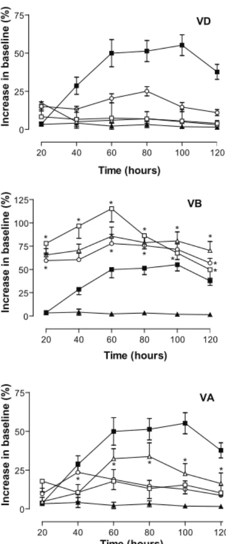
antinociceptive action of morphine lasted about 120 min. Dichloromethane extract (VD) had similar curves to all doses tested, and reached maximal response 80 minutes after administration. Although VD induced an increase
in the baseline, this was not a signiicant effect. VB
extract developed a dose-response curve, with very little differences between each tested dose (10, 30, and 100 mg/
kg). Twenty minutes after VB administration a signiicant
increase in the baseline could already be observed with all doses. At 120 min, the antinociceptive effect of morphine began to decline, whereas the effect of VB continued at high levels within 120 min, and lasted 2 h after a single oral administration (data not shown). The response to VA
was quite different. Signiicant antinociceptive activity
was observed at the dose of 100 mg/kg. This effect appeared after 60 minutes of administration and was maintained until the end of the experiment.
Figure 2. Effects of Vitex cymosa leaf extracts in the formalin test. Animals were pre-treated with different doses of extracts or vehicle by oral administration. Groups are: vehicle ( ▓ ), morphine – 1.7mg/kg (■) or extracts at different doses (□). The results are presented as mean ± S.D. (n=10) of time, in seconds, that the animal spent licking the injected paw calculated as described on methods section. Statistical signiicance was calculated by ANOVA followed by Bonferroni’s test.*p<0.05 when compared with vehicle-treated mice.
of writhings. At the doses of 30 and 100 mg/kg there was
signiicant effect to all three fractions, VD, VA and VB
(Figure 1).
Figure 1. Effects of Vitex cymosa leaf extracts on acetic acid-induced writhings. Animals were pre-treated with extracts or vehicle by oral administration. Groups are: vehicle (■), VD (Δ), VB (□), or VA (○). The results are presented as mean±SD; (n=10) of total writhings calculated as described on methods section. *p<0.05 ANOVA followed by Bonferroni’s test
Formalin test
Intraplantar injection of formalin evoked a characteristic biphasic licking response. For the control
group, the duration of licking in the 1st phase was
48.8±11.7 s, and for the 2nd phase was 141.6±33.0 s. As
shown in Figure 2, pre-treatment with different doses of
VD, VA or VB had no signiicant effect against the 1st
phase. However, 2nd phase was markedly reduced by VD,
with maximal effect observed with 100 mg/kg. VA and
VB developed signiicant effect on the 2nd phase only at
doses of 30 and 100 mg/ kg. However, VA effects were lesser pronounced than those of VB or VD. Results
obtained demonstrated that VD was the most eficient
in reducing the time that the animal spent licking the formalin-injected paw.
Tail-lick and hot plate tests
In view of the fact that V. cymosa extracts
demonstrated signiicant antinociceptive activity on
acetic acid-induced writhing and formalin tests, we decided to test its action on spinal and supraspinal
models of pain - tail lick and hot plate tests. As shown in
the Figure 3, all three extracts induced an increase in the time to withdrawal the tail from the bath (demonstrated as increase in baseline). Morphine (2.3 mg/kg) produced its maximum antinociceptive effect between 60 and 100 min after oral administration, with maximal values reaching 50% increase in baseline at 100 min. The
10 100
50 75 100
Log Dose (mg/kg)
Number of writhings
*
*
*
*
*
*
0 50 100 150 200
* *
1st phase 2nd phase
VD
*
V M 10 30 100 V M 10 30 100
*
*
Dose (mg/kg)
Licking (seconds)
0 50 100 150 200
* *
1st phase 2nd phase
VB
V M 10 30 100 V M 10 30 100
*
*
Dose (mg/kg)
Licking (seconds)
0 50 100 150 200
* *
1st phase 2nd phase
VA
*
*
Dose (mg/kg)
Licking (seconds)
Figure 3. Effects of Vitex cymosa leaf extracts in the tail lick
response. Animals were pre-treated with different doses of extracts or vehicle by oral administration. Groups are: vehicle (▲), morphine (2.3 mg/kg, ■), extracts at 10 (□), 30 (○), or
100 mg/kg (Δ). The results are presented as mean±SD. (n=10)
of increase in baseline (%) calculated as described on methods section. Statistical signiicance was calculated by ANOVA followed by Bonferroni’s test.*p<0.05 when compared with vehicle-treated mice. Where no error bars are shown is because they are smaller than the symbol.
To test the hypothesis that V. cymosa could have
supraspinal analgesic effect, the hot plate model was
used. Upon oral administration of V. cymosa extracts (at
100 mg/kg), no antinociceptive effect was observed (data not shown).
Mechanism of action
In order to elucidate the mechanism by which
V. cymosa extracts induce antinociceptive effect, animals were pre-treated with the non-selective opioid antagonist, naloxone. Figure 4 shows that naloxone (5
mg/kg) completely reverted the morphine (2.3 mg/kg)
antinociceptive effect in tail lick model. Naloxone also
reverted the antinociceptive activity of all V. cymosa
extracts tested (at 30 mg/kg). Results of naloxone against
V. cymosa extracts were comparable to those obtained with naloxone against morphine on the spinal model of analgesia.
Figure 4. Effect of naloxone on the antinociceptive activity of Vitex cymosa leaf extracts. Animals were pre-treated with naloxone (5 mg/kg, i.p.) 15 min before oral administration of extracts (30 mg/kg). Groups are: vehicle (◊), morphine (2.3 mg/ kg, ▼), morphine + naloxone ( ), VD (▲), VD + naloxone (Δ), VA (●), VA + naloxone (○), VB (■), VB + naloxone (□). The results are presented as mean±S.D. (n=10) of increase in baseline (%) calculated as described on methods section. Statistical signiicance was calculated by ANOVA followed by Bonferroni’s test. *p<0.05 when comparing extracts+naloxone or morphine+naloxone with extracts or morphine-treated mice. Where no error bars are shown is because they are smaller than the symbol.
Discussion
The purpose of this paper was to establish a
scientiic basis for one of the traditional uses of Vitex cymosa, which is against rheumatic pains. All tested
extracts signiicantly inhibited the abdominal contortions
induced by acetic acid in mice. Acetic acid causes an
increase in peritoneal luid levels of prostaglandins,
involving in part, peritoneal receptors (Deraedt et al.,
1980) and inlammatory pain by inducing capillary
permeability (Amico-Roxas et al., 1984). Although the
writhing test has poor speciicity (Le Bars et al, 2001),
it is a very sensitive method of screening antinociceptive effects of compounds. The acetic acid-induced writhing method is able to determine antinociceptive effects of compounds and dose levels that might appear inactive in other methods (Gene et al., 1998) This results indicate
that Vitex cymosa presents antinociceptive activity.
Despite the assayed extracts possess very different chemical compositions, they all consist of a varied class
20 40 60 80 100 120 0
25 50 75
VD
Time (hours)
Increase in baseline (%)
20 40 60 80 100 120 0
25 50 75 100
125 VB
*
*
* *
* * *
* *
* *
*
Time (hours)
Increase in baseline (%)
20 40 60 80 100 120 0
25 50
75 VA
* *
* *
*
Time (hours)
Increase in baseline (%)
20 40 60 80 100 120
0 25 50 75 100
Time (hours)
Increase in baseline (%)
of compounds for which some kind of antinlammatory
and/or analgesic activity has been described in literature. Flavonoids, for example, were detected in all the assayed
Vitex cymosa extracts (VD, VA and VB). Several studies
have reported the antinociceptive effects of lavonoids
(Thirugnanasambantham et al., 1990; Pathak et al., 1991; Ramesh et al., 1998; Toker et al., 2004), and, of particular interest is that one reporting the antinociceptive activity of luteolin (Block et al., 1998), which was isolated from VD extract. The antinociceptive activity of this
lavonoid has been evaluated by Block et al. (1998) in
the acetic acid-induced abdominal constriction model in mice. Pharmacological analysis revealed that luteolin
caused gradual and signiicant inhibition of abdominal
writhings. When compared to acetylsalicylic acid, acetaminophen, dipyrone and indomethacin, by using the same pharmacological procedure, luteolin was about 8-16-fold more active than these standard drugs (Block et al., 1998). Luteolin was also detected, among other
lavonoids, in extract of Vitex rotundifolia fruits (Emi et al., 1998). Thirugnanasambantham et al. (1990) showed
that various synthetic lavone derivatives, structurally related to luteolin, displayed signiicant analgesic
activity and suggested that some of them could be acting through an opiate-like mechanism. However, whether or not the antinociceptive action of luteolin involves such mechanism requires further investigations. Luteolin, together with apigenin, orientin and vitexin - which are
present in Vitex cymosa extracts, were also isolated in
a bioassay-guided fractionation of an antinociceptive
extract from Zea mays stigmas (corn silk), which activity
was evaluated by the p-benzoquinone-induced writhings
and the formalin-induced edema models (Abdel-Wahab et al., 2002).
Concerning the isolated triterpenes, they are all hydroxyl-derivatives of ursolic and oleanolic acids, for which antinociceptive activity has been demonstrated previously by our group (Costa et al., 2003).
The presence of iridoids in the active extracts is also noteworthy. Although there are some reports on the
anti-inlammatory activity for this class of compounds
(Ghisalberti, 1998; Recio et al., 1994; Singh, et al., 1993)
including a report from Vitex genus (Suksamrarn et al.,
2002) pharmacological data on analgesic activity for this class of compounds is scarce. There is one report on the analgesic properties of agnuside (Okuyama et al., 1998) but no reports for the iridoids tarumal or viteoid
II. It is interesting to note that Vitex cymosa is used in
the Brazilian traditional medicine to treat rheumatoid arthritis. Also noteworthy, is the fact that one of the most prescribed phytomedicines to treat degenerative diseases of the musculoskeletal system nowadays is the South
African plant Harpagophytum procumbens D.C., known
as Devil’s Claw, which active principles are claimed to
be iridoid glucosides (Stewart & Cole, 2005; Baghdikian et al., 1997).
The neurogenic and inlammatory pain was
evaluated using formalin test. Drugs that act primarily as central analgesics inhibit both phases while peripherally acting drugs inhibit only the phase 2 (Rosland et al.,
1990). We observed that none of V. cymosa extracts was
able to decrease the time that the animal spent licking the
injected paw on the irst phase, while the second phase
was inhibited by all tested extracts, specially VD. The presence of dihydroxy- and trihydroxy derivatives of oleanolic and ursolic acids in this extract is noteworthy, since, in a previous work from our group, we have demonstrated the analgesic potential of a mixture of ursolic, oleanolic and micrometric acids, in the same models reported here (Costa et al., 2003). Inhibition of the second phase alone, (Rosland et al, 1990), suggests a
peripheral antinociceptive activity of Vitex cymosa. These
results corroborate with the inhibitory effect of V. cymosa
extracts on the acetic acid-induced writhing response. Several studies demonstrated that opioids can produce analgesia through peripheral mechanisms after
inlammation of tissues (Stein et al., 1989, 1996). V. cymosa extracts produced antinociceptive effect in the formalin test in mice after oral administration. These
indings are in agreement with others studies where
antinociceptive effects of peripherally acting µ opioid agonist, loperamide, were detected in the formalin test after subcutaneous administration (Shannon & Lutz, 2002). The peripheral antinociceptive activity produced by opioid system is based on the opening of potassium channels, causing hyperpolarization in nociceptive afferents. (Milan, 1999).
In order to verify if V. cymosa extracts could
develop central antinociceptive effect, the models of
tail lick (spinal analgesia) and hot plate (supra-spinal
analgesia) were used. Extracts from the leaves of V.
cymosa were able to increase latency time in the tail lick.
In this model, after the thermal stimulus applied to the
tail, supericial tissue layers are broadly stimulated and a tail withdrawal relex is evoked, which characterizes
spinal mechanism and involves sensory and motor components (Green & Young, 1951). The rapid onset with an early maximum effect, which warred very shortly after drug administration, is characteristic of the
time course of action of opioid agonists (e.g. morphine,
fentanyl and etorphine) that mediate analgesia via central mechanisms and were described under both normal and
inlammatory conditions (Aceto et al., 1997; Millan et
al., 1987). Administration of VD, VB, and VA produced different time course of action. VD did not evoke a
signiicant effect and, the antinociceptive effect of VA
the centrally acting opioids, with higher effects. In this
way, these results presented in the tail lick test show
spinal antinociceptive activity by the V. cymosa extracts.
The spinal antinociceptive activity is associated with a reduction in the release of excitatory neurotransmitters (glutamate, substance P etc.) at the spinal cord, and hyperpolarization of ascending neurons (Besson & Chaouch, 1987). In the hot plate test, the antinociceptive
activity was not observed probably due to the dificulty in
crossing the blood-brain barrier by the active molecules of V. cymosa extracts, revealing a probable supraspinal antinociceptive effect if the active molecules were administered by intracerebroventricular route. (Grisel & Mogil, 2000). The supraspinal antinociceptive activity is related to activation of the endogenous inhibitory control of pain. (Saadé & Jabbur; 2008).
The reduction of the antinociceptive effect of the extracts produced by the prior administration of naloxone, a non-selective opioid antagonist, shows the participation of the opioid system in the mechanism
of action of extracts of Vitex cymosa, proving the
involvement of this system in the peripheral, spinal and supraspinal antinociceptive actions. Together, these data are consistent with the interpretation that systemically administered morphine exerts its analgesic effects by interacting with both central and peripheral opioid receptors (Perrot et al., 2001; Shannon & Lutz, 2002).
Conclusions
The lactone (+)-trans
-4-hydroxy-6-propyl-1-oxocyclohexan-2-one which was determined to be
the active principle of V. cymosa dichloromethane bark
extracts could not be isolated from the dichloromethane extracts from the leaves. The exact mechanism of action and the active principles responsible for the analgesic
activities described here remain to be conirmed, but
taking all these in vivo results together, it can be suggested
that Vitex cymosa leaf extracts were able to promote
peripheral and central antinociceptive activity mediated by the opioid system.
Acknowledgements
This work was supported by FAPERJ and CNPq. We are indebted to Dr. Vali Pott for plant collection and
identiication.
References
Abdel-Wahab SM, El-Tanbouly ND, Kassem HA, Mohamed EA 2002. Phytochemical and biological study of corn silk (styles and stigmas of Zea mays L.). Bull Fac Pharm 40: 93-102.
Aceto MD, Harris LS, Bowman ER 1997. Etorphines: µ-opioid
receptor-selective antinociception and low physical dependence capacity. Eur J Pharmacol 338: 215-223. Agrawal PK 1989 Carbon-13 NMR of Flavonoids. Elsevier
Science Publishers B.V., New York, USA.
Amico-Roxas M, Caruso A, Trombadore S, Scifo R, Scapagnini U 1984. Gangliosides antinociceptive effects in rodents.
Arch Internation Pharmacodyn Ther 272: 103-117. Baghdikian B, Lanhers MC, Fleurentin J, Ollivier E, Maillard C,
Balansard G, Mortier F 1997. An analytical study, anti-inlammatory and analgesic effects of Harpagophytum procumbens and Harpagophytum zeyheri. Planta Med 63: 171-176.
Besson JM, Chaouch A 1987. Peripheral and spinal mechanisms of nociception. Physiol Rev 67: 67-186
Bhargava SK 1989. Antiandrogenic effects of a lavonoid-rich fraction of Vitex negundo seeds: a histological and biochemical study in dogs. J Ethnopharmacol 27: 327-339.
Block LC, Santos ARS, Souza MM, Scheidt C, Yunes RY, Santos MA, Delle Monache F, Filho VC 1998. Chemical and pharmacological examination of antinociceptive constituents of Wedelia paludosa. J Ethnopharmacol 61: 85-89.
Boros CA, Stermitz FR 1990. Iridoids. An updated review. Part I. J Nat Prod 53: 1055-1147.
Brito NRS 1992. Contribuição à farmacognosia de Verbenaceae I. O gênero Vitex L. Rev Bras Farm 73: 19-23. Esse trabalho não é da RBFAR
Chawla AS, Sharma AK, Handa SS 1991. Chemical investigation
and antiimlamatory activity of Vitex negundo seeds:
Part I. Indian J Chem 30B: 773-776.
Correa MP 1926. Dicionário das Plantas Úteis do Brasil e das Exóticas Cultivadas. Imprensa Nacional, Rio de Janeiro, Brasil.
Costa VB, Coube CS, Marinho BG, Matheus ME, Leitão SG, Fernandes PD 2003. Anti-imlammatory and analgesic activity of Bouchea luminensis. Fitoterapia 74: 364-371.
Dayrit FM, Lapid MRG, Cagampang JV, Lagurin LG 1987. Phytochemical studies on the leaves of Vitex negundo L., (“Lagundi”) I. Investigations of the bronchial relaxing constituents. Philippine J Sci 116: 403-410.
Dayrit FM, Lagurin LG 1994. Identiication of iridoids in the pharmacologically-active fraction of Vitex negundo L.
Philippine J Sci 123: 292-299.
Deraedt R, Jougney S, Delevalcee F, Falhout M 1980. Release of prostaglandin E and F in an algogenic reaction and its inhibition. Eur J Pharmacol 51: 17-24.
Emi O, Kuniharu S, Mikio Y 1998. Pharmacologically active components of Viticis Fructus (Vitex rotundifolia). I. The components having vascular relaxation effects. Nat Med 52: 218-225.
Vitex polygama Cham. J Chromatogr B 832: 36-40. Gallo MBC, Vieira PC, Fernandes JB, Silva MFGF, Salimena
FR 2008. Compounds from Vitex polygama active against kidney diseases. J Ethnopharmacol 115: 220-222.
Gene RM, Segura L, Adzet T, Marin E, Inglesias J 1998.
Heterotheca inuloides: antiinlammatory and analgesic
effects. J Ethnopharmacol 60: 157-62.
Ghisalberti EL 1998. Biological and pharmacological activity of naturally occurring iridoids and secoiridoids.
Phytomedicine 5: 147-163.
Gomes NM, Rezende CM, Fontes SP, Matheus ME, Fernandes PD 2007. Antinociceptive activity of Amazonian copaiba oils. J Ethnopharmacol 109: 486-492.
Gonçalves JLS, Leitão SG, Miranda MMFS, Santos MGM, Romanos MTV, Delle Monache F, Wigg MD 2001. In vitro antiviral effect of lavonoid-rich extracts of Vitex polygama (Verbenaceae) against aciclovir-resistant herpes simplex virus type1. Phytomedicine 8: 477-480. Green AF, Young PAA 1951. A comparison of heat and pressure
analgesiometric methods in rats. Brit J Pharmacol 6: 572-87.
Grisel JE, Mogil JS 2000. Effects of supraspinal orphanin FQ/ nociceptin. Peptides 21: 1037-1045.
Le Bars D, Gozariu M, Cadden S 2001. Animal models of nociception. Pharmacol Rev 53: 597-652.
Leitão SG, Delle Monache F 1998. 2’’-O-caffeoylorientin from
Vitex polygama. Phytochemistry 49: 2167-2169. Leitão SG, Fonseca EN, Santos TC, França F, Delle Monache
F 2008. Caffeoylquinic acid derivatives from two Brazilian Vitex species. Biochem Syst Ecol 36: 312-315.
Lorke D 1983. A new approach to practical acute toxicity testing. Arch Toxicol 54: 275-287.
Mahato R, Kundu A 1994. 13C-NMR spectra of pentacyclic triterpenoids - a compilation and some salient features.
Phytochemistry 37: 1517-1575.
Matheus ME, Berrondo LF, Vieitas EC, Menezes FS, Fernandes PD 2005. Evaluation of the antinociceptive properties from Brillantaisia palisotii Lindau stems extracts. J Ethnopharmacol 102: 377-381.
Milan MJ 1999.The induction of pain: an integrative review.
Prog Neurobiol 57: 1-164.
Millan MJ, Czlonkowski A, Pilcher CWT, Almeida OFX, Millan MH, Colpaert FC, Herz A 1987. A model of chronic pain in the rat: functional correlates of alterations in the activity of opioid systems. J Neurosci 7: 77-87. Miranda LSM, Marinho BG, Costa JS, Leitão SG, Santos TC,
Delle Monache F, Fernandes PD, Vasconcellos MLAA, Pereira VLP 2010. Structural determination Vitex cymosa Bertero active principle: Diastereoselective synthesis of (±)-trans -4-hydroxy-6-propyl-1-oxocyclohexan-2-one and its antinociceptive activity.
Bioorg Chem 28: 181-185.
Mills S, Bone K 2000. Principles and practice of phytotherapy -
Modern herbal medicine. 1st ed., Churchill Livingstone, London.
Okuyama E, Fujimori S, Yamazaki M, Deyama T 1998. Pharmacologically active components of Viticis fructus (Vitex rotundifolia). II. The components having analgesic effects. Chem Pharm Bull 46: 655-662. Pathak D, Pathak K, Sigla AK 1991. Flavonoids as medicinal
agents: recent advances. Fitoterapia 62: 371-388. Perrot S, Guilbaud G, Kayser V 2001. Differential behavioral
effects of peripheral and systemic morphine and naloxone in a rat model of repeated acute inlammation. Anesthesiol 94: 870-875.
Ramesh M, Nageshwar Rao Y, Appa Rao AVN, Prabhakar MC, Rao CS, Muralidhar N, Reddy BM 1998. Antinociceptive and anti-inlammatory activity of a lavonoid isolated from Caralluma attenuate. J Ethnopharmacol 62: 63-66.
Recio MD, Giner RM, Manez S, Rios JL 1994. Structural considerations on the iridoids as antiinlammatory agents. Planta Med 60: 232-234.
Rosland JH, Tjolsen A, Maehle B, Hole DK 1990. The formalin test in mice. Effect of the formalin concentration. Pain 42: 235-42.
Saadé NE, Jabbur SJ 2008. Nociceptive behavior in animal models for peripheral neuropathy: Spinal and supraspinal mechanisms. Prog Neurobiol 86: 22-47. Sá Barreto LCL, Xavier HS, Barbosa-Filho JM,
Braz-Filho R 2005. Ecdisteróide e iridóide glicosilado de
Vitex gardneriana Schauer (Verbenaceae). Rev Bras Farmacogn 15: 51-54.
Santos TC, Schripsema J, Delle Monache F, Leitão SG 2001. Iridoids from Vitex cymosa. J Braz Chem Soc 12: 763-766.
Seto T, Tanaka T, Tanaka O, Naruhashi N 1984. Glucosyl esters of 19α-hydroxyursolic acid derivatives in leaves of
Rubus species. Phytochemistry 23: 2829-2834.
Singh GB, Bani S, Singh S, Khajuria A, Sharma Ml, Gupta BD, Banerjee SK 1993. Antiinlammatory activity of the iridoids kutkin, picroside-1 and kutkoside from
Picrorhiza kurrooa. Phytother Res 7: 402-407.
Shannon HE, Lutz EA 2002. Comparison of the peripheral and central effects of the opioid agonists loperamide and morphine in the formalin test in rats. Neuropharmacol 42: 253-261.
Sidhartha PS, Srinivasa-Aithal K, Srinivasan KK 1990. Anti-inlammatory and wound healing activities of the crude
alcoholic extract and lavonoids of Vitex leucoxylon.
Fitoterapia 61: 263-265.
Stein C, Millan MJ, Shippenberg TS, Peter K, Herz A 1989. Peripheral opioid receptors mediating antinociception in inlammation. Evidence for involvement of µ, δ and
κ receptors. J Pharmacol Exper Ther 248: 1269-1275.
Stein C, Plüger M, Yassaouridis A, Hoelzl J, Lehrberger K, Welte C 1996. No tolerance to peripheral analgesia in
Clin Invest 98: 793-799.
Stewart KM, Cole D 2005. The commercial harvest of devil's claw (Harpagophytum spp.) in southern Africa: The devil's in the details. J Ethnopharmacol 100: 225-236. Suksamrarn A, Kumpun S, Kirtikara K, Yingyongnarongkul
B, Suksamrarn S 2002. Iridoids with anti-inlammatory activity from Vitex peduncularis. Planta Med 68: 72-73.
Thirugnanasambantham P, Viswanathan S, Mythirayee C, Krishnamurty V, Santa Ramachandran, Karmeswaran L 1990. Analgesic activity of certain lavone derivatives: a structure-activity study. J Ethnopharmacol 28: 207-214.
Toker G, Küpeli E, Memisoglu M, Yesilada E 2004. Flavonoids with antinociceptive and anti-inlammatory activities from the leaves of Tilia argentea (silver linden). J
Ethnopharmacol 95: 393-397.
You KM, Son KH, Chang HW, Kang SS, Kim HP 1998.
Vitexicarpin, a lavonoid from Vitex rotundifolia,
inhibits mouse lymphocyte proliferation and growth of cell lines in vitro. Planta Med 64: 546-550.
*Correspondence
Suzana Guimarães Leitão
Departamento de Produtos Naturais e Alimentos, Faculdade de Farmácia, Universidade Federal do Rio de Janeiro
CCS, Bloco A, 2o andar salas 4 e 10, Ilha do Fundão, 21941-590, Rio de Janeiro-RJ, Brazil