Avaliação da função barreira da pele utilizando a modelação matemática das curvas de
PTEA: diferenças relacionadas com o envelhecimento cutâneo
1 2 2 2
Felipe de Freitas Nunes , Pedro Contreiras Pinto , Luis Monteiro Rodrigues , Catarina Rosado 1
Laboratório de Farmacotécnica, Universidade Federal do Paraná, Av. Pref. Lothário Meissner 632, 80210-170 Curitiba, PR – Brasil
2
Experimental Dermatology Unit, Universidade Lusófona de Humanidades e Tecnologias, Campo Grande 376, 1749-024 Lisboa – Portugal
E-mail: [email protected]
_________________________________________________________________________________
_Resumo
O presente estudo procurou investigar alterações na função de barreira cutânea causadas pelo envelhecimento, através da metodologia POST (plastic occlusion stress test), e aplicando um modelo matemático compartimental aos dados de PTEA (perda transepidérmica de água) obtidos após remoção da oclusão.
Participaram no estudo 32 voluntários de ambos os sexos, sem patologias cutâneas. Estes foram divididos em dois grupos: I- idade inferior a 30 anos; II- mais de 30 anos. Foi aplicado no antebraço ventral de cada voluntário um penso oclusivo, e após a remoção dessa oclusão mediu-se a PTEA por 30 min. Aplicou-se um modelo bicompartimental matemático a estes dados, calculando-se o t1/2evap - tempo de semi-vida de evaporação- e a DWM – massa dinâmica de água.
Os resultados indicam diferenças discretas entre os parâmetros cinéticos obtidos nos dois grupos, o que sugere que o equilíbrio hídrico e a função de barreira da pele não sofrem uma alteração significativa nas faixas etárias estudadas. No entanto, observaram-se diferenças ligeiras na velocidade de decaimento da PTEA, sendo mais rápida e acentuada no grupo jovem e decrescendo a um ritmo mais lento com o envelhecimento.
Conclui-se, a partir dos resultados obtidos, que através da metodologia dinâmica empregada foi possível detectar diferenças discretas na dinâmica da água da pele senil, indicando potencial de aplicação na área de testes de eficácia de produtos de aplicação cutânea.
Palavras chave: função barreira; POST; PTEA; envelhecimento cutâneo.
___________________________________________________________________________________________________
Abstract
________________________________________________________________________________________________ Rev. Lusófona de Ciências e Tecnologias da Saúde,
Versão electrónica: http://revistas.ulusofona.pt/index.php/revistasaude
Assessment of skin barrier function using mathematical modeling of
TEWL-desorption curves: age related differences
The study aimed to investigate changes in the cutaneous barrier function caused by ageing using the POST (plastic occlusion stress test) methodology followed by mathematical modeling of the TEWL data obtained after removal of the occlusion.
32 healthy volunteers participated in the study. They were separated into two groups: one of volunteers under and the other of subjects over 30 years old. An occlusive patch was applied to the ventral forearm of each volunteer for 2 hours. After removal of the patch, the TEWL was measured for 30 minutes. A bi-compartmental mathematical model was fitted to the data and t1/2evap – evaporation half-life- and DWM – dynamic water mass- were calculated.
Discrete differences were found between the kinetic parameters obtained for the two groups, which seems to indicate that the water balance and skin barrier were not severely altered in the age ranges included in the study. Nevertheless, the desorption curves of the under 30 volunteers presented a sharp decay, whereas in senile skin the decay occurred at a much slower pace.
It can be concluded that the dynamic methodology was able to establish a mild differences in the water dynamics of aged skin, which can potentially be applied to the efficacy and safety testing of skin products.
Key words: barrier function; POST; TEWL; ageing.
Aceite em 08/10/2010 2010; (7) 2: 240 248
-Introdução
Está hoje em dia completamente estabelecido que é a pele o orgão responsável pela limitação da entrada de xenobióticos nocivos e pela retenção de material endógeno. Esta divide-se histologicamente em três camadas interdependentes ( epiderme, derme e hipoderme), mas a função de barreira é particularmente dependente do Stratum corneum (SC), a camada mais externa da epiderme. Por ser constituído por numerosas camadas de corneócitos imersos numa matriz lipídica, forma uma barreira semipermeável, controlando a transferência de substâncias à derme e regulando a
[1-8]
perda de água do organismo .
Uma das formas mais simples de avaliar quantitativamente a função de barreira cutânea é através da medição da Perda Transepidérmica de Água (PTEA), o fluxo de água livre que atravessa o SC
[5, 9-12]
continuamente, através de difusão passiva . Esta estratégia permitiu confirmar que a epiderme do recém nascido, particularmente do prematuro, é pouco competente, e sofre maturação nas primeiras 2 a 4
[13]
semanas de vida . No entanto, ao contrário daquilo que seria esperado, tem sido muito mais complicado estabelecer diferenças marcadas na pele senescente, encontrando-se mesmo resultados antagónicos na literatura. A grande maioria das investigações, baseadas em medições basais de PTEA, encontraram pouca
[14-18]
correlação entre esta e a idade . No entanto, existem algumas pesquisas que reportaram diferenças pequenas
[19-22]
na perda de água em voluntários de idade avançada . Estas discrepâncias entre os resultados podem ser explicadas, por um lado, pelas diferentes regiões anatómicas consideradas, e, por outro, pelas variadas gamas de idades dos voluntários incluídos.
A metodologia de quantificação da PTEA através de evaporimetria é consensualmente considerada um procedimento válido e útil para a caracterização cutânea, porém actualmente sabe-se que é limitada por fornecer apenas dados instantâneos, tornando-se sujeita
[23-26]
a grande variabilidade . Com o fim de superar estas dificuldades, novas estratégias dinâmicas têm sido desenvolvidas. A partir de estímulos, avalia-se a resposta da pele e caracteriza-se o balanço hídrico dinâmico, o que permite analisar a função barreira e outros factores que influenciam o teor de água nas
[23, 27-30]
diferentes camadas da pele . O teste de oclusão
[31-33]
plástica, POST, é uma destas abordagens . A oclusão prolongada é um teste de esforço que promove alterações na distribuição de água nas diferentes
[34-37]
camadas . Esta estratégia tem sido aplicada em várias áreas, como a da avaliação da eficácia de
[38-40]
cosméticos , e estudos anteriores realizados pelos autores indicam a aplicabilidade desta metodologia à detecção de danos na barreira cutânea, bem como à
[23, 31]
avaliação da pele seca .
Introduction
It is generally accepted that the skin is the organ responsible for keeping xenobiotics out and that limits the exit of endogenous materials. It can be divided into three layers (epidermis, dermis and hypodermis), but the barrier function is mostly performed by the Stratum corneum (SC), the most impermeable layer of the epidermis. It is formed by numerous layers of corneocytes immersed in a lipid matrix, forming a semi-permeable barrier that controls the transfer of substances to the dermis and the loss of water from the
[1-8]
body .
One of the simplest ways of quantifiying the barrier function is through the measurement of transepidermal water loss (TEWL), the continuous flow of free water
[5, 9-12]
through the SC . This strategy was able to establish that the barrier of newborn babies, specially of the preterm infants, is impaired and matures significantly
[13]
in the first 2 to 4 weeks . Nevertheless, and despite what would be expected, it has been a lot more difficult to find significant differences in senescent skin and the literature contains antagonist results. Most studies based on TEWL measurements could not establish any
[14-18]
link between this parameter and age . On the other hand, other authors have reported slight differences in the TEWL of volunteers that were older than 60 years
[19-22]
. Such discrepant results can be explained by the different anatomical regions that were sampled, and by the varied age ranges of the volunteers.
Measurements of TEWL by evaporimetry are considered the gold standard, but can be criticized by providing only instantaneous data and being prone to
[23-26]
inter and intra-variability . Numerous strategies have been developed to overcome such difficulties, based in dynamic challenges to the skin, that enable a complete characterization of the hydric balance and a deeper analysis of the factors that influence the water
[23, 27-30]
content in the different layers of the skin . The plastic occlusion stress test ,POST, is one of such
[31-33]
dynamic approaches . The prolonged occlusion of the skin causes stress and changes in the water
[34-37]
distribution in the different layers . This methodology has been extensively employed in
[38-40]
efficacy studies , but previous studies conducted by the authors have shown the applicability to the
[23-31]
assessment of skin barrier function and dry skin . The purpose of this investigation is to test the sensitivity of the methodology to the study of changes in the barrier function and water dynamics caused by ageing. This study is an innovative attempt to find parameters that correlate skin ageing and barrier impairment, since, as previously mentioned, there are conflicting reports in the literature.
TEWL-desorption curves: age related differences
O objetivo do presente trabalho é testar a aplicabilidade da metodologia ao estudo de alterações na dinâmica hidrica e função de barreira causadas pelo envelhecimento. Esta investigação é uma nova tentativa de encontrar parâmetros representativos da barreira cutânea associados ao envelhecimento, pois, como foi exposto acima, existe uma grande divergência entre as pesquisas feitas nesta área.
Material e Métodos
Participaram deste estudo 32 voluntários saudáveis, de ambos os sexos, selecionados de acordo com os critérios de inclusão, com idades compreendidas entre 20 e 64 anos, sem história de doenças dermatológicas. Antes de assinarem o termo de consentimento, foram plenamente informados da natureza da pesquisa e dos procedimentos envolvidos. Um total de 16 voluntários representou no estudo o grupo I, com faixa etária igual ou inferior a 30 anos (idade média de 23,9 ± 3,1 anos) e 16 voluntários foram incluídos no grupo II com idade superior a 30 anos (idade média de 48,0 ± 12,6 anos). Todo o estudo foi conduzido no antebraço ventral, tendo os voluntários sido instruídos a não aplicar nenhum produto que contivesse ingredientes hidratantes nessa região anatómica.
Os procedimentos adotados seguiram os princípios gerais relativos à experimentação em humanos, respeitando todas as normas éticas previstas segundo a
[41]
Declaração de Helsínquia .
Todo o trabalho foi realizado em ambiente climatizado (Temperatura: 20ºC ± 2?ºC; Humidade Relativa: 40-50%), assegurando a adaptação prévia dos voluntários às condições ambientais, de forma a minimizar os
[42]
interferentes na qualidade dos resultados . A medição da PTEA, expressa em g/m2.h, foi efectuada com um Tewameter® TM300, Courage + Khazaka, Colónia, Alemanha, respeitando as guidelines presentes na
[42]
literatura .
A área anatómica escolhida para o estudo foi a região central da face anterior do antebraço, em local equidistante do pulso, para evitar maior variabilidade interindividual.
Foi aplicado no antebraço de cada voluntário um sistema oclusivo ocupando cerca de 4cm2 de área,
[23]
durante um período de 2h . Este consiste em camadas sobrepostas de película aderente, Parafilm® (Pechiney Plastic Packaging, Chicago, IL, E.U.A.) e gaze, que foram então aplicadas sobre a pele utilizando adesivo cirúrgico (Leukoplast, Beiersdorf SA, Hamburgo, Alemanha). Foram feitas medições da PTEA basal em local próximo do penso oclusivo. Adicionalmente, a PTEA foi registada imediatamente após a remoção do sistema oclusivo, durante um período de 30 min. Os dados de PTEA foram analisados utilizando um
Material and Methods
32 healthy male and female volunteers participated in this study, selected according to the inclusion criteria, aged between 20 and 64 years, with no history of skin diseases. The volunteers were fully informed of the nature of the study and procedures involved. A total of 16 volunteers represented group I, aged less than or equal to 30 years (mean age 23.9 ± 3.1 years) and the other 16 volunteers formed group II, aged over 30 years (mean age 48.0 ± 12.6 years). The entire study was conducted at the ventral forearm, where before the measurements, the volunteers were instructed not to apply any product containing moisturizing ingredients. The procedures followed were in accordance with the general principles relating to human experimentation and with the ethical standards of the Helsinki
[41]
Declaration .
The study was conducted in a controlled environment (temperature: 20ºC ± 2ºC Relative Humidity: 40-50%), ensuring the acclimatization of the volunteers, to minimize interferences that would affect the quality of
[42]
results . TEWL measurements, expressed in g/m2.h, were performed with a Tewameter® TM300 (Courage + Khazaka, Koln, Germany), following published
[42]
guidelines. .
The anatomical area chosen for the study was the central region of the anterior forearm, at a site equidistant from the wrist, to avoid greater variability. An occlusive patch was applied to the forearm of each
[23]
volunteer for 2h . The patch, occupying about 4cm2, consisted of a layer of cling film sandwiched between Parafilm® (Pechiney Plastic Packaging, Chicago, IL, USA) and covered with gauze, which was then applied to the skin using surgical adhesive (Leukoplast, Beiersdorf S.A., Hamburg, Germany). Basal TEWL measurements were made in a site adjacent to that where the patch was applied. Additionally, the TEWL was recorded immediately after removal of the occlusive system, for a period of 30 minutes.
The TEWL data were analyzed using a mathematical model adapted to a macro from MS EXCEL 2003®. The calculated relevant parameters for the study were t1/2evap (half-life of evaporation) and DWM
[23, 26]
(dynamic water mass) . t1/2evap is expressed in minutes and is equivalent to the time required for the TEWL to be reduced by half. DWM quantifies, in physiological terms, the water mass involved in the process of restoration of the basal water content. In numerical terms the DWM, expressed in g.m-2, is defined as the area under the curve from the tmax time
[13]
modelo matemático adaptado a uma macro de MS EXCEL 2003®. Os parâmetros calculados considerados relevantes para o estudo foram o t1/2evap (tempo de semi-vida de evaporação) e DWM (massa de
[23, 26]
água dinâmica) . O t1/2evap. é expresso em minutos e equivale ao tempo necessário para que a PTEA seja reduzida para metade. A DWM quantifica, em termos fisiológicos, a massa de água dinâmica envolvida no processo de hidratação cutâneo através da capacidade do organismo em restabelecer seu conteúdo hídrico. Em termos numéricos a DWM, expressa em g.m-2, é definida como a área sob a curva desde o tempo tmax até
[13]
ao fim do processo de análise .
Resultados e Discussão
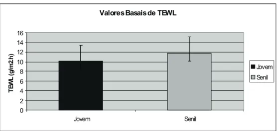
Na figura 1 pode observar-se a comparação entre as PTEA basais obtidas nos dois grupos de voluntários (I, com idade inferior a 30 anos; II, com mais de 30 anos). Os valores observados mostraram-se relativamente próximos, não tendo sido encontradas diferenças estatisticamente significativas entre a pele jovem e a pele senil (p=0,117).
A fim de descrever e avaliar quantitativamente a dinâmica da água na pele e da função de barreira cutânea, aplicou-se a modelação matemática ao perfil da PTEA após o teste de oclusão nos diferentes grupos para extrair parâmetros com correspondência
[26, 32]
fisiológica . Através desta, são calculados o t1/2evap ( tempo de semi-vida de evaporação) e a
Results and Discussion
Figure I illustrates a comparison between the basal TEWL measured in the two groups (I- below 30 years of age; II- older than 30). It is apparent that the differences in this variable are minute (p=0.117).
The plastic occlusion stress test has been developed as a dynamic approach to the study of the skin water dynamics, where the response of the skin to a determined stimulum is evaluated and the factors that influence the skin hydric content in the deeper layers
[26, 32]
can be more thoroughly investigated . Two parameters can be calculated after the POST: t1/2evap
Valores Basais de TEWL
0 2 4 6 8 10 12 14 16 Jovem Senil T E W L (g /m 2. h ) Jovem Senil
Figura 1- Gráfico comparativo dos valores basais de PTEA medidos nas duas faixas
etárias (valores médios ±SD)
Figure 1- Comparison of the basal TEWL values obtained in the two
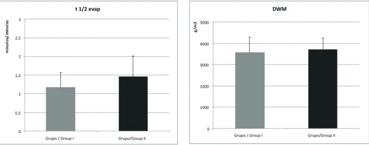
DWM (massa de água dinâmica). As curvas de desorção obtidas após uma oclusão de 2h, estão representadas nas Figuras 2a) e b). É possível notar que no grupo I o decaimento dos valores de PTEA obtidos após remoção da oclusão é rápido e bastante abrupto. Por outro lado, na pele senil o decaimento ocorre a um ritmo bastante mais lento.O grupo II proporcionou valores mais elevados de t1/2evap, cerca de 1,5 minutos, no entanto não tiveram uma diferença estatisticamente significativa em relação aos do grupo I (p = 0,187) (Fig.3). A DWM foi aproximada entre os dois grupos (p=0.243) (Fig. 4).
(evaporation half-life) and DWM (dynamic water mass). The desorption curves obtained after a 2 hours occlusion are represented in Figs. 2. a) and b). It can be observed that in group I the decay in TEWL obtained after occlusion is sharp and abrupt. On the other hand, in the results obtained for senile skin, the decay occurs at a slower pace.
Group II provided slightly higher values of t1/2evap, around 1.5 minutes, however this result was not statistically different from the values calculated for the younger group (p = 0.187) (Fig.3). The DWM was also similar between the two groups (p=0.243) (Fig. 4).
Grupo / Group I 0 5 10 15 20 25 30 35 40 45 50 0 10 20 30 time (min) T E W L (g /m 2. h ) Observado / Observed Calculado / Calculated Grupo / Group II 0 5 10 15 20 25 30 35 40 45 50 0 10 20 30 time (m in) T E W L (g /m 2 .h ) Observado / Observed
Cal cul ado / Cal cul ated
Figuras 2 a) e b)- Gráficos das curvas comparativas da perda transepidérmica de água pelo tempo, obtidas após
uma oclusão de 2h dos Grupos I (a)) e II (b)) (valores médios ±SD).
Figures 2 a) and b)- Graphs of the comparative curves of transepidermal water loss over time, obtained after a
two hours occlusion, of Groups I (a)) and II (b)) (mean values ±SD)
a)
b)
É consensual que o envelhecimento causa um conjunto de alterações que, entre outras, levam à perda de competência na função de barreira. Por outro lado, a determinação da PTEA é tradicionalmente considerada a metodologia de referência para avaliação in vivo da função de barreira cutânea, tendo sido extensamente aplicada com este fim. No entanto, apesar de se terem observado boas correlações entre a PTEA e a idade cronológica na comparação entre bebés prematuros e
[43]
recém-nascidos de gravidezes de termo , não foram estabelecidas de forma conclusiva diferenças neste parâmetro entre adultos e indivíduos de idade
[39,44,45]
avançada . No presente estudo também ficou estabelecido que os valores basais de PTEA não variam significativamente com a idade.
[46]
O estudo de Guy et al recorreu a uma abordagem dinâmica com oclusões de 12 horas, tendo sido possível determinar que a dinâmica de retenção de água diferiu de forma estatisticamente significativa entre os voluntários do grupo de média de idades maior e menor. Os voluntários mais velhos apresentavam valores mais elevados de PTEA 30 segundos após a remoção da oclusão e foram obtidos rácios PTEA 15 minutos/ PTEA basal mais baixos nos voluntários mais novos. A presente investigação utilizou a modelação matemática para obter parâmetros cinéticos representativos da dinâmica hídrica através do POST: t1/2evap e DWM. Tal como no estudo referido acima, foi possível notar que no grupo I o decaimento dos valores de PTEA obtidos após remoção da oclusão foi
It is widely recognized that ageing causes changes that, amongst other consequences, lead to a loss of skin barrier function. On the other hand, TEWL is normally considered the gold standard in the in vivo assessment of cutaneous barrier. Nevertheless, even though good correlations between TEWL and chronological age were established between premature infants relative to
[43]
that of full term infants , from adulthood through old age baseline TEWL does not appear to change
[39,44,45]
considerably with increasing age . The present study also confirmed this trend.
[46]
The study by Guy et al employed a dynamic approach using 12 hours occlusion and was able to establish statistically significant differences in the water dynamics of the young and old subjects. The older volunteers exhibited higher TEWL 30 seconds post-occlusion and lower 15 minutes post-occlusion to baseline TEWL rates were observed in the other group. The current investigation used mathematical modelling to generate kinetic parameters that reflected the hydric dynamics caused by a POST : t1/2evap and DWM. Akin to the study cited above, it could be observed that in Group I the TEWL values post-occlusion deceased in a fast and abrupt manner, whereas in senile skin the decay occurred at a much slower pace. Nevertheless, no statistically significant differences were established in the calculated kinetic parameters, even though t1/2evap was considerably higher in the older volunteers, i.e., there were no marked alterations in the barrier function within the t 1/2 evap 0 0,5 1 1,5 2 2,5 3
Grupo / Group I Grupo/Group II
m in u to s/ m in u te s DWM 0 1000 2000 3000 4000 5000
Grupo / Group I Grupo/Group II
g
/m
2
Figura 3- Comparação entre os t1/evap obtidos nos
dois grupos estudados (valores médios ±SD)
Figure 3- Comparison between the t1/2evap obtained
in groups I and II (mean values ±SD)
Figura 4- Comparação entre as DWM obtidas nos dois
grupos estudados (valores médios ±SD)
Figure 4- Comparison between the DWM obtained in
rápido e bastante abrupto, enquanto que na pele senil o decaimento ocorreu a um ritmo bastante mais lento. No entanto, não foi possível estabelecer diferenças estatisticamente significativas entre os parâmetros cinéticos calculados, embora o t1/2evap fosse mais elevado no grupo dos voluntários mais velhos, ou seja, não foram reveladas alterações muito marcadas na barreira cutânea nas faixas etárias estudadas. Este facto estará provavelmente ligado à relativamente baixa média de idades do grupo II (48,0 ± 12,6 anos) e seria recomendável alargar o estudo a voluntários com idade mais avançada (acima dos 60 anos) por forma a aumentar o fosso entre os dois grupos.
Conclusões
O trabalho realizado traz uma nova visão para o estudo do envelhecimento da pele. A medição da PTEA constitui uma valiosa ferramenta, sobretudo quando utilizada em conjunto com outras técnicas, no entanto, como mencionado anteriormente, é limitada na avaliação do estado da função barreira da pele. A modelação matemática das curvas de PTEA resultantes de um POST evita estas desvantagens ao fornecer informações adicionais a respeito da capacidade de retenção de água da pele.
A análise da dispersão dos resultados da presente investigação indica que o método tem boa reprodutibilidade e corrobora o estudo anterior que estabeleceu uma diminuição da função de barreira no envelhecimento cutâneo.
Concluindo, este estudo demonstrou que a metodologia apresenta uma boa capacidade discriminativa e valida os objetivos propostos, confirmando que também pode ser aplicada ao estudo de envelhecimento da pele. Além disso, indicia que pode ser estendido à caracterização de diferentes patologias cutâneas ou ser usado em ensaios de eficácia e segurança de medicamentos ou cosméticos.
two age groups. Such fact is probably linked to the relatively “young” age of the volunteers included in group II (48.0 ± 12.6 years), which seems to indicate that it would be advisable to broaden the study to older subjects (over 60 years old) in order to expand the age gap between the groups.
Conclusions
This study brings a new insight into the study of skin ageing. TEWL measurements are an invaluable tool, especially when combined with other techniques, however, as previously mentioned, it has limitations in the assessment of skin barrier function. The mathematical modeling of the TEWL desorption curves obtained after a POST can overcome such difficulties since it provides additional information on water accumulation capacity.
An analysis of the dispersion of the results indicates that the method provided a good reproducibility and the results corroborated the findings by other authors, that were able to find age dependant changes in the skin barrier.
In conclusion, this investigation illustrates the good discriminative capacity of the methodology and confirms its applicability to the study of ageing. It also indicates that it can be employed in the characterization of cutaneous pathologies, as well as in the assessment of the efficacy and safety of drugs and cosmetics.
Referências / References
[1]. Fresnel M, Hernandez M, Madeleine M. Manual de Cosmetologia. 3ª ed. Rio de Janeiro: Revinter; 1996. [2]. Kede MP, Sabatovich O. Dermatologia Estética. São Paulo: Editora Atheneu; 2004.
[3]. Viglioglia PA, Rubin J. Cosmetria II – Ciencia que comprende la atención cosmética de la piel sana o enferma. Buenos Aires: Americana de Publicaciones SA.; 1991.
[4]. Proksch E, Brandner J, Jensen J. The skin: an indispensable barrier. Exp Dermatology 2008; 17: 1063-72.
[5]. Ritcher T, Peuckert C, Sattler M, Koenig K, Riemann I, Hintze U, Wittern KP, Wiesendanger R, Wepf R. Dead but highly dynamic - the Stratum corneum is divided into three hydration zones. Skin Pharmacol Physiol 2004;17:246-57.
[6]. Cuzzi-Maya T, Piñeiro-Maceira J. Dermopatologia: Bases para Diagnóstico Morfológico. São Paulo :Editora Roca; 2001. [7]. Dal´Belo SE. Avaliação da eficácia fotoprotetora, penetração cutânea e segurança de formulações cosméticas contendo extratos de chá verde e Ginkgo biloba. PhD Thesis, Faculdade de Ciências Farmacêuticas de Ribeirão Preto, Universidade de São Paulo, Ribeirão Preto, 2008.
[8]. Smith, W. Stratum corneum barrier integrity controls skin homeostasis. Int J of Cosmet Sci 1999; 21(2):99-106.
[9]. Leveque, J.L. Measurement of transepidermal water loss in Cutaneous Investigation in Health and Disease - Noninvasive Methods and instrumentation. New York: Marcel Dekker; 1989.
[10]. Barel AO, Clarys P. Study of the stratum corneum barrier function by transepidermal water loss measurements: comparison between two commercial instruments: Evaporimeter and Tewameter. Skin Pharmacol. 1995;8(4):186-95.
[11]. Rosado C, Rolim MI, Rodrigues LM. In Vivo Study of the performance of two models of evaporimeters in controlled conditions: Static and Dynamic Measurements. Rev. Lusófona de Ciências e Tecnologias da Saúde, 2005; (2) 2: 89-93.
[12]. Machado M, Salgado TM, Hadgraft J, Lane ME. The relationship between transepidermal water loss and skin permeability. Int J Pharm. 2010; 384: 73–7.
[13]. Wilson D, Maibach HI. TEWL and the newborn. In: Bioengineering of the skin: water and the Stratum corneum. Elsner P, Berardesca E, Maibach HI. editors London: CRC Press; 1994 p.115-132.
[14]. Rougier A, Lotte C, Corcuff P, Maibach HI. Relationship between skin permeability and corneocyte size according to anatomical site, age and sex in man. J. Soc. Cosmet. Chem 1988; 39:15-26.
[15]. Tupker RA, Coenraads PJ, Pinnagoda J, Nater JP. Baseline transepidermal water loss (TEWL) as a prediction of susceptibility to sodium lauryl sulphate. Contact Dermatitis 1989;20: 265-269.
[16]. Baker H. Deperdition d'eau par voie trans-epidermique. Ann Dermatol Syphiligr 1971;98: 289-296.
[17]. Grice KA. Transepiderma water loss, in The Physiology and Pathophysiology of the Skin, Jarret A ed. Academic Press, London, 1980, 2115-2127
[18]. Roskos KV, Maibach HI, Guy RH. The effect of aging on percutaneous absorption in man, J Pharm Biopharm, 1989;17:617-630.
[19]. Wilhelm KP, Cua AB, Maibach HI. Skin aging. Effect on transepidermal water loss, stratum corneum hydration, skin surface pH, and casual sebum content. Arch Dermatol. 1991;127(12):1806-9
[20]. Leveque JL, Corcuff P, De Rigal J, Agache P. In vivo studies of the evolution of physical properties of the human skin with age. Int. J. Dermatol. 1984;23:322-329.
[21]. Jemec G, Agner T, Serup J. Transonychial water loss. Relation to sex, age and nailplate thickness. Br J Dermatol 1989;121:443-446.
[22]. Thune P, Nilsen T, Hanstad K., Gustavsen T, Dahl HL. The water barrier function of the skin in relation to the water content of Stratum corneum, pH and skin lipids. Acta Dermatovenereologica: 1988;68:277- 283.
[23]. Rosado C, Rodrigues LM. Assessment of dry skin using dynamic methods. J Appl Cosmetol 2006; 24: 139-157.
[24]. Chilcott RP, Dalton CH, Emanuel AJ. The effects of physical damage on TEWL. In: The essential stratum corneum Marks R, Leveque JL, Voegeli R. editors. London: Martin Dunitz Ltd; 2002. p 287–302.
[25]. Chilcott RP, Dalton CH, Emanuel AJ, Allen CE, Bradley ST. Transepidermal water loss does not correlate with skin barrier function in vitro. J Invest Dermatol 2002; 118: 871–875.
[26]. Bashir SJ, Chew AL, Anigbogu A, Dreher F, Maibach HI. Physical and physiological effects of stratum corneum tape stripping. Skin Res Technol 2001; 7 (1):40–48.
[27]. Berardesca E, Herbst R, Maibach HI. Plastic occlusion stress test as a model to investigate the effects of skin delipidization on the stratum corneum water holding capacity in vivo. Dermatology 1993; 187 (2): 91–94.
[28]. Rodrigues LM, Pinto PC, Pereira LM. Quantitative description of human skin water dynamics by a disposition decomposition analysis (DDA) of trans-epidermal water loss and epidermal capacitance. Skin Res Technol 2003; 9:24–30. [29]. Berardesca E, Maibach HI. The plastic occlusion stress test (POST) as a model to investigate skin barrier function. In: Dermatologic Research Techniques MAIBACH, H.I. editors Boca Raton: CRC PRESS 1996, p.179-186.
[30]. Elsner P, Berardesca E, Maibach HI. Bioengineering of the skin: Water and Stratum corneum. Boca Raton: CRC Press;1994.
[31]. Rosado C, Pinto P, Rodrigues LM. Modeling TEWL- desorption curves: a new practical approach for the quantitative in vivo assessment of skin barrier. Exp Dermatol, 2005;14 (5):386-390.
using the plastic occlusion stress test (POST). Contact Dermatitis 1990; 23 (1):1–5.
[33]. Berardesca E, Fideli D, Borroni G, Rabiosi G, Maibach HI. In vivo hydration and water retention capacity of the stratum corneum in clinically uninvolved skin in atopic and psoriatic patients. Acta Derm Venereol (Stockh) 1990; 70:400–404.
[34]. Rodrigues LM, Pinto PC, Galego N, Da Silva PA, Pereira LM. Transepidermal water loss kinetic modeling approach for the parameterization of skin water dynamics. Skin Res Technol 1999; 5: 72–82.
[35]. Pinto PC, Pereira LM, Minhós R, Rodrigues LM. Testing the discriminative capacity of compartmental modelling for the analysis of the in vivo epidermal water content changes following topical application under occlusion. IFSCC Magazine 2002; 5: 1–6.
[36]. Rosado C, Pinto P, Rodrigues LM. Assessment of moisturizers and barrier function restoration using dynamic methods. Skin Res Technol 2009; 15: 77–83.
[37]. Berardesca E, Elsner P. Dynamic measurements: the plastic occlusion stress test (POST) and the moisture accumulation test (MAT). In: Bioengineering of the skin: water and the stratum corneum. Elsner P, Berardesca E, Maibach HI. editors. London: CRC Press;1994.p.97–102.
[38]. Berardesca E, Maibach HI. Monitoring the water holding capacity in visually non irritated skin by plastic occlusion stress test (POST). Clin Exp Dermatol 1990; 15:107–110. 1990.
[39]. Fluhr JW, Lazzerini S, Distante F, Gloor M, Berardesca E. Effects of prolonged occlusion on stratum corneum barrier function and water holding capacity. Skin Pharmacol Appl Skin Physiol 1999; 12: 193–198.
[40]. GRICE, K.A.; BETTLEY, F.R.; Skin water loss and accidental hypothermia in psoriasis, ichthyosis and erythrodermia. Br Med J. p. 195-201. 1967.
[41]. Goodyear MD, Krleza-Jeric K, Lemmens T. The Declaration of Helsinki, BMJ 2007; 335:624-625.
[42]. Rogiers, V. EEMCO Guidance for the assessment of transepidermal water loss in cosmetic sciences. Skin Pharmacol Appl Skin Physiol. 2001; 14:117–128.
[43]. Hammarlund K, Sedin G.Transepidermal water loss in newborn infants. III. Relation to gestational age. Acta Paediatr Scand. 1979;68(6):795-801.
[44]. Grice KA, Bettley FR. Skin water loss and accidental hypothermia in psoriasis, ichthyosis, and erythroderma. Br Med J. 1967 ;28;4(5573):195-8.
[45]. Menon GK, Kligman AM. Barrier functions of human skin: a holistic view. Skin Pharmacol Physiol. 2009;22(4):178-89. [46]. Roskos KV, Guy RH Assessment of skin barrier function using transepidermal water loss: effect of age. Pharm Res. 1989;6(11):949-53.