Dynamic
11C-Choline PET / CT for the primary diagnosis of
prostate cancer
_______________________________________________
Shay Golan
1, Meital Nidam
2, Hanna Bernstine
2,Jack Baniel
1, David Groshar
21 Institute of Urology, Rabin Medical Center – Beilinson Hospital, Petach Tikva, Sackler Faculty of Medicine, Israel; 2 Department of Nuclear Medicine, Rabin Medical Center – Beilinson Hospital, Petach Tikva, Sackler Faculty of Medicine, Israel
INTRODUCTION
Prostate cancer is the second most common cancer in men worldwide. An estimated 1.1 million new cases were recorded in 2012 (1). The diagnosis is based on direct systematic sampling of the pros-tate guided by transrectal ultrasound. This invasive method is necessary because gray-scale ultrasound itself is unreliable in detecting cancerous lesions
(2). Nevertheless, the standard 12-core biopsy can miss up to one-third of cancers, resulting in repea-ted and more extensive biopsies (3).
Efforts have been made to develop new imaging modalities for the diagnosis and cha-racterization of prostate cancer. This has become of paramount importance, given the emergen-ce of new therapeutic approaches for localized prostate cancer (4).
ABSTRACT
Objectives: To test the ability of dynamic 11C-PET / CT to discriminate cancerous tissue
from background tissue in patients with localized prostate cancer.
Materials and Methods: Twenty-four consecutive patients with prostate cancer were prospectively evaluated with dynamic 11C-choline PET / CT prior to radical
prostatec-tomy. The PET / CT scan was divided into 18 sequences of 5 seconds each, followed by 9 sequences of 60 seconds each. Whole-mount sections of harvested prostates served as reference standards. Volumes of interest were positioned on the dynamic PET / CT images and the following quantitative variables were calculated: perfusion coefficient (K1), washout constant (K2), area under the curve (AUC) at 175 and 630 seconds, and average and maximum standardized uptake values (SUVavg, and SUVmax). Wilcoxon signed-ranks test was used to compare benign and cancerous areas of the prostate.
Results: Areas of cancerous tissue were characterized by higher SUVavg and SUVmax than areas of benign tissue (3.67 ± 2.7 vs. 2.08 ± 1.3 and 5.91 ± 4.4 vs. 3.71 ± 3.7, re-spectively, P < 0.001), in addition to a higher K1 (0.95 ± 0.58 vs. 0.43 ± 0.24, P < 0.001) and greater cumulative tracer uptake, represented by the AUC at 175 and 630 seconds (P <0.001). No associations were found between dynamic parameters and preoperative prostate specific antigen level or Gleason score.
Conclusions: In this pilot study, 11C-choline PET / CT demonstrated increased tracer
uptake with higher values of static and dynamic parameters in areas of prostate can-cer compared to areas of benign tissue. Larger studies are warranted to validate these results and examine the potential applicability of 11C-choline dynamic PET / CT for the
diagnosis of prostate cancer.
ARTICLE INFO
Keywords:
Positron-Emission Tomography; Tomography, X-Ray Computed; Prostatic Neoplasms
Int Braz J Urol. 2018; 44: 900-5
_____________________ Submitted for publication: January 18, 2018 _____________________ Accepted after revision: May 31, 2018
Active surveillance and focal therapy are being applied more often in patients with low-risk localized prostate cancer, requiring improved tu-mor identification and classification. Better deli-neation of tumor extent would also assist surgical planning in patients with high-risk disease under-going radical prostatectomy.
At present, patients with a suspicion of prostate cancer and negative biopsy findings are referred for multiparametric magnetic resonance imaging which has shown good sensitivity for hi-gh-grade tumors and an ability to detect anterior tumors, sometimes missed by systematic biopsy (5). However, its accuracy is often poor for low--volume or low-grade tumors (6), and despite the introduction of a scoring system (PIRADS), inter--reader variability remains a concern (7).
In addition, positron emission tomography (PET) has been examined as a potential means of visualizing primary prostate cancer and evalua-ting nodal extensions. By using a radiotracer for cancer diagnosis, clinicians can obtain informa-tion not only on the morphological appearance of the tumor but also on its biological behavior. Attention has recently been directed at 11
C-cho-line PET / computed tomography (CT). ChoC-cho-line is a precursor of phosphatidylcholine, an essential element of cell membrane phospholipids. It has therefore been postulated that the ability of 11
C--choline PET to demonstrate cancerous lesions is related to the increased activity of choline kina-se in tumor cells (8). Studies of the accuracy of
11C-choline PET / CT for the primary diagnosis
of prostate cancer reported a sensitivity of 72% to 87% and a specificity of 62% to 84% (9-12). However, the vast majority evaluated radiotra-cer uptake semi-quantitatively, based on the standardized uptake value (SUV) at a single time point following injection. We speculated that tis-sue delineation could be improved by adding a dynamic (blood perfusion) phase to the PET / CT scan, which would allow for the assessment of multiple reactions involving choline uptake.
The aim of the present feasibility stu-dy was to examine the value of stu-dynamic 11
C--choline PET / CT for the primary diagnosis of prostate cancer in patients undergoing radical prostatectomy.
MATERIALS AND METHODS
Patients
A prospective case-series design was used. The study group consisted of consecutive patients with localized biopsy-proven adenocarcinoma of the prostate who underwent robot-assisted radical prostatectomy performed by a single surgeon (J.B.) at a tertiary medical center in 2012-2014. The local institutional review board approved the study. All patients provided written informed consent to parti-cipate in the study.
Data were collected on demographics, pros-tate-specific antigen (PSA) values, and preoperative biopsy results. Standard 11C-choline PET / CT was
performed 2 months or less before surgery and at le-ast 3 months after prostate biopsy.
PET / CT protocol
Images were obtained using an integrated 8-slice PET-CT scanner (Discovery ST, GE Medical Systems, and Milwaukee, WI). The protocol included a dynamic PET acquisition limited to a single bed po-sition and centered on the prostate, starting with the injection of 10-20mCi (370-740 MBq) of 11C-choline
with a low-dose CT scan. After a scout view of the pelvis was obtained, the study centered on the pros-tate, with PET coverage of 15.3 cm. A non-diagnostic low-dose (30 mA) CT scan was acquired. 11C-choline
was injected as a rapid bolus flushed with 50 cc sa-line 0.9% at a rate of 5.0 mL / sec using an automa-tic power injector (Dual-shot, Nemoto, Japan). The dynamic study was acquired as a 3D scan (matrix size 64 x 64, 3.27-slice thickness) consisting of 18 sequential frames of 5 seconds each followed by 9 frames of 60 seconds each. PET emission data after attenuation correction was reconstructed with a 3D OSEM algorithm (2 iterations, 20 subsets).
Data processing and kinetic analysis
within the frame (Figure-1). Time activity curves were generated for the mean activities of each VOI and fitted by linear regression function (Fi-gure-2). A one-compartment model was used to assess tracer kinetics.
In pharmacokinetic modeling, tracer kine-tics are separated into compartments and their in-teractions are represented by rate constants. The one-compartment pharmacokinetic model is used to simplify drug disposition in plasma and highly per-fused tissue, under the assumption that there is an instantaneous mixing (drug behaves uniformly in a single unit) when the drug reaches the bloodstream. Changes in plasma concentration are equivalent to changes in tissue concentrations; dosing, sampling, and elimination occur from the central compartment. Total drug distribution is calculated as the ratio be-tween the rate constant from the peripheral to the central compartment (K1) and the rate constant from the central to the peripheral compartment (K2).
The following parameters were used to eva-luate suspected prostate tumor against background prostate tissue: transport constant (perfusion) K1 (mL / cm / min), rate constant K2 (l / min), total distribu-tion volume (VT = K1 / K2, mL / ccm), area under the curve for tracer uptake during 175 seconds (AUC175) and 630 seconds (AUC630), SUVavg ,and SUVmax.
Standard of reference
The surgical specimens served as the stan-dard of reference. The harvested prostates were submitted for whole-mount histology. Axial sec-tions of 3-5 mm were obtained in a plane per-pendicular to a theoretical line between the base and the apex. Orientation was preserved by inking right and left margins with different colors (right--black, left-red) and by anatomical landmarks (i.e., seminal vesicle and urethra). Whole-mount sections of 4 µm each were stained with hematoxylin and eosin according to standard procedure. A
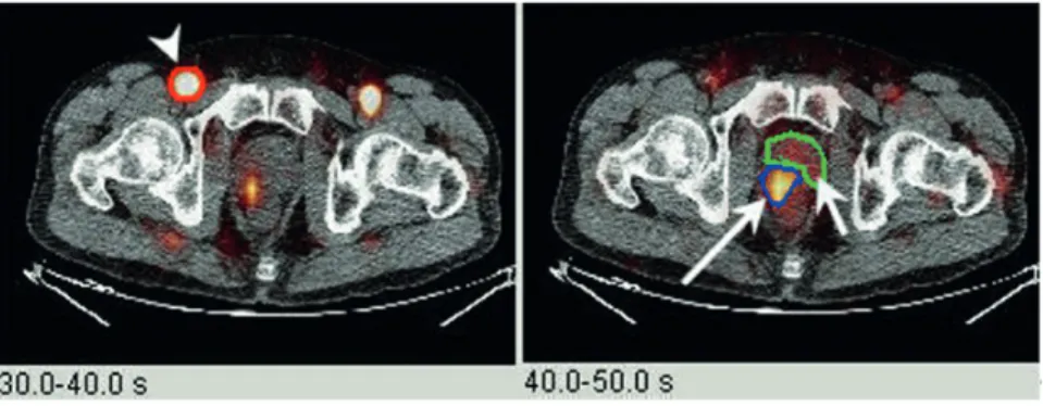
Board-Figure 1 - 11C-choline PET / CT dynamic blood flow. Axial fused PET / CT sequential blood flow images (left: 30-40 seconds after
injection; right: 40-50 seconds after injection) showing preferential arterial supply to the prostate tumor relative to background prostate tissue (arrowhead = iliac artery; large arrow = prostate cancer; small arrow = background prostate tissue).
Figure 2 - Linear fitted time-activity curves for prostate cancer (lighter line) and background prostate tissue (darker line).
Prostate cancer
Background prostate
minutes -26 [10,5, 24,8]
-certified uropathologist (S.Z.) reviewed the slides and identified cancer foci by laterality and prostatic areas (base, mid-gland, and apex) and with respect to anatomic landmarks, as previously described.
Matching PET / CT and histopathology
A single investigator (H.B), specialized in nuclear medicine, assessed the correspondence between the PET / CT images and histopathology findings. The most evident locations of the prosta-te tumor and normal prostaprosta-te tissue segment were visually identified on the PET / CT images. The lo-cation of the prostate tumor was then confirmed from the pathological report with respect to anato-mic landmarks. Thereafter, an oval region of interest (mean size 15 mm) was placed on the most evident tumor and in an area free of tumor.
Statistical analysis
Continuous parameters are given as mean ± SD Static (SUVmax, SUVavg) and dynamic (K1, K2, AUC and Vt) parameters of 11C choline PET / CT were
com-pared between cancerous and non-cancerous regions using the Wilcoxon signed-rank test. A p value of < 0.05 was considered statistically significant.
RESULTS
The study cohort included 24 patients of mean age 64 ± 5.4 years. The average interval from PET / CT scanning to surgery was 14 days (range, 3 to 61 days). Mean PSA level was 7.5 ± 5.3 ng / mL. On final surgical pathology, 16 patients (66%) had tumor confined to the prostate (AJCC stage II). Glea-son score was 6 in 6 patients (25%), 7 in 16 patients (66%), and 9 in 2 patients (9%). A summary of the clinical and pathologic characteristics is presented in Table-1.
Dynamic PET / CT parameters
Tracer uptake was observed in all 24 pa-tients. SUVavg and SUVmax were significantly higher in areas of cancerous tissue than areas of benign tissue (3.67 ± 2.7 vs. 2.08 ± 1.3 and 5.91 ± 4.4 vs. 3.71 ± 3.7, respectively, P < 0.001). There was a statistically significant difference in mean dyna-mic parameters of AUC175, AUC630, and K1 between
tumor and non-tumor zones (P < 0.001). Findings for constant of tracer washout from the tissue (K2) achieved borderline significance (P = 0.076). Table-2 shows the dynamic PET / CT parameters in benign and malignant tissue, and Figure-2 shows the linear fitted time-activity curves for prostate cancer and background prostate tissue. No associations were found between either Gleason scores or PSA levels and PET / CT parameters (P = 0.18 and P = 0.47, respectively).
DISCUSSION
The present pilot study demonstrates the robust ability of dynamic 11C-choline PET / CT to
differentiate cancerous from benign prostatic tissue. Statistically significant differences were found in both static and dynamic PET / CT parameters.
The moderate-range accuracy previous-ly reported for diagnostic 11C-choline PET / CT in
prostate cancer (9-12) was largely based on static parameters. However, while static parameters repre-sent the sum of multiple reactions that occur during tracer uptake in a tissue, dynamic descriptors add sequential information that may assist in
charac-Table 1. Clinical and pathologic characteristics of 24 patients with prostate cancer
Characteristic Value
Age (yr), mean (SD) 64.1 (5.4)
PSA ( ng/ml), mean (SD) 7.5 (5.3)
Prostate volume (cm3), mean (SD) 52.7 (28.3)
Final pathologic grade, n (%)
Gleason (3+3)=6 Gleason (3+4)=7 Gleason (4+3)=7 Gleason (4+5)=9 Gleason (5+4)=9
6 (25) 13 (55) 3 (12) 1 (4) 1 (4)
Final pathologic stage, n (%)
pT2a pT2b pT2c pT3a pT3b
5 (21) 1 (4) 10 (42)
6 (25) 2 (8)
terizing the examined tissue. Two recent publica-tions examined the diagnostic value of dynamic
18F-choline PET / CT for prostate cancer. In a study
of 64 patients with prostate cancer, 19 with a pri-mary diagnosis and 45 after biochemical recurrence, Takesh (13) reported a significant increase in mean dynamic constants (transport and rate) as well as in maximal SUV in cancerous areas compared to benign tissue. Similarly, Mathieu et al. (14) evalu-ated 39 patients with prostate cancer (18 with an initial diagnosis before any therapy) and found that tracer uptake was more rapid and intense in areas of cancerous lesions than in benign tissue. Although these studies provided encouraging results for the diagnostic ability of dynamic PET / CT, they were limited by a retrospective design, heterogenic pa-tient sample (primary and post-treatment disease), and non-uniform standard reference.
The present study is distinctive in that it prospectively evaluated a cohort of consecutive pa-tients, all with a diagnosis of localized prostate can-cer who were primarily treated with radical pros-tatectomy. Whole-mount sections of the surgical specimen served as the reference standard for each patient. Furthermore, we used 11C choline as the
ra-diotracer. 11C choline is harder to handle than 18
F--choline but it has a shorter half-life. We believe this is an important advantage because the rapid clea-rance in blood of 11C choline and its rapid uptake
by prostate cells make it possible for clinicians to obtain early PET images without significant
excre-tion of the tracer into the urine. The higher urinary excretion of 18F choline is a potential confounder as
it may mask background activity (15).
We observed fast and intense uptake of 11C
choline in cancerous tissue, with higher values of K1 in malignant regions in the prostate and a higher cumulative amount of tracer in the cancerous tissue, as demonstrated by the AUC. Contractor et al. (8) showed a relationship between increased 11
C-choli-ne uptake and overexpression of choliC-choli-ne kinase. In their study, malignant lesions overexpressed choline kinase in both the cytoplasm and nucleus, and both SUVavg and SUVmax were strongly associated with choline kinase staining.
Despite the rapid 11C-choline uptake and
malignant nature of the tissue, we found no as-sociation of tracer uptake with the preoperative PSA level or Gleason score. This finding might be explained by the small size of the cohort and the small number of patients (n = 2) who had a high Gleason score on final pathologic analysis. Pre-vious studies, using static 11C-choline PET / CT,
showed limited ability to discriminate cancerous areas from areas of prostatitis (16). Dynamic 11
C--choline PET / CT appears to better differentiate prostatitis from cancer, however the small number of patients with prostatitis in our study (n = 3) mandates further evaluation of this aspect.
Indeed, the small number of patients is the main limitation of our study. Another limitation is the fact that a single radiologist analyzed and
in-Table 2 - Comparison of 11C choline PET/CT parameters between prostate cancer and benign prostatic tissue.
Parameter (mean±SD) Benign prostatic tissue Prostate cancer 95% CI P value
SUVavg 2.08±1.3 3.67±2.7 0.90-2.25 <0.001
SUVmax 3.66±3.7 5.91±4.4 1.65-2.83 <0.001
AUC175 (mL/ccm/min) 187.2±134 386.4±332 108.02-290.32 <0.001
AUC630 (mL/ccm/min) 964.8±644 1954.5±1734 488.44-1490-89 <0.001
Perfusion coefficient (K1) (l/ccm/min) 0.43±0.24 0.95±0.58 0.31-0.71 <0.001
Washout constant (K2) (1/min) 0.18±0.09 0.22±0.13 (-0.00-0.08) 0.076
Vt (K1/K2) (mL/ccm) 2.50±1.11 4.44±1.38 1.37-2.49 <0.001
terpreted the PET / CT images. Importantly, while novel imaging modalities using radiolabeled tracers with PSMA have shown promising results, the exact role of 68Ga-PSMA-PET / CT in primary prostate
cancer is not yet entirely clear (17). Our preliminary results could potentially lead to a change in the way choline is being used as a tracer and improve the focal delineation of primary prostate cancer.
In conclusion, Dynamic 11C-choline PET /
CT successfully distinguishes areas of prostate can-cer from benign tissue based on values of static and dynamic parameters. Larger studies are warranted to validate our results and to examine the applicabi-lity of 11C-choline dynamic PET / CT in the primary
diagnosis of prostate cancer.
CONFLICT OF INTEREST
None declared.
REFERENCES
1. Ferlay J SI, Ervik M, Dikshit R, Eser S, Mathers C, Rebelo M, Parkin DM, Forman D, Bray, F. GLOBOCAN 2012 v1.0, Cancer Incidence and Mortality Worldwide: IARC CancerBase No. 11 2013. Available at. <http://globocan.iarc.fr>. Accessed may 12, 2017.
2. Kuligowska E, Barish MA, Fenlon HM, Blake M. Predictors of prostate carcinoma: accuracy of gray-scale and color Doppler US and serum markers. Radiology. 2001;220:757-64.
3. Eichler K, Hempel S, Wilby J, Myers L, Bachmann LM, Kleijnen J. Diagnostic value of systematic biopsy methods in the investigation of prostate cancer: a systematic review. J Urol. 2006;175:1605-12.
4. Cooperberg MR, Carroll PR. Trends in Management for Patients With Localized Prostate Cancer, 1990-2013. JAMA. 2015;314:80-2.
5. N. Mottet JB, E. Briers, R.C.N. van den Bergh, M. Bolla, N.J. van Casteren, P. Cornford, et al. EAU Guidelines on Prostate Cancer 2015 [cited 2015]. Available at. <http://uroweb.org/wp-content/ uploads/EAU-Guidelines-Prostate-Cancer-2015-v2.pdf>. 6. Bratan F, Melodelima C, Souchon R, Hoang Dinh A,
Mège-Lechevallier F, Crouzet S, et al. How accurate is multiparametric MR imaging in evaluation of prostate cancer volume? Radiology. 2015;275:144-54.
7. Vaché T, Bratan F, Mège-Lechevallier F, Roche S, Rabilloud M, Rouvière O. Characterization of prostate lesions as benign or malignant at multiparametric MR imaging: comparison of three scoring systems in patients treated with radical prostatectomy. Radiology. 2014;272:446-55.
8. Contractor K, Challapalli A, Barwick T, Winkler M, Hellawell G, Hazell S, et al. Use of [11C]choline PET-CT as a noninvasive method for detecting pelvic lymph node status from prostate cancer and relationship with choline kinase expression. Clin Cancer Res. 2011;17:7673-83.
9. Farsad M, Schiavina R, Castellucci P, Nanni C, Corti B, Martorana G, et al. Detection and localization of prostate cancer: correlation of (11)C-choline PET/CT with histopathologic step-section analysis. J Nucl Med. 2005;46:1642-9.
10. Giovacchini G, Picchio M, Coradeschi E, Scattoni V, Bettinardi V, Cozzarini C, et al. [(11)C]choline uptake with PET/CT for the initial diagnosis of prostate cancer: relation to PSA levels, tumour stage and anti-androgenic therapy. Eur J Nucl Med Mol Imaging. 2008;35:1065-73.
11. Testa C, Schiavina R, Lodi R, Salizzoni E, Corti B, Farsad M, et al. Prostate cancer: sextant localization with MR imaging, MR spectroscopy, and 11C-choline PET/CT. Radiology. 2007;244:797-806.
12. Scher B, Seitz M, Albinger W, Tiling R, Scherr M, Becker HC, et al. Value of 11C-choline PET and PET/CT in patients with suspected prostate cancer. Eur J Nucl Med Mol Imaging. 2007;34:45-53.
13. Takesh M. Kinetic Modeling Application to (18) F-fluoroethylcholine Positron Emission Tomography in Patients with Primary and Recurrent Prostate Cancer Using Two-tissue Compartmental Model. World J Nucl Med. 2013;12:101-10. 14. Mathieu C, Ferrer L, Carlier T, Colombié M, Rusu D,
Kraeber-Bodéré F, et al. Assessment of Lymph Nodes and Prostate Status Using Early Dynamic Curves with (18)F-Choline PET/CT in Prostate Cancer. Front Med (Lausanne). 2015;2:67.
15. Pantaleo MA, Nannini M, Maleddu A, Fanti S, Ambrosini V, Nanni C, et al. Conventional and novel PET tracers for imaging in oncology in the era of molecular therapy. Cancer Treat Rev. 2008;34:103-21.
16. Souvatzoglou M, Weirich G, Schwarzenboeck S, Maurer T, Schuster T, Bundschuh RA, et al. The sensitivity of [11C]choline PET/CT to localize prostate cancer depends on the tumor configuration. Clin Cancer Res. 2011;17:3751-9.
17. Udovicich C, Perera M, Hofman MS, Siva S, Del Rio A, Murphy DG, et al. (68)Ga-prostate-specific membrane antigen-positron emission tomography/computed tomography in advanced prostate cancer: Current state and future trends. Prostate Int. 2017;5:125-9.
_______________________ Correspondence address: