Submitted 4 May 2014 Accepted 25 August 2015 Published17 September 2015
Corresponding author Christophe Hendrickx,
christophe.hendrickx@hotmail.com
Academic editor Andrew Farke
Additional Information and Declarations can be found on page 29
DOI10.7717/peerj.1245
Copyright 2015 Hendrickx et al.
Distributed under
Creative Commons CC-BY 4.0
OPEN ACCESS
The non-avian theropod quadrate I:
standardized terminology with an
overview of the anatomy and function
Christophe Hendrickx1,2,6, Ricardo Ara ´ujo2,3,4,5and Oct´avio Mateus1,2 1Departamento de Ciˆencias da Terra, Universidade Nova de Lisboa, GeoBioTec, Faculdade de
Ciˆencias e Tecnologia, Caparica, Portugal 2Museu da Lourinh˜a, Lourinh˜a, Portugal
3Huffington Department of Earth Sciences, Southern Methodist University, Dallas, TX, USA 4Instituto Superior T´ecnico, Universidade de Lisboa, Lisboa, Portugal
5Museum f¨ur Naturkunde, Berlin, Germany
6Current affiliation: Evolutionary Studies Institute, Center of Excellence in Palaeosciences, University of the Witwatersrand, South Africa
ABSTRACT
The quadrate of reptiles and most other tetrapods plays an important morpho-functional role by allowing the articulation of the mandible with the cranium. In Theropoda, the morphology of the quadrate is particularly complex and varies importantly among different clades of non-avian theropods, therefore conferring a strong taxonomic potential. Inconsistencies in the notation and terminology used in discussions of the theropod quadrate anatomy have been noticed, including at least one instance when no less than eight different terms were given to the same structure. A standardized list of terms and notations for each quadrate anatomical entity is proposed here, with the goal of facilitating future descriptions of this important cranial bone. In addition, an overview of the literature on quadrate function and pneumaticity in non-avian theropods is presented, along with a discussion of the inferences that could be made from this research. Specifically, the quadrate of the large majority of non-avian theropods is akinetic but the diagonally oriented intercondylar sulcus of the mandibular articulation allowed both rami of the mandible to move laterally when opening the mouth in many of theropods. Pneumaticity of the quadrate is also present in most averostran clades and the pneumatic chamber—invaded by the quadrate diverticulum of the mandibular arch pneumatic system—was connected to one or several pneumatic foramina on the medial, lateral, posterior, anterior or ventral sides of the quadrate.
Subjects Paleontology
Keywords Quadrate, Terminology, Anatomy, Theropod, Dinosaur, Mandibular articulation
INTRODUCTION
pneumatic sinuses, and vascular passages (e.g.,Witmer, 1990;Witmer, 1997;Bakker, 1998; Sedlmayr, 2002;Kundr´at & Jan´aˇcek, 2007;Holliday & Witmer, 2008;Tahara & Larsson, 2011; seeAppendix S1).
Although the outward morphology of the quadrate is relatively simple, it varies signif-icantly among theropods in the structure of its head, mandibular articulation, quadra-tojugal contact and the presence of pneumatic openings, quadrate foramen, and lateral process (e.g.,Holtz, 2003;Therrien, Henderson & Ruff, 2005;Hone & Rauhut, 2010;Zanno & Makovicky, 2011). Variation in the quadrate morphology in the derived theropod group Aves has long been used as a mean of systematic significance (e.g.,Lowe, 1926;Samejima & Otsuka, 1987;Barbosa, 1990;Elzanowski, Paul & Stidham, 2001;Elzanowski & Stidham, 2010). Similarly, but to a lesser degree, the systematic potential of the quadrate bone has also been noted for non-avian theropods (Marya´nska & Osm´olska, 1997;Currie, 2006), highlighting the importance that should be given to the description of this bone in the liter-ature on non-avian theropod anatomy. Nevertheless, the terminology and abbreviations of the quadrate anatomy has been inconsistent in non-avian theropods, and several different anatomical terms for the same quadrate sub-entity are often used (seeAppendix S2). Al-though a list of anatomical terms has been given byBaumel & Witmer (1993),Elzanowski, Paul & Stidham (2001)andElzanowski & Stidham (2010)for the avian quadrate, the terminology proposed by these authors has not been applied to the description of the non-avian theropod quadrate hitherto. Indeed, the quadrate of birds has greatly changed in its morphology throughout the evolution of this clade and hence displays many features absent in more primitive theropods. Thus, many anatomical terms coined byBaumel & Witmer (1993),Elzanowski, Paul & Stidham (2001)andElzanowski & Stidham (2010) cannot be applied to the non-avian theropod quadrate. Moreover, some quadrate entities such as the quadrate foramen and the lateral process observable in non-avian theropods are absent in their avian descendants and do not appear in the list made by these authors.
The work presented here has two major aims. First, we propose a standardization of the anatomical terms for the quadrate sub-units, each associated with a two to four letters abbreviation and followed by a definition, in order to facilitate future descriptions of this bone in the literature. Second, we present and discuss the current knowledge on the function and pneumaticity of this important bone in non-avian theropods. A comprehensive study on the anatomy and phylogenetic potential of the non-avian theropod quadrate through cladistic and phylogenetic morphometric analyses will be provided in a companion article that will be published later.
Theropod classification
PROPOSED TERMINOLOGY OF THE QUADRATE
ANATOMY
Favored terminology
The anatomical terms of the theropod quadrate were grouped in five main sections: quadrate body, quadrate head, mandibular articulation, pterygoid flange, and pneumatic openings. The terms for each quadrate sub-units were selected by their relevance, signifi-cance and importance in the non-avian theropod literature. The non-standardized tradi-tional Romerian directradi-tional and anatomical terms (Romer, 1956;Wilson, 2006) were, there-fore, favored over the terminology of theNomina Anatomica Veterinaria(NAV) updated by theICVGAN (2012)and theNomina Anatomica Avium(NAA) provided byBaumel (1993) as Romerian terms are the most commonly used in the non-avian theropod literature (e.g.,Eddy & Clarke, 2011; C Hendrickx, pers. obs., 2015). Consequently, ‘anterior’ and ‘posterior’ are used as directional terms in lieu of the veterinarian alternatives ‘cranial’ and ‘caudal,’ respectively. Because non-avian archosaurs are the direct ancestors of birds,Harris (2004)recommended to adopt the NAA as the standardized nomenclature to describe all archosaurs (and even diapsids), yet we favorWilson’s (2006) opinion to retain Romerian terms for non-avian dinosaurs. As noted byWilson (2006), the Romerian nomenclature is the lingua franca for most of the dinosaur/archosaur literature. In addition, standard terminologies using Romerian terms are often proposed to describe the saurischian anatomy (e.g.,Wilson, 1999;Wilson et al., 2011;Hendrickx & Mateus, 2014;Hendrickx, Mateus & Ara´ujo, 2015). Comparison between the NAA nomenclature and the Romerian terminology here proposed for the quadrate anatomy is provided inFig. 1andTable 1.
Romer’s (1956) terminology of the quadrate is limited. He only expanded the vocabulary to describe this bone in reptiles to six terms, namely: the main body, quadrate shaft, quadrate foramen, quadrate head, quadrate flange and articular termination. Three terms were kept as such in the proposed terminology of the quadrate (i.e., quadrate shaft, quadrate foramen, and quadrate head) and the three others were slightly modified. The quadrate body (instead of “main body of [the] quadrate” sensuRomer, 1956: p. 640), mandibular articulation (instead of “articular termination” sensuRomer, 1956: p. 632) and pterygoid flange (instead of “quadrate flange” sensuRomer, 1956: p. 146) were chosen not only because they are more commonly used in the theropod literature currently describing the quadrate (C Hendrickx, pers. obs., 2015), but are also more specific of the loci of the anatomical sub-entity described. It should be noted that the pterygoid flange ofRomer (1956)describes a wing-like process of the pterygoid and not the anteriorly projected ramus of the quadrate.
Quadrate body
Quadrate body (qb)
Table 1 Proposed terminology and abbreviations of the non-avian theropod quadrate. Standardized terminology and abbreviations of the non-avian theropod quadrate and comparison with the terminology of the avian quadrate based onBaumel & Witmer (1993),Elzanowski, Paul & Stidham (2001)andElzanowski & Stidham (2010).
Non-avian theropod quadrate Avian theropod quadrate
Quadrate q Os quadratum (Quadratum)
Quadrate body qb Corpus quadrati
Quadrate shaft qs /
Quadrate ridge qr /
Quadrate ridge groove qrg /
Quadrate foramen qf /
Lateral process lpq /
Quadratojugal contact qjc Cotyla quadratojugalis
Ventral quadratojugal contact vqjc /
Dorsal quadratojugal contact dqjc /
Quadratojugal process qjp /
Ventral projection of the dorsal quadratojugal contact vpdq / Dorsal projection of the ventral quadratojugal contact dpvq /
Squamosal contact sqc /
Posterior fossa pfq /
Quadrate head qh Caput quadrati
Otic capitulum oca Capitulum oticum
Squamosal capitulum sca Capitulum squamosum
Intercapitular sulcus icas Incisura/Vallecula intercapitularis
Mandibular articulation mar Pars/Processus mandibularis
Ectocondyle ecc Condylus (mandibularis) lateralis
Entocondyle enc Condylus (mandibularis) medialis
Mediocondyle mec Condylus caudalis
Intercondylar sulcus ics Sulcus/Vallecula intercondylaris
Anterior intercondylar notch ain /
Posterior intercondylar notch pin /
Pterygoid flange pfl Processus orbitalis
Pterygoid contact ptc Condylus pterygoideus/Facies articularis pterygoidea
Medial fossa mfq Fossa basiorbitalis
Ventral shelf vsh /
Quadrate pneumatic foramen qpf /
Dorsal pneumatic foramen dpf /
Medial pneumatic foramen mpf Foramen pneumaticum basiorbitale
Posterior pneumatic foramen ppf Foramen pneumaticum caudomediale
Anterior pneumatic foramen apf Foramen pneumaticum rostromedial
Ventral pneumatic foramen vpf Foramen pneumaticum adventitium
Posterior pneumatic fossa ppfo /
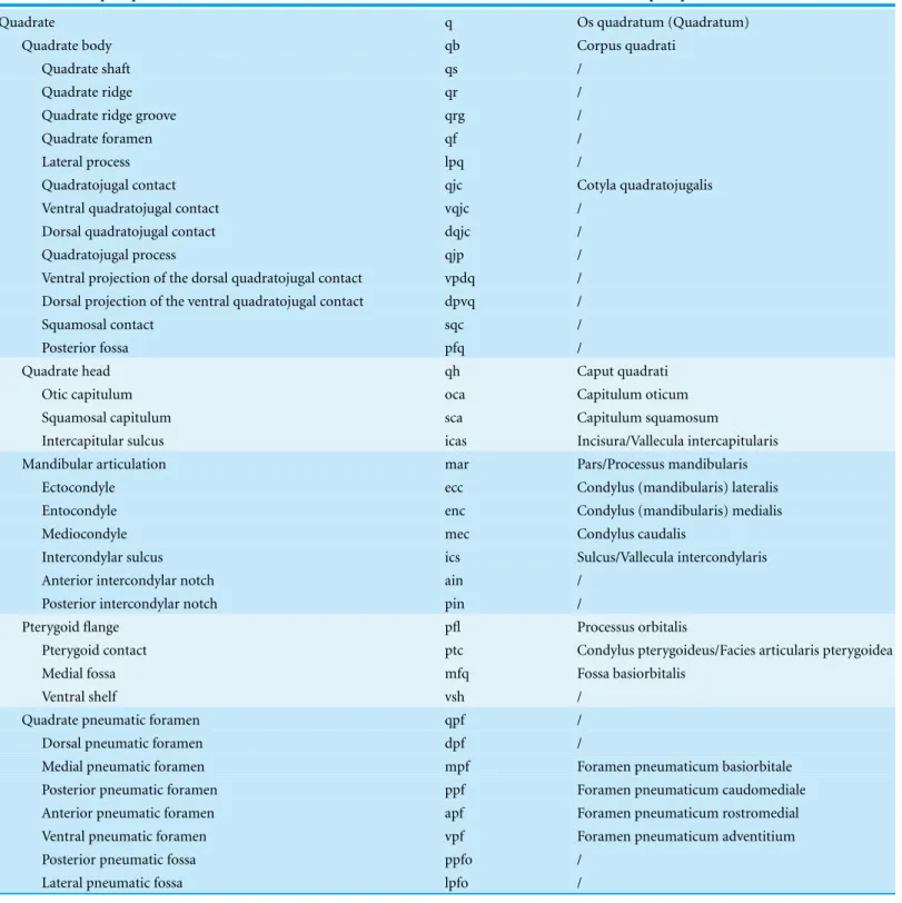
Figure 2 Anatomy of non-avian theropod quadrates. Line drawings of the right (A–E) quadrate of
Tsaagan mangas(IGM 100-1015) in (A) anterior, (B) lateral, (C) posterior, (D) medial and (E) ventral views; left (F–I) and right (J–K) quadrates (F) of Baryonyx walkeri (NHM R9951), (G) Aerosteon riocoloradensis(MCNA-PV-3137), (H) an indeterminate Oviraptoridae (IGM A;Marya´nska & Osm´olska, 1997), (I)Tyrannosaurus rex(BHI 3333;Larson & Carpenter, 2008), (J)Allosaurus‘jimmadseni’ (SMA 0005), and (K)Majungasaurus crenatissimus(FMNH PR 2100) in (F–I) posterior and (J–K) ventral views. Abbreviations: ain, anterior intercondylar notch; dqjc, dorsal quadratojugal contact; ecc, ectocondyle; enc, entocondyle; ics, intercondylar sulcus; lpq, lateral process of the quadrate; mar, mandibular articula-tion; mfq, medial fossa of the quadrate; oca, otic capitulum; pfl, pterygoid flange; pfq, posterior fossa of the quadrate; pin, posterior intercondylar notch; ppf, posterior pneumatic foramen; qb, quadrate body; qf, quadrate foramen; qh, quadrate head; qj, quadratojugal; qjp, quadratojugal process; qr, quadrate ridge; qrg, quadrate ridge groove; qs, quadrate shaft; sqc, squamosal contact; sca, squamosal capitulum; vqjc, ventral quadratojugal contact; vpdq, ventral projection of the dorsal quadratojugal contact; vsh, ventral shelf.
and sometimes by the medial margin of the quadrate foramen, the dorsal margin of the mandibular articulation, the ventral margin of the quadrate head, and a medial margin mostly formed by the quadrate shaft and the medial fossa of the pterygoid flange. The quadrate body is equivalent to the ‘Corpus ossis quadrati’ ofBaumel & Witmer (1993), and the ‘Corpus quadrati’ ofElzanowski, Paul & Stidham (2001)andElzanowski & Stidham (2010)for avian theropods (Fig. 1A).
Quadrate shaft (qs)
Quadrate ridge (qr)
Ventrodorsally elongated column, ridge or crest located on the quadrate body and visible in posterior view (Figs. 2C,2F–2K). Although the quadrate ridge is present in the large majority of non-avian theropods, a description of the structure is often omitted in the literature. The quadrate ridge is referred as a ‘column’ byWelles (1984), a ‘ridge-like mediodorsal edge’ byCarr (1996), a ‘prominent rounded ridge’ bySmith et al. (2007), a ‘columnar ridge’ byRauhut, Milner & Moore-Fay (2010), a ‘robust ridge’ byBrusatte, Carr & Norell (2012), a ‘ridge’ or ‘pillar’ byChoiniere et al. (2014a), and a ‘bulging ridge’ by Lautenschlager et al. (2014).
Quadrate ridge groove (qrg)
Groove dividing the quadrate ridge in two different units at two-thirds, or more dorsally, of the quadrate height (Fig. 2G). A quadrate ridge groove exists in some allosauroid theropods.
Quadrate foramen (qf)
Aperture in the quadrate body or concavity on the lateral margin of the quadrate body and delimited ventrally by the ventral quadratojugal contact and dorsally by the dorsal quadratojugal contact and its ventral projection in some theropod taxa (Figs. 2A, 2E–2Gand2I). Most authors usually refer to this perforation as the quadrate foramen (e.g., Welles, 1984;Sereno & Novas, 1994;Charig & Milner , 1997;Marya´nska & Osm´olska, 1997;Currie & Carpenter, 2000;Coria & Currie, 2006;Currie, 2006;Norell et al., 2006; Choiniere et al., 2010;Choiniere et al., 2014a;Choiniere et al., 2014b;Zanno, 2010;Brusatte, Carr & Norell, 2012). Yet, it can be also called the ‘paraquadratic foramen’ (e.g.,Barsbold & Osm´olska, 1999;Kobayashi & L¨u, 2003;Kobayashi & Barsbold, 2005), the ‘paraquadrate foramen’ (Sampson & Witmer, 2007;Dal Sasso & Maganuco, 2011;Lautenschlager et al., 2014), the ‘paraquadrate fenestra’ (Smith et al., 2007) or the ‘quadrate fenestra’ (e.g.,Carr , 1996;Sereno et al., 1998;Currie, 2003;Eddy & Clarke, 2011). A quadrate foramen exists in all non-avian theropods but Ceratosauria and Megalosauridae.
Lateral process (lpq).
Lateral or anterolateral projection of the lateral margin of the quadrate body (Fig. 2B). Also known as the ‘dorsal wing’ (Welles, 1984;Currie, 2006), the ‘anterolateral wing’ (Madsen & Welles, 2000), the ‘lateral lamina’ (Coria & Salgado, 1998) and the ‘lateral ramus’ (Sampson & Witmer, 2007), this process can contact the quadratojugal and/or the squamosal and therefore either be referred to the ‘quadratojugal ramus’ (Sampson & Witmer, 2007) or the ‘squamosal ramus’ (Norell et al., 2006).
Quadratojugal contact (qjc)
Ventral quadratojugal contact (vqjc)
Ventral area of contact of the quadrate with the quadratojugal (Figs. 2B,2Fand2H). The ventral quadratojugal contact of the quadrate always receives the quadratojugal bone.
Dorsal quadratojugal contact (dqjc)
Dorsal area of contact of the quadrate with the quadratojugal (Figs. 2Band2F). The ventral quadratojugal contact of the quadrate can either receive the quadratojugal or both quadratojugal and squamosal in some theropod taxa.
Ventral projection of the dorsal quadratojugal contact (vpdq)
Small projection of the dorsal quadratojugal contact delimiting the laterodorsal margin of the quadrate foramen (Fig. 2I).
Dorsal projection of the ventral quadratojugal contact (dpvq)
Small projection of the ventral quadratojugal contact delimiting the lateroventral margin of the quadrate foramen.
Quadratojugal process (qjp)
Anterior projection of the ventral quadratojugal contact of the quadrate (Fig. 2B). Also known as the ‘quadratojugal lamina’ (Lautenschlager et al., 2014).
Lateroventral process (lvp)
Lateromedially oriented ventral projection of the ventral quadratojugal contact of the quadrate that bounds the quadratojugal ventrally (Fig. 2H). The lateroventral process is similar to the ‘lateral process’ ofMarya´nska & Osm´olska (1997).
Squamosal contact (sqc)
Contact on the lateral margin of the quadrate with the squamosal (Fig. 2B).
Posterior fossa (pfq)
Depression or concavity situated on the posterior side of the quadrate body and dorsal to the mandibular articulation, ventral to the quadrate head and lateral to the quadrate ridge (Fig. 2B). The posterior fossa can include or exclude the quadrate foramen.
Quadrate head
Quadrate head (qh)
‘Caput quadrati’ ofElzanowski, Paul & Stidham (2001)andElzanowski & Stidham (2010), and roughly equivalent to the ‘Processus oticus’ (Baumel & Witmer, 1993). In birds, the ‘Processus oticus’ (Baumel & Witmer, 1993), and the ‘Pars oticus’ ofElzanowski, Paul & Stidham (2001)andElzanowski & Stidham (2010)also includes several sub-units that are either absent in non-avian theropods (e.g., ‘Crista Tympanica’, ‘Tuberculum subcapitulare’), or here included in the quadrate body (e.g., ‘Sulcus pneumaticus’, ‘Foramen pneumaticum rostromediale’). The bistylic quadrate head present in some tyrannosaurids, alvarezsauroids, oviraptorids and avian theropods is divided into otic and squamosal capitula.
Otic capitulum (oca)
Medial capitulum of the quadrate head articulating with the braincase (Fig. 2H). The otic capitulum is referred as the ‘capitulum (condylus) oticum’ byBaumel & Witmer (1993), Elzanowski, Paul & Stidham (2001)andElzanowski & Stidham (2010)for avian theropods (Fig. 1A).
Squamosal capitulum (sca)
Lateral capitulum of the quadrate head articulating with the squamosal (Fig. 2H). The squamosal capitulum is similar to the ‘capitulum (condylus) squamosum’ ofBaumel & Witmer (1993),Elzanowski, Paul & Stidham (2001)andElzanowski & Stidham (2010)for avian theropods (Fig. 1C).
Intercapitular sulcus (icas)
Groove separating the ootic capitulum from the squamosal capitulum on the dorsal surface of the quadrate head (Fig. 2H). The intercapitular sulcus (Witmer, 1990) is equivalent to the ‘incisura intercapitularis’ ofBaumel & Witmer (1993), and the ‘vallecula intercapitularis’ ofElzanowski, Paul & Stidham (2001)andElzanowski & Stidham (2010) for avian theropods (Fig. 1E).
Mandibular articulation
Mandibular articulation (mar)
Ectocondyle (ecc)
Lateral condyle of the mandibular articulation (Fig. 2). The ectocondyle is equivalent to the ‘condylus (mandibularis) lateralis’ ofBaumel & Witmer (1993),Elzanowski, Paul & Stidham (2001)andElzanowski & Stidham (2010)for avian theropods (Fig. 1F).
Entocondyle (enc)
Medial condyle of the mandibular articulation (Fig. 2). The entocondyle has been referred as the ‘condylus (mandibularis) medialis’ byBaumel & Witmer (1993),Elzanowski, Paul & Stidham (2001)andElzanowski & Stidham (2010)for avian theropods (Fig. 1F).
Mediocondyle (mdc)
Posterior condyle of the mandibular articulation located between the ecto- and ento-condyles. The mediocondyle is referred as the ‘third condyle’ byClark, Perle & Norell (1994)andXu & Wu (2001), the ‘accessory condyle’ byKobayashi & L¨u (2003)and Lautenschlager et al. (2014), and the ‘condylus caudalis’ ofBaumel & Witmer (1993)and Elzanowski, Paul & Stidham (2001)for avian theropods.
Intercondylar sulcus (ics)
Groove separating the ectocondyle from the entocondyle and articulated with the interglenoid ridge of the articular (Figs. 2Eand2K). The intercondylar sulcus, a term also used byCarrano, Loewen & Sertich (2011), can be referred as a ‘groove’ (e.g., Madsen, 1976;Britt, 1991;Madsen & Welles, 2000;Currie, 2006), ‘swelling’ (Charig & Milner , 1997), ‘sulcus’ (e.g., Kobayashi & L¨u, 2003;Norell et al., 2006;Sadleir, Barrett & Powell, 2008), ‘trochlea’ (Brochu, 2003;Brusatte et al., 2010), ‘trochlear surface’ (Brusatte et al., 2010; Brusatte, Carr & Norell, 2012), and ‘intercondylar bridge’ (Zanno, 2010). The intercondylar sulcus is similar to the ‘sulcus intercondylaris’ (Baumel & Witmer, 1993) and the ‘vallecula intercondylaris’ (Elzanowski, Paul & Stidham, 2001;Elzanowski & Stidham, 2010) of the quadrate of avian theropods (Fig. 1F).
Anterior intercondylar notch (ain)
Notch located between the ectocondyle and entocondyle, on the anterior margin of the mandibular articulation (Fig. 2K).
Posterior intercondylar notch (pin)
Notch located between the ectocondyle and entocondyle, on the posterior margin of the mandibular articulation, and being referred as the ‘pit’ byBakker (1998)(Fig. 2J).
Pterygoid flange
Pterygoid flange (pfl)
2012), the ‘pterygoid ramus’ (e.g., Sereno & Novas, 1994;Sampson & Witmer, 2007; Choiniere et al., 2010;Choiniere et al., 2014a;Choiniere et al., 2014b), the ‘pterygoid wing’ (e.g., Welles, 1984;Madsen & Welles, 2000;Eddy & Clarke, 2011), the ‘pterygoid ala’ (e.g., Currie, 2003;Currie, 2006;Sadleir, Barrett & Powell, 2008;Dal Sasso & Maganuco, 2011), the ‘pterygoid process’ (Molnar, 1991;Carr , 1996;Sereno et al., 2008), the ‘optic wing’ (Balanoff& Norell, 2012), the ‘orbital process’ (Clark, Perle & Norell, 1994;Chiappe, Norell & Clark, 2002), and the ‘processus orbitalis’ (Baumel & Witmer, 1993;Elzanowski, Paul & Stidham, 2001;Elzanowski & Stidham, 2010) for avian theropods (Fig. 1B).
Pterygoid contact (ptc)
Area of contact with the pterygoid on the medial margin of the pterygoid flange or the quadrate body (Fig. 2D). In avian theropods, the pterygoid contact is homologous to the ‘facies pterygoidea’ inElzanowski, Paul & Stidham (2001)and the ‘facies articularis pterygoidea’ inElzanowski & Stidham (2010). It is also homologous to the ‘condylus pterygoideus,’ located on the quadrate body, inBaumel & Witmer (1993),Elzanowski, Paul & Stidham (2001), andElzanowski & Stidham(2010;Fig. 1D).
Medial fossa (mfq)
Depression or concavity located on the medial surface of the pterygoid flange, typically on its posteroventral extremity (Fig. 2D). The medial fossa is delimited by the quadrate shaft and the ventral shelf in some theropod taxa. The medial fossa is similar to the ‘fossa corporis quadrati’ ofFuchs (1954), and the ‘fossa basiorbitalis’ ofElzanowski, Paul & Stidham (2001)andElzanowski & Stidham (2010)for avian theropods (Fig. 1D).
Ventral shelf (vsh)
A medial or posteromedial fold of the ventral margin of the pterygoid flange (Figs. 3A,3G and3M). The term ‘shelf ’ was employed bySereno & Novas (1994)and ventral shelf was used bySampson & Witmer (2007),Eddy & Clarke (2011)andCarrano, Loewen & Sertich (2011).
Pneumatic foramina and fossae
Quadrate pneumatic chamber (qpc)
Internal chamber within the quadrate, either fully contained within the bone or communi-cating externally by one or several pneumatic foramina. The quadrate pneumatic chamber hosts the quadrate sinus/diverticulum and, in some cases, includes several interconnected chambers separated by thin bony lamellae within the quadrate body and pterygoid flange (Kundr´at & Jan´aˇcek, 2007;Tahara & Larsson, 2011;Gold, Brusatte & Norell, 2013).
Dorsal pneumatic foramen (dpf)
Aperture located on the anterodorsal surface of the quadrate, just ventral to the quadrate head.
Medial pneumatic foramen (mpf)
Figure 3 (...continued)
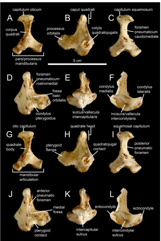
Left quadrate (M–R) ofBaryonyx walkeri (NHM R9951) in (M) anterior, (N) lateral, (O) posterior, (P) medial, (Q) dorsal, and (R) ventral views. Right quadrate (S–W) ofEustreptospondylus oxoniensis
(OUMNH J.13558; reversed) in (S) anterior, (T) lateral, (U) posterior, (V) medial and (W) ventral views (courtesy of Paul Barrett). Abbreviations: afq, anterior fossa; ain, anterior intercondylar notch; dqjc, dorsal quadratojugal contact; ecc, ectocondyle; enc, entocondyle; ics, intercondylar sulcus; lpq, lateral process; mfq, medial fossa; pfq, posterior fossa; pfl, pterygoid flange; qf, quadrate foramen; qh, quadrate head; qjp, quadratojugal process; qr, quadrate ridge; vpdq, ventral projection of the dorsal quadratojugal contact; vqjc, ventral quadratojugal contact; vsh, ventral shelf of the pterygoid flange.
foramen is homologous to the ‘foramen pneumaticum’ ofBaumel & Witmer (1993), and the ‘foramen pneumaticum basiorbitale’ ofElzanowski, Paul & Stidham (2001)and Elzanowski & Stidham (2010)for avian theropods.
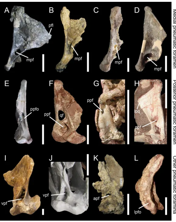
Posterior pneumatic foramen (ppf)
Aperture or recess on the posterior surface of the quadrate body, typically at mid-height of the quadrate (Figs. 2Gand5). The posterior pneumatic foramen is similar to and likely homologous to the ‘foramen pneumaticum caudomediale’ ofElzanowski & Stidham (2010)for avian theropods (Fig. 1C).
Anterior pneumatic foramen (apf)
Aperture or recess on the anterior surface of the quadrate body, typically at mid-height of the quadrate (Fig. 5K). The anterior pneumatic foramen is likely homologous to the ‘foramen pneumaticum medial’ ofElzanowski, Paul & Stidham (2001), and the ‘foramen pneumaticum rostromediale’ ofElzanowski & Stidham (2010).
Ventral pneumatic foramen (vpf)
Aperture or recess on the ventral surface of the quadrate. The ventral pneumatic foramen is equivalent to the ‘foramen pneumaticum adventitium’ (or ‘ectopic pneumatic foramen’) of Elzanowski & Stidham (2010)for avian theropods (Figs. 5Iand5J).
Posterior pneumatic fossa (ppfo)
Shallow and well-delimited pneumatic recess on the posterior surface of the quadrate body, at mid-height of the bone and medial to the quadrate foramen (Fig. 5E).
Lateral pneumatic fossa (lpfo)
Shallow and poorly-delimited pneumatic recess on the ventral portion of the lateral surface of the quadrate, directly dorsal to the ectocondyle (Fig. 5L).
MORPHOLOGICAL VARIATION IN QUADRATE
SUB-UNITS
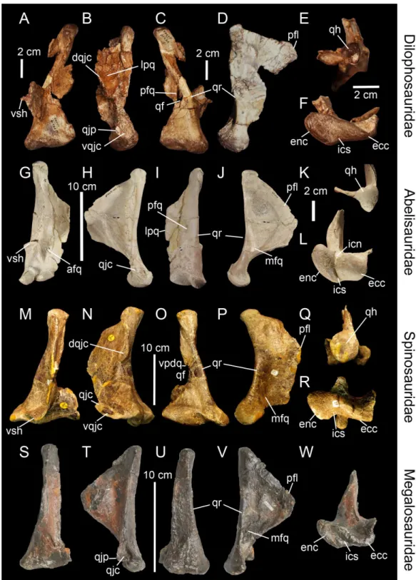
Figure 4 (...continued)
(GM F10004-1; reversed) in ventral views (courtesy of Stephen Brusatte). Right quadrate (M–Q) of
Falcarius utahensis(UMNH VP 14559; reversed) in (M) anterior, (N) lateral, (O) posterior, (P) medial, and (Q) ventral views (courtesy of Lindsay Zanno). Left quadrate (R–W) ofBambiraptor feinbergi
(AMNH 30556) in (R) anterior, (S) lateral, (T) posterior, (U) medial, (V) dorsal, and (W) ventral views. Abbreviations: afq, anterior fossa; dqjc, dorsal quadratojugal contact; ecc, ectocondyle; enc, entocondyle; ics, intercondylar sulcus; lpfo, lateral pneumatic fossa; lpq, lateral process; mfq, medial fossa; mpf, medial pneumatic foramen; pfq, posterior fossa; ppf, posterior pneumatic foramen; pfl, pterygoid flange; qf, quadrate foramen; qh, quadrate head; qjp, quadratojugal process; qr, quadrate ridge; vpdq, ventral pro-jection of the dorsal quadratojugal contact; vpf, ventral pneumatic foramen; vqjc, ventral quadratojugal contact; vsh, ventral shelf of the pterygoid flange.
The quadrate ridge is easily distinguishable in many theropod taxa such as Dilophosaurus wetherilli(Welles, 1984;Fig. 3C),Aerosteon riocoloradensis(MCNA-PV 3137;Fig. 4C) andProceratosaurus bradleyi(NHM R.4860) but the demarcation of this structure may be only subtly developed, as inNoasaurus leali(PVL 4061),Majungasaurus crenatissimus(FMNH PR 2100;Fig. 3I), andEustreptospondylus oxoniensis(OUMNH J.13558;Fig. 3U). The quadrate ridge is developed as a ‘columnar ridge’ in many theropod taxa such asDilophosaurus wetherilli(Welles, 1984),Allosaurus‘jimmadseni’ (SMA 0005; Allosaurus‘jimmadseni’ sensuChure, 2000;Loewen, 2010) andEotyrannus lengi(MIWG 1997.550) but also forms a thin crest as in Tyrannosauridae (AMNH 5027;Carr , 1996; Brusatte, Carr & Norell, 2012). Although the ventral portion of the quadrate ridge is usually demarcated just above the entocondyle of the mandibular articulation, its dorsal termination is more variable. The dorsal termination can reach the quadrate head like in Acrocanthosaurus atokensis(NCSM 14345) or flatten at the mid-height of the quadrate such as inAlbertosaurus sarcophagus(Currie, 2003: Fig. 10B). The quadrate ridge can be divided into two ridges by a deep groove as inAllosaurus fragilis(AMNH 600) and Allosaurus europaeus(ML 415). The quadrate ridge can also flare at the second dorsal third of the quadrate, and reappears slightly more dorsally, as observed in some derived Spinosauridae (Hendrickx, Ara´ujo & Mateus, 2014). Likewise, the ventral portion of the quadrate ridge can also dichotomize into two crests separated by a concavity such as in the tyrannosauridsAlbertosaurus sarcophagus,Daspletosaurussp. (Currie, 2003: Figs. 10 and 28) andTyrannosaurus rex(AMNH 5027).
separated by a deep pneumatic foramen facing ventrally (e.g.,Alioramus altai;Fig. 5I; Tyrannosaurus rexFMNH PR2081). In anterior view, the pterygoid flange can be straight and only projected anteriorly, as in the carcharodontosauridShaochilong maortuensis (Brusatte et al., 2010: Fig. 7A), or anteromedially recurved. The anteroventral margin of the pterygoid flange can either be straight, or medially and/or dorsally deflected, forming a horizontally oriented or dorsally inclined shelf-like structure here referred as the ventral shelf, as inMajungasaurus crenatissimus(FMNH PR 2100;Fig. 3G),Carnotaurus sastrei (MACN-CH 894) andAllosaurus fragilis(Madsen, 1976: plate 3d).
The medial fossa of the quadrate (Fig. 2D, mfq) is easily homologized between taxa as it is always situated on the pterygoid flange, typically on its ventromedial surface. This fossa is posteriorly delimited by the quadrate body in non-avian theropods and sometimes by the ventral shelf of the pterygoid flange. The medial fossa can be of variable depth (deep inCryolophosaurus; FMNH PR1821; shallow inEustreptospondylus; OUMNH J.13558), pneumatized (e.g.,Falcarius; UMNH VP 14559;Fig. 4P), and situated in the ventralmost part of the pterygoid flange (e.g.,Tsaagan; IGM 100-1015) or at mid-height of it and directly dorsal to a large pneumatic recess like inMapusaurus roseae(MCF PVPH-108.102).
The posterior fossa of the quadrate (Fig. 2B, pfq) can be located either in between the quadrate and the quadratojugal, being confluent with the quadrate foramen (e.g.,Mapusaurus; MCF PVPH-108.102), or in the middle of the quadrate shaft and between the quadrate ridge and the lateral limit of the quadrate shaft (e.g., ‘Syntarsus’; MNA V2623),Tsaagan(Norell et al., 2006),Majungasaurus(Sampson & Witmer, 2007;Fig. 3I). The posterior fossa can either be strongly ventrodorsally elongated like in the carchar-odontosauridAcrocanthosaurus(NCSM 14345), or form an oval concavity lateromedially wide (e.g.,Majungasaurus;Sampson & Witmer, 2007). Similarly to the medial fossa, the posterior fossa can host a pneumatic foramen positioned dorsally (e.g.,Sinornithomimus; IVPP V11797–10) or ventrally (e.g.,Garudimimus; IGM 100-13) inside the fossa.
Due to the highly variable morphology of the quadrate foramen, this structure deserves special attention. It can be completely absent (e.g.,Carnotaurus,Torvosaurus, Eustreptospondylus;Fig. 3U), or form a very small aperture (e.g., ‘Syntarsus’;Tykoski, 2005) to a large opening (e.g.,Bambiraptor;Fig. 4T). In most non-avian theropods, only a small portion of the lateral margin of the quadrate foramen is delimited by the quadratojugal (e.g.,Sinraptor;Currie, 2006) while in some non-avian theropods, the majority of the lateral margin is formed by the quadratojugal (e.g.,Dromaeosaurus). Finally, in a few theropods, the foramen can be completely enclosed in the quadrate (e.g.,Aerosteon;Sereno et al., 2008;Fig. 4C).
line of variable width along the lateral margin of the quadrate. Where separated by the quadrate foramen, the ventral and dorsal contacts can display a wide variety of surface and outlines. Both quadratojugal contacts may face laterally, anteriorly or posteriorly, and their articulating surface can be smooth, irregular or deeply grooved by several radiating ridges, as inAllosaurus fragilis(Madsen, 1976). The ventral quadratojugal contact is typically D-shaped or ovoid in lateral view. Its anterior margin can extend far anteriorly, forming the quadratojugal process (Norell et al., 2006), and its ventral margin can project far laterally, as in Oviraptoridae (Marya´nska & Osm´olska, 1997). The dorsal quadratojugal contact varies from a very thin line to a broad surface in lateral or posterior views and its dorsal extension can reach the quadrate head or terminate well ventral to it. A ventral projection of this contact may be present, and such projection delimiting part of the lateral border of the quadrate can either be short, like inDaspletosaurussp. (Currie, 2003: Fig. 28A) andBaryonyx walkeri(Fig. 3O), or form an elongated ramus, like in the therizinosaurid Falcarius utahensis(Zanno, 2010: Fig. 1H) and the basal coelurosaurZuolong salleei (Choiniere et al., 2010: Fig. 3B).
In some basal theropods, ceratosaurs and dromaeosaurids, the lateral process of the quadrate (Fig. 2B, lpq) forms a wing-like projection similar to the pterygoid flange. This process is an extension of the quadrate body laterally so it is difficult to delimit. Such process is present inAllosaurus‘jimmadseni’ (SMA 0005),Sinraptor dongi(Currie, 2006: Fig. 1D), andErlikosaurus andrewsi(Clark, Perle & Norell, 1994: Fig. 7). The lateral process also varies in shape and size, as it can be lateromedially short and parabolic in posterior view (e.g.,Carnotaurus; MACN-CH 894), or lateromedially elongated and subtriangular in posterolateral view (e.g.,Dilophosaurus; UCMP 37302;Fig. 3B). Its ventral border can also extend to the quadrate foramen (e.g.,Bambiraptor; AMNH 30556;Fig. 4T) or more ventrally, sometimes reaching the lateral condyle of the mandibular articulation (e.g.,Ilokelesia,Majungasaurus; MCF PVPH 35, FMNH PR 2100;Fig. 3I).
This condition has also been observed in the dromaeosauridMahakala omnogovae(Turner, Hwang & Norell, 2007) butTurner, Pol & Norell(2011: Fig. 4) later reconsidered the head of the quadrate as not being bistylic. The morphology of the quadrate head is variable in dorsal view; it is subtriangular in most basal theropods (Sereno & Novas, 1994) likeDilophosaurus(UCMP 37302;Fig. 3E),Erlikosaurus(Lautenschlager et al., 2014) andBambiraptor(AMNH 30556;Fig. 4V), oval to subcircular in megalosaurids likeAfrovenator(UC OBA1) andTorvosaurus(BYUVP 9246), and allosauroids such as Aerosteon(MCNA-PV-3137;Fig. 4E),Sinraptor(IVPP 10600) andShaochilong(IVPP V2885.3), or subquadrangular in some Spinosaurinae such asIrritator(SMNS 58022). While the dorsal surface of the quadrate head is either convex or flattened in posterior view in most non-avian theropods, the quadrate head of some allosaurids (Bakker, 1998: Fig. 5C) and derived tyrannosaurids (FMNH PR208) shows a well-marked concavity on the dorsal margin. The quadrate head can also be conical in posterior view, as in Oviraptoridae (Marya´nska & Osm´olska, 1997: Fig. 1B). Despite this variability, the quadrate head can be easily homologized inter-taxically due to the obvious location of this structure.
With the exception of the therizinosaurErlikosaurus andrewsiand the ornithomimosaur Sinornithomimus dongiwhich both seem to have an autapomorphical tricondylar condi-tion on the mandibular articulacondi-tion (Clark, Perle & Norell, 1994;Kobayashi & L¨u, 2003; Lautenschlager et al., 2014), all other non-avian theropods have two mandibular condyles. The presence of three mandibular condyles was also noted in the oviraptorosaurAvimimus portentosus(Chatterjee, 1995) and the dromaeosauridSinornithosaurus millenii(Xu & Wu, 2001). However,Vickers-Rich, Chiappe & Kurzanov (2002)only found two condyles in Avimimusand our observations confirm that the third condyle ofSinornithosaurusseems to be part of the much broader lateral condyle (Xu & Wu, 2001: Fig. 4D).
The intercondylar sulcus (Fig. 2E, ics) varies in orientation, size and depth. It can be large, shallow and sub-perpendicular to the long axis passing through the mandibular articulation as inTyrannosaurus rex(FMNH PR2081), or narrow, deep and strongly lateromedially-oriented as in some derived spinosaurids (e.g., MHNM.KK376).
In posterior view, the shape of the mandibular articulation (Fig. 2C, mar) can vary from the biconvex condition known in most theropods, to the W-shaped articulation typical ofCitipati osmolskae(Clark, Norell & Rowe, 2002: Fig. 6) or a single convex articulation seen in some dromaeosaurids such asTsaagan mangas(IGM 100/1015). InTsaagan, the convex outline of the mandibular articulation in posterior view results from a poor delimitation of the ecto- and entocondyle and the separation of these two condyles by a shallow intercondylar sulcus; yet this morphology might be due to the bad preservation of the mandibular condyle. A posterior intercondylar notch (Fig. 2J, pin) was observed inAllosaurus(Bakker, 1998: Fig. 5B, C; SMA 0005) andSuchomimus tenerensis (MNN GAD 502) whereas an anterior intercondylar notch (Fig. 2K, ain) is present in the abelisauridsMajungasaurus crenatissimus(FMNH PR 2100;Fig. 3L) andCarnotaurus sastrei(MACN-CH 894).
homologies is difficult to assess because these structures have very diverse interspecific variability. Nevertheless, as in other saurischian taxa (Schwarz, Frey & Meyer, 2007), these pneumatic structures have phylogenetic signal (e.g., Gold, Brusatte & Norell, 2013; Hendrickx, Ara´ujo & Mateus, 2014; see below). These openings can appear on different sides and portions of the quadrate. The medial and posterior pneumatic foramina (Fig. 2G, ppf) usually occur in the medial and posterior fossa respectively, and their position inside these fossae is quite variable. Pneumatic foramina can also be located in a pneumatic recess outside the medial fossa and directly ventral to it such as in the carcharodontosaurids Mapusaurus roseae(Coria & Currie, 2006) andAcrocanthosaurus atokensis(Eddy & Clarke, 2011). In the latter, the pneumatic aperture is divided by a septum.
REVIEW OF THE QUADRATE FUNCTION AND
PNEUMATICITY IN NON-AVIAN THEROPODS
Function of the quadrate
In all archosaurs, and all amniotes except Mammaliaformes, the main function of the quadrate is the articulation of the cranium with the mandible, yet this bone also plays an important role in the mobility of the skull in many extant theropods. Streptostyly is a fundamental property of all avian theropods, and quadrate kinesis in birds, known already in the beginning of the 19th century (Nitzsch, 1816), has been extensively studied over the past sixty years (e.g.,Fisher, 1955;Bock, 1964;Bock, 1999;Bock, 2000;B¨uhler, 1981;Zusi, 1984;Zusi, 1993;B¨uhler , 1985;B¨uhler, Martin & Witmer, 1988;Chatterjee, 1991;Chatterjee, 1997;Hoese & Westneat, 1996;Zweers, Vanden Berge & Berkhoudt, 1997;Zweers & Vanden Berge, 1998;Bout & Zweers, 2001;Gussekloo & Bout, 2005;Meekangvan et al., 2006). Streptostyly consists of the rotation of the quadrate at its dorsal articulation against the squamosal which typically leads to a transverse movement, although a lateral movement of the quadrate around an anteroposteriorly directed axis occurs in some lepidosaur taxa (Metzger, 2002). Cranial kinesis in avian theropods with a streptostylic quadrate includes upward (protraction) and downward (retraction) rotation of the rostrum relative to the braincase. Three main types of kinesis, in which the role of the quadrate is relatively equivalent, are recognized relative to the position of the dorsal flexion zone of the cranium and the nature of the nasal opening in modern theropods (Bock, 1964;B¨uhler, 1981;Zusi, 1984;Meekangvan et al., 2006). In prokinesis, flexion occurs at the nasofrontal joint and the upper jaw thereby moves as one unit; in amphikinesis, flexion occurs in two zones of flexibility and the upper jaw and its tip are bent upward; in rhynchokinesis, flexion occurs forward from the nasofrontal joint, allowing its anterior part to be moved (Zusi, 1984).
Although the synovial quadrate head joint existing in theropods, and all other archosaurs, is necessary to infer cranial kinesis, its presence in akinetic taxa such as crocodiles demonstrates that the synovial joint cannot be considered alone as an argument for cranial kinesis. Synovial joints have actually been interpreted as growth zones rather than articular surfaces of mobile joints based on the presence of very thin articular cartilage covering the end of this joint (Holliday & Witmer, 2008). According toHolliday & Witmer (2008), “articular cartilage persists in loading environments that exert hydrostatic pressures (which result in a change in volume but not shape) but exert low shear stresses.” Indeed, one of the key centers of deformation during normal biting is the quadrate-squamosal contact, which would have experienced large shear stresses associated with torque and asymmetrical loading during biting (Rayfield, 2005), and the presence of a minimal amount of cartilage between the quadrate and squamosal would therefore suggest that the synovial zone was rather a growth zone than a mobile one. A streptostylic quadrate in Tyrannosaurus rex(Molnar, 1991;Molnar, 1998),Nanotyrannus lancensis(Larson, 2013), Oviraptor philoceratops(Smith, 1992),Heyuannia huangi(L¨u, 2005) andDromiceiomimus brevitertius(Russell, 1972) based on the saddle joint between the quadrate and squamosal only is therefore unlikely.
Nevertheless, and more convincingly, a streptostylic quadrate was also proposed in the alvarezsauridShuvuuia desertibyChiappe, Norell & Clark (1998). In this taxon, the quadratojugal/jugal? (n.b.,Dufeau (2003)considers the quadratojugal to be absent in Shuvuuia deserti), instead of being firmly sutured to the quadrate as in other non-avian theropods, would have contacted the lateral surface of the quadrate through a movable joint (Chiappe, Norell & Clark, 1998;Chiappe, Norell & Clark, 2002). According to Chiappe, Norell & Clark (1998), the absence of a laterodorsal contact of the quadrate with the quadratojugal/jugal, as well as a lateroventral process of the squamosal, would have permitted the quadrate to pivot anteroposteriorly, and the upper jaw to rotate ventrodorsally due to this transversal movement. These authors have implied the existence of a bending zone between the frontals and the nasal–preorbital bones inS. deserti, allowing the flexion of the snout as a single unit when the quadrate displaced forward, like in prokinetic birds. Nevertheless, the complex contacts between the nasal, frontal and prefrontal illustrated bySereno(2001: Fig. 12B) makes assessment ofChiappe, Norell & Clark’s (1998) hypothesis dubious (Holliday & Witmer, 2008).Holliday & Witmer (2008) also note that the maxillojugal and palatal flexion zones necessary to allow a true prokinesis in alvarezsaurids are still not clearly defined. Likewise, the contact between the pterygoid flange of the quadrate and the pterygoid also needs to be better documented in order to imply any specific movement of the quadrate inside the cranium ofS. deserti.
quadratojugal, it is unlikely that the skull of these two oviraptorids could display avian-like kinesis. As in other non-avian theropods, the oviraptorid quadrate was an immovable bone (Barsbold, 1977;Marya´nska & Osm´olska, 1997) so that the quadratojugal, if kinetic, could only pivot either ventrodorsally or mediolaterally from the quadratojugal contact of the quadrate. Yet, the quadratojugal of at leastNemegtomaiadoes not seem to have a loose articulation with the jugal given that the articulating surface between the two bones is anteroposteriorly extensive (L¨u et al., 2004: Fig. 2), disallowing mobility between the jugal and quadratojugal. Consequently, we consider unlikely that movement was possible between the quadrate and quadratojugal inHeyuanniaandNemegtomaiaand, unlike Barsbold (1977), see the oviraptorosaur skull as akinetic.
Quadrate articulation with the mandible and orientation of the intercondylar sulcus are highly variable among non-avian theropods, therefore suggesting some variation in the movement of the mandibular rami when the jaw opened. The helical intercondylar sulcus present in many non-avian theropods (C Hendrickx, pers. obs., 2015) was noticed byBakker (1998)in basal theropod dinosaurs, byHendrickx & Buffetaut (2008)in spinosaurids, and byMolnar (1991)andLarson (2008) inTyrannosaurus rex. These authors suggested that the spiral groove of the mandibular articulation constrained the diagonal ridge of the articular glenoid fossa, which fitted into the intercondylar sulcus, to slide laterally. This would force the mandibular rami of the mandible to displace laterally when the lower jaw was depressed, enlarging the width of the larynx in order to swallow large-size prey items (Hendrickx & Buffetaut, 2008).
In Allosaurus, the enlargement of the mandibular condyles associated with the posteroventral inclination of the ventral part of the quadrate, and the intercondylar notch, were interpreted byBakker (1998) as joint-stabilization zones. According to Bakker (1998), the anteroposterior enlargement of the articulating surface would improve the stability of the mandibular articulation when the mouth was widely opened, whereas the intercondylar notch, morphologically convergent to the depression of knee joints in crocodiles and birds, would be hosting one or several ligaments within the quadrate-mandibular articulation (Bakker, 1998). An intercondylar notch is present in the abelisauridsCarnotaurus sastrei(MACN-CH 894) andMajungasaurus crenatissimus (FMNH PR 2100), and the spinosauridSuchomimus tenerensis(MNN GAD 502), perhaps implying similar jaw mechanics of the mandibular articulation as inAllosaurus. Yet,Bakker’s (1998) jaw mechanics hypotheses based on the shape of the mandibular articulation and the presence of an intercondylar notch require further investigation with modern functional analysis methods such as FEA to be tested.
Pneumaticity in the quadrate
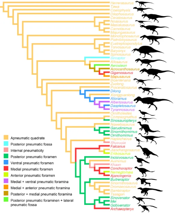
Fig. 4). The presence of one or several pneumatic foramina has indeed been recorded in carcharodontosaurids (e.g.,Coria & Currie, 2006;Eddy & Clarke, 2011), megaraptorans (Sereno et al., 2008), tyrannosauroids (e.g.,Molnar, 1991;Brochu, 2003;Currie, 2003; Xu et al., 2004;Witmer & Ridgely, 2010;Brusatte, Carr & Norell, 2012;Gold, Brusatte & Norell, 2013), compsognathids (Currie & Chen, 2001), alvarezsauroids (J Choiniere, pers. comm., 2014), therizinosaurs (Clark, Perle & Norell, 1994;Zanno, 2010), oviraptorids (e.g.,Marya´nska & Osm´olska, 1997;L¨u, 2003;Kundr´at & Jan´aˇcek, 2007;Balanoff& Norell, 2012), ornithomimosaurs (e.g.,Witmer, 1997;Tahara & Larsson, 2011), dromaeosaurids (Makovicky, Apestegu´ıa & Agnol´ın, 2005) and troodontids (Barsbold, Osm´olska & Kurzanov, 1987;Currie & Zhao, 1993;Varricchio, 1997;Xu et al., 2002;Xu & Norell, 2004). An incipient development of a pneumatic recess, the posterior pneumatic fossa, also exists in the basal allosauroidSinraptor dongi(Currie, 2006), suggesting that quadrate pneu-maticity may be an avetheropod synapomorphy. Yet, external manifestation of quadrate pneumaticity only occurs in derived members of Allosauroidea, Tyrannosauroidea, and Ornithomimosauria, and an apneumatic quadrate exists in the basal members of each of these clades (i.e.,SinraptorandAllosaurusfor Allosauroidea (Currie, 2006; C Hendrickx, pers. obs., 2011),TanycolagreusandProceratosaurusfor Tyrannosauroidea (Carpenter, Miles & Cloward, 2005;Rauhut, Milner & Moore-Fay, 2010), andNqwebasaurus for Ornithomimosauria; see Choiniere, Forster & de Klerk’s (2012) codings of their datamatrix). Pneumatic foramina have not been reported for any alvarezsauroid taxa, but are present in basalmost members of Therizinosauria, Oviraptorosauria and Paraves. This suggests that external quadrate pneumaticity occurred independently in several basal avetheropod clades and is a possibly synapomorphy of the clade Therizinosauria
+Pennaraptora (Fig. 6).
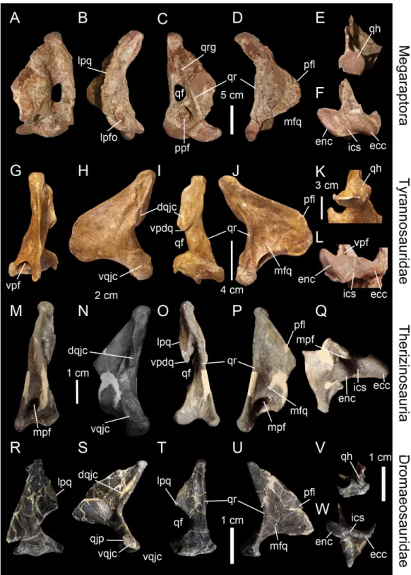
Figure 5 Morphology and position of pneumatic openings in the quadrate of non-avian Theropoda.Right quadrate (A) of the carcharodontosauridAcrocanthosaurus atokensis(NCSM 14345; reversed) in medial view. Left quadrate (B) of the carcharodontosauridMapusaurus roseae (MCF-PVPH-108) in medial view. Left quadrate (C) of the carcharodontosauridGiganotosaurus carolinii(MUCPv CH 1) in medial view. Right quadrate (D) of the therizinosaurFalcarius utahensis(UMNH VP 14559; reversed) in medial view (courtesy of Lindsay Zanno). Right quadrate (E) of the metriacanthosaurid
Figure 5 (...continued)
Garudimimus brevipes (IGM 100–13) in posterior view (courtesy of Yoshitsugu Kobayashi). Right quadrate (H) of the dromaeosauridBuitreraptor gonzalezorum(MPCA 245; reversed) in posterior view. Right quadrate (I) of the tyrannosauridAlioramus altai(IGM 100–844) in ventral view (courtesy of Mick Ellison). Left quadrate (J) of the tyrannosauridTyrannosaurus rex(FMNH PR2081; cast, reversed) in ventral view. Left quadrate (K) of the carcharodontosauridMapusaurus roseae(MCF-PVPH-108) in anterior view. Left quadrate (L) of the neovenatoridAerosteon riocoloradensis(MCNA PV 3137) in lateral view (courtesy of Mart´ın Ezcurra). Abbreviations: apf, anterior pneumatic foramen; lpq, lateral process; lpfo, lateral pneumatic fossa; mpf, medial pneumatic foramen; ppf, posterior pneumatic foramen; ppfo, posterior pneumatic fossa; qf, quadrate foramen; vpf, ventral pneumatic foramen. Scale bars=10 cm (A–C, J, K), 5 cm (E–G, L), 1 cm (D, H, I).
Figure 6 Distribution of quadrate pneumaticity in Theropoda.Cladogram of non-avian theropods based on the theropod classification summarized byHendrickx, Mateus & Ara´ujo (2015)and showing the phylogenetic distribution of quadrate pneumaticity and the different quadrate pneumatic foramina in theropod dinosaurs. Silhouettes by Funkmonk (Dilophosaurus, Shuvuuia, Dromaeosauroides, and
Suzhousaurus), M. Martyniuk (OrnitholestesandSimilicaudipteryx), T. Michael Keesey (Deinocheirus),
Choiniere et al.(2010;Zuolong; modified) and S. Hartman (all others).
2003;Witmer & Ridgely, 2010;Fig. 5J). In non-tyrannosaurid tyrannosauroids, such a ventral pneumatic foramen is present inDilong paradoxus(Gold, Brusatte & Norell, 2013) but was not observed in the closely related taxaGuanlong wucaii,Proceratosaurus lengi, andXiongguanlong baimoensis(Gold, Brusatte & Norell, 2013). It is also not clearly present inEotyrannus lengi(contraGold, Brusatte & Norell, 2013; C Hendrickx, pers. obs., 2011). A ventral pneumatic foramen of the quadrate is most likely synapomorphic of non-proceratosaurid Tyrannosauroidea (Fig. 6). A pneumatic foramen can also be seen on the anterior surface of the quadrate, as inMapusaurus roseae(Coria & Currie, 2006; Fig. 5K),Heyuannia huangi(L¨u, 2005),Erlikosaurus andrewsi(Lautenschlager et al., 2014), Troodon formosus(Currie & Zhao, 1993), and perhapsTyrannosaurus rex(Molnar, 1991). More rarely, a pneumatic fossa can be situated on the lateral and posterior surface of the quadrate body, as inAerosteon riocoloradensis(MCNA-PV 3137;Fig. 5L) andSinraptor dongi(Currie, 2006;Fig. 5E), respectively. The presence of an anterior pneumatic foramen, a lateral pneumatic fossa, or a posterior pneumatic fossa is an autapomorphy in each of these taxa.
Carcharodontosauridae (Coria & Currie, 2006;Eddy & Clarke, 2011) and Tyrannosauri-dae (Molnar, 1991;Brochu, 2003) possess several pneumatic openings which perforate different sides of the quadrate and sometimes intercommunicate (Brochu, 2003). The pneumatic foramina usually enter a large pneumatic chamber within the quadrate bone as inTyrannosaurus rex(Molnar, 1991;Brochu, 2003;Witmer & Ridgely, 2010),Alioramus altai(Gold, Brusatte & Norell, 2013),Conchoraptor gracilis(Kundr´at & Jan´aˇcek, 2007) orOrnithomimus edmontonicus(Tahara & Larsson, 2011). The neovenatoridAerosteon riocoloradensisalso possesses a large posterior pneumatic foramen leading to a pneumatic chamber (Sereno et al., 2008).
These pneumatic foramina and the pneumatic chamber associated with them are invaded by the quadrate diverticulum of the mandibular arch pneumatic system which, together with the periotic pneumatic system, forms the tympanic sinus of archosaurs (Dufeau, 2011;Tahara & Larsson, 2011). The mandibular arch pneumatic system includes the quadrate and/or the articular diverticulum which both have their embryological origins as parts of the first pharyngeal (=mandibular) arch, like the middle ear sac
Larsson, 2011). InTyrannosaurus rex, however, the siphoneal diverticulum does not pass through the quadrate, and the quadrate diverticulum only enters the ventral opening of the pterygoid flange, and then passes with or without the siphoneal diverticulum along the medial fossa of the pterygoid flange (Tahara & Larsson, 2011). Likewise, the quadrate diverticulum only pneumatizes two distinct regions of the quadrate inAcrocanthosaurus atokensisandMapusaurus roseae(Tahara & Larsson, 2011).
CONCLUSIONS
Here we propose a revised nomenclature of the quadrate bone and a corresponding set of abbreviations that provide a standard set of terms for describing this cranial bone in non-avian theropod dinosaurs. The quadrate can be divided into five regional categories—the quadrate body, quadrate head, mandibular articulation, pterygoid flange, and pneumatic foramina and fossae—and many anatomical sub-units such as—the quadrate shaft, quadrate head, quadrate ridge, quadrate foramen, lateral process, quadratojugal contact, squamosal contact, pterygoid contact, mandibular articulation, medial fossa, and posterior fossa. Although they are highly variable in shape, all quadrate entities, with perhaps the exception of the posterior fossa, are easy to homologize across taxa, and a description of their morphology should be provided in the literature.
The quadrate of the large majority of non-avian theropods is akinetic, and it is unlikely that a streptostylic quadrate is present in the derived alvarezsauroidsShuvuuia deserti, as was previously thought. A lateral movement of the rami while the mandible was depressed occurred in various theropods (e.g., spinosaurids). This lateral movement of the rami was due to a helicoidal and diagonally oriented intercondylar sulcus of the mandibular articulation. The presence of an intercondylar notch in allosaurids is interpreted as a joint-stabilization zone that would improve the stability of the mandibular articulation when the mouth was widely opened. However, this assumption needs further investigation from modern functional morphology techniques.
A pneumatic quadrate is present in members of most non-avian avetheropod clades, in which a pneumatic foramen is seen in the ventral part of the pterygoid flange and in the medial and lateral fossae. Pneumatic foramina invading the quadrate seem to be independently acquired by allosauroids, tyrannosauroids, compsognathids, and ornithomimosaurs throughout their evolution. The presence of pneumatic foramina in the quadrate of basalmost members of therizinosaurs, oviraptorosaurs, troodontids and dromaeosaurids suggests that quadrate pneumaticity is a synapomorphy of the clade Therizinosauria+Pennaraptora. Although the pneumatic recess invaded by the
quadrate diverticulum of the mandibular arch pneumatic system is linked to a single pneumatic foramen in most avetheropods, the presence of several pneumatic openings perforating different sides of the quadrate has been recorded in Carcharodontosauridae and Tyrannosauridae.
Institutional Abbreviations
AMNH American Museum of Natural History, New York, USA
BYUVP Brigham Young University Vertebrate Paleontology, Provo, Utah, USA
CMNH Carnegie Museum, Pittsburgh, Pennsylvania, USA
FMNH Field Museum of Natural History, Chicago, Illinois, USA
GM Ganzhou Museum, Ganzhou City, Jiangxi Province, China
IGM Mongolian Institute of Geology, Ulaan Bataar, Mongolia
IVPP Institute for Vertebrate Paleontology and Paleoanthropology,
Beijing, China
MACN Museo Argentino de Ciencias Naturales, Buenos Aires,
Argentina
MCF PVPH Museo Municipal Carmen Funes, Paleontologia de Vertebra-dos, Plaza Huincul, Argentina
MCNA Museo de Ciencias Naturales y Antropol ´ogicas de Mendoza,
Mendoza, Argentina
MIWG Dinosaur Isle, Isle of Wight Museum Services, Sandown, UK
ML Museu da Lourinh˜a, Lourinh˜a, Portugal
NCSM North Carolina Museum of Natural Sciences, Raleigh, North
Carolina, USA
MNHN Mus´eum national d’Histoire Naturelle, Paris, France
MNA Museum of Northern Arizona, Flagstaff, Arizona, USA
MNN Mus´ee National du Niger, Niamey, Niger
MPCA Museo Provincial Carlos Ameghino, Cipolletti, R´ıo Negro,
Argentina
MSNM Museo di Storia Naturale di Milano, Milan, Italy
MUCPv Museo de Ciencias Naturales de la Universidad Nacional de Comahue, Neuqu´en, Argentina
NH Horniman Museum & Gardens, London, UK
NHM The Natural History Museum, London, UK
OUMNH Oxford University Museum, Oxford, UK
PVL Fundaci ´on ‘Miguel Lillo,’ San Miguel de Tucum´an, Argentina
PVSJ Instituto y Museo de Ciencias Naturales, San Juan, Argentina
SMA Sauriermuseum Aathal, Aathal, Switzerland
SMNS Staatliches Museum f¨ur Naturkunde, Stuttgart, Germany;
RTMP Royal Tyrrell Museum of Palaeontology, Drumheller, Alberta,
Canada
UCMP University of California Museum of Paleontology, Berkeley,
California, USA
UC University of Chicago Paleontological Collection, Chicago,
USA;
ACKNOWLEDGEMENTS
We thank editor Andrew Farke (Raymond M. Alf Museum of Paleontology) and reviewers Jonah Choiniere (Uni. Witwatersrand) and Federico Agnolin (MACN) who kindly provided insightful comments that greatly improved this paper. The quadrate of many non-avian theropods were examined first hand in several institutions and we thank P Sereno (Uni. Chicago), P Makovicky (FMNH), W Simpson (FMNH), M Lamanna (CMNH), A Henrici (CMNH), M Carrano (NMNH), M Brett-Surman (NMNH), S Chapman (NHM), P Barrett (NHM), P Jeffery (OUMNH), S Hutt (MIW), R Allain (MNHN), R Schoch (SMNS), H-J Siber (SMA), C Dal Sasso (MSNM), A Kramarz (MACN), F Novas (MACN), R Barbieri (MPCA), L Salgado (MUCPv), J Ignacio Canale (MUCPv-CH), R Coria (MCF PVPH), C Succar (MCF PVPH), J Calvo (CePaLB), R Mart´ınez (PVSJ), C Mehling (AMNH), M Norell (AMNH), D Krauze (SBU), J Groenke (SBU), P Brinkman (NCSM), and L Zanno (NCSM) for access to specimens in their care. Photographs of theropod quadrates were kindly shared by M Lamanna (CMNH), M Ezcurra (MACN), R Delcourt (Uni. S˜ao Paulo), M Carrano (USNM), E Buffetaut (CNRS), M Ellison (AMNH), L Witmer (Uni Ohio), S Brusatte (Uni. Edinburgh), R Benson (Uni. Oxford), C Foth (BSPG), P Currie (Uni. Alberta), J Canale (MUCPv-CH), P Barrett (NHM), J Choiniere (Uni. Witwatersrand), D Eddy (Uni. Texas), P Viscardi (Horniman), S Nesbitt (Uni. Texas), Y Kobayashi (HUM), R Tahara (McGill Uni.), R Pei (AMNH), C Dal Sasso (MSNM), P Sereno (Uni. Chicago), C Abraczinskas (Uni. Chicago), N Smith (Uni. Chicago), L Zanno (FMNH), R Tykoski (MNSD), D Burnham (Uni. Kansas), P Asaroff(MACN), R Irmis (UMNH), V Shneider (NCMNS), C Brochu (Uni. Iowa), S Lautenschlager (Uni. Bristol), M Mortimer, K Peyer (MNHN), and R Molnar (MNA), and the authors would like to address their sincere thanks to all of these people. We acknowledge the use of Phylopic for the theropod silhouettes, and thank Scott Hartman, Funkmonk, M Martyniuk, and T Michael Keesey for providing their artworks on Phylopic. A special thank goes to Paolo Viscardi for taking photos of the ostrich quadrate at the Horniman Museum & Gardens, and D Dufeau for sharing his MSc thesis onShuvuuia. We also thank Isabel Torres for giving a final check in the English. CH dedicates this paper to the memory of Roger Bec.
ADDITIONAL INFORMATION AND DECLARATIONS
Funding
This research was supported by the Fundac¸˜ao para a Ciˆencia e a Tecnologia (FCT) scholarships (Minist´erio da Ciˆencia, Tecnologia e Ensino superior, Portugal) SFRH/BD/ 62979/2009 (CH) and SFRH/BPD/96205/2013 (RA). The funders had no role in study design, data collection and analysis, decision to publish, or preparation of the manuscript.
Grant Disclosures
The following grant information was disclosed by the authors:
Competing Interests
The authors have declared that no competing interests exist.
Author Contributions
• Christophe Hendrickx conceived and designed the experiments, performed the
experiments, analyzed the data, contributed reagents/materials/analysis tools, wrote the paper, prepared figures and/or tables, reviewed drafts of the paper.
• Ricardo Ara ´ujo conceived and designed the experiments, performed the experiments,
analyzed the data, contributed reagents/materials/analysis tools, wrote the paper, reviewed drafts of the paper.
• Oct´avio Mateus conceived and designed the experiments.
Supplemental Information
Supplemental information for this article can be found online athttp://dx.doi.org/ 10.7717/peerj.1245#supplemental-information.
REFERENCES
Bakker RT. 1998.Brontosaur killers: late Jurassic allosaurids as sabre-tooth cat analogues.Gaia 15:145–158.
BalanoffAM, Norell MA. 2012.Osteology ofKhaan mckennai(Oviraptorosauria: Theropoda).
Bulletin of the American Museum of Natural History372:1–77DOI 10.1206/803.1.
BalanoffAM, Xu X, Kobayashi Y, Matsufune Y, Norell MA. 2009. Cranial osteology of the theropod dinosaurIncisivosaurus gauthieri(Theropoda: Oviraptorosauria).American Museum
Novitates3651:1–35DOI 10.1206/644.1.
Barbosa A. 1990.Identification key of Iberian waders (Charadriiformes) based on the os quadratum.Miscell`ania Zool`ogica14:181–185.
Barsbold R. 1977.Kinetism and peculiarity of the jaw apparatus of oviraptors (Theropoda, Saurischia).Soviet-Mongolian Paleontological Expedition, Trudy4:37–47 (in Russian).
Barsbold R, Osm ´olska H. 1999.The skull ofVelociraptor(Theropoda) from the late Cretaceous of Mongolia.Acta Palaeontologica Polonica44(2):189–219.
Barsbold R, Osm ´olska H, Kurzanov SM. 1987.On a new troodontid (Dinosauria, Theropoda) from the early Cretaceous of Mongolia.Acta Palaeontologica Polonica32(1–2):121–132.
Baumel JJ. 1993.Handbook of avian anatomy: nomina anatomica avium. 2nd edition. Vol. 23. Cambridge: Nuttall Ornithological Club. 779pp.
Baumel JJ, Witmer LM. 1993.Osteologia. In: Baumel JJ, ed.Handbook of avian anatomy: nomina
anatomica avium. Cambridge: Nuttall Ornithological Club, 45–132.
Benson RBJ. 2010.A description ofMegalosaurus bucklandii(Dinosauria: Theropoda) from the Bathonian of the UK and the relationships of middle Jurassic theropods.Zoological Journal of
the Linnean Society158(4):882–935DOI 10.1111/j.1096-3642.2009.00569.x.
Benson R, Carrano M, Brusatte S. 2010.A new clade of archaic large-bodied predatory dinosaurs (Theropoda: Allosauroidea) that survived to the latest Mesozoic.Naturwissenschaften 97(1):71–78DOI 10.1007/s00114-009-0614-x.
Bock WJ. 1964. Kinetics of the avian skull. Journal of Morphology 114(1):1–41
DOI 10.1002/jmor.1051140102.
Bock WJ. 1999.Avian cranial kinesis revisited.Acta Ornitologica34(2):115–122.
Bock WJ. 2000.The evolution of avian cranial kinesis. In: Zhou Z, Zhang F, eds.Proceedings of the 5th symposium of the society of avian paleontology and evolution, vol. 1. Beijing, 191–201.
Bout RG, Zweers GA. 2001.The role of cranial kinesis in birds.Comparative Biochemistry and Physiology Part A: Molecular & Integrative Physiology131(1):197–205
DOI 10.1016/S1095-6433(01)00470-6.
Britt BB. 1991.Theropods of Dry Mesa Quarry (Morrison Formation, late Jurassic), Colorado, with emphasis on the osteology ofTorvosaurus tanneri.Brigham Young University Geology Studies37:1–72.
Brochu CA. 2003.Osteology ofTyrannosaurus rex: insights from a nearly complete skeleton and high-resolution computed tomographic analysis of the skull.Society of Vertebrate Paleontology
Memoir7:1–138DOI 10.2307/3889334.
Brusatte SL. 2012.Dinosaur paleobiology. Hoboken: Wiley-Blackwell, 336pp.
Brusatte SL, Carr TD, Norell MA. 2012.The osteology ofAlioramus, a gracile and long-snouted tyrannosaurid (Dinosauria: Theropoda) from the late Cretaceous of Mongolia.Bulletin of the
American Museum of Natural History366:1–197DOI 10.1206/770.1.
Brusatte SL, Chure DJ, Benson RBJ, Xu X. 2010.The osteology ofShaochilong maortuensis, a carcharodontosaurid (Dinosauria: Theropoda) from the late Cretaceous of Asia.Zootaxa 2334:1–46.
B¨uhler P. 1981.Functional anatomy of the avian jaw apparatus.Form and Function in Birds 2:439–468.
B¨uhler P. 1985.On the morphology of the skull ofArchaeopteryx. In: Hecht MK, Ostrom JH, Viohl G, Wellnhofer P, eds.The beginnings of birds. Eichst¨att: Freunde des Jura-Museums Eichst¨att, 135–140.
B¨uhler P, Martin LD, Witmer LM. 1988.Cranial kinesis in the late Cretaceous birdsHesperornis
andParahesperornis.The Auk105(1):111–122.
Burnham DA. 2004.New information onBambiraptor feinbergi(Theropoda: Dromaeosauridae) from the late Cretaceous of Montana. In: Currie PJ, Koppelhus EB, Shugar MA, Wright JL,
eds.Feathered dragons: studies on the transition from dinosaurs to birds. Bloomington: Indiana
University Press, 67–111.
Carpenter K, Miles C, Cloward K. 2005.New small theropod from the upper Jurassic Morrison formation of Wyoming. In: Carpenter K, ed.The carnivorous dinosaurs. Bloomington: Indiana University Press, 23–48.
Carr TD. 1996.Cranial osteology and craniofacial ontogeny of Tyrannosauridae (Dinosauria: Theropoda) from the dinosaur park formation (Judith River Group, upper Cretaceous, Campanian) of Alberta. MSc dissertation, University of Toronto, Toronto, Ontario, Canada. 358 pp.
Carrano MT, Benson RBJ, Sampson SD. 2012. The phylogeny of Tetanurae (Dinosauria: Theropoda). Journal of Systematic Palaeontology 10(2):211–300
DOI 10.1080/14772019.2011.630927.
Carrano MT, Loewen MA, Sertich JJW. 2011.New materials ofMasiakasaurus knopfleri
Sampson, Carrano, and Forster, 2001, and implications for the morphology of the Noasauridae (Theropoda: Ceratosauria).Smithsonian Contributions to Paleobiology95:1–53