258 Radiol Bras. 2017 Jul/Ago;50(4):258–262
Spondyloarthropathy: diagnostic imaging criteria
for the detection of sacroiliitis
Espondiloartropatias: critérios de ressonância magnética na detecção da sacroileíte
Moacir Ribeiro de Castro Jr.1, Sonia de Aguiar Vilela Mitraud2, Marina Celli Francisco3, Artur da Rocha Corrêa
Fernandes4, Eloy de Ávila Fernandes5
Castro Jr MR, Mitraud SAV, Francisco MC, Fernandes ARC, Fernandes EA. Spondyloarthropathy: diagnostic imaging criteria for the detection of sacroiliitis. Radiol Bras. 2017 Jul/Ago;50(4):258–262.
Abstract
Resumo
Diagnostic imaging is crucial to the diagnosis and monitoring of spondyloarthropathies. Magnetic resonance imaging is the most relevant tool for the early detection of sacroiliitis, allowing the institution of therapeutic strategies to impede the progression of the disease. This study illustrates the major criteria for a magnetic resonance imaging-based diagnosis of spondyloarthropathy. The cases selected here present images obtained from the medical records of patients diagnosed with sacroiliitis over a two-year period at our facility, depicting the active and chronic, irreversible forms of the disease. Although computed tomography and conventional radiography can also identify structural changes, such as subchondral sclerosis, erosions, fat deposits, and ankylosis, only magnetic
resonance imaging can reveal active inlammatory lesions, such as bone edema, osteitis, synovitis, enthesitis, and capsulitis.
Keywords: Sacroiliitis; Magnetic resonance imaging; Spondyloarthropathies; Ankylosing spondylitis; Computed tomography; Radi-ography.
A avaliação por imagem é fundamental para o diagnóstico e acompanhamento clínico dos pacientes com espondiloartropatias. A ressonância magnética é o método de imagem mais importante para a detecção precoce de sacroileíte, permitindo a instituição de
terapias que podem impedir a progressão da doença. Este estudo ilustra os principais critérios de ressonância magnética na dei -nição de sacroileíte nas espondiloartropatias, com imagens selecionadas dos prontuários dos pacientes diagnosticados no nosso serviço, demonstrando tanto os achados da doença em atividade como as alterações crônicas de caráter irreversível. Embora a
tomograia computadorizada e a radiograia convencional possam identiicar lesões estruturais crônicas, tais como esclerose sub
-condral, erosões, depósitos de gordura e anquilose, apenas a ressonância magnética é capaz de demonstrar lesões inlamatórias
ativas, tais como edema ósseo, osteíte, sinovite, entesite e capsulite.
Unitermos: Sacroileíte; Ressonância magnética; Espondiloartropatias; Espondilite anquilosante; Tomograia computadorizada; Ra
-diograia.
Study conducted in the Department of Diagnostic Imaging of the Escola Paulista de Medicina da Universidade Federal de São Paulo (EPM-Unifesp), São Paulo, SP, Brazil.
1. Doctoral Student in the Graduate Program in Clinical Radiology, Collaborat-ing Physician in the Department of Diagnostic ImagCollaborat-ing of the Escola Paulista de Medicina da Universidade Federal de São Paulo (EPM-Unifesp), São Paulo, SP, Brazil.
2. PhD, Coordinator of the Computed Tomography Sector at the Hospital São Paulo – Escola Paulista de Medicina da Universidade Federal de São Paulo (EPM--Unifesp), São Paulo, SP, Brazil.
3. MD, Radiologist at Clínica IMED – Clínica Médica e Diagnóstico por Imagem, Curitibanos, SC, Brazil.
4. PhD, Associate Professor in the Department of Diagnostic Imaging of the Es-cola Paulista de Medicina da Universidade Federal de São Paulo (EPM-Unifesp), São Paulo, SP, Brazil.
5. PhD, Advising Professor for the Graduate Program in Clinical Radiology of the Escola Paulista de Medicina da Universidade Federal de São Paulo (EPM-Unifesp), São Paulo, SP, Brazil.
Mailing address: Dr. Moacir Ribeiro de Castro Jr. Departamento de Diagnóstico por Imagem – EPM-Unifesp. Rua Napoleão de Barros, 800, Vila Clementino. São Paulo, SP, Brazil, 04024-002. E-mail: [email protected].
Received November 15, 2015. Accepted after revision May 16, 2016.
INTRODUCTION
Seronegative spondyloarthropathies include a group of chronic systemic inlammatory diseases, characterized
by the absence of rheumatoid factor in serum, common clinical indings (such as inlammatory arthritis and en-thesitis), inlammatory low back pain, and the presence of human leukocyte antigen(1,2). This group of diseases includes ankylosing spondylitis, psoriatic arthritis, reac-tive arthritis, inlammatory enteropathic arthritis, and undifferentiated spondyloarthropathy.
Patients with seronegative spondyloarthropathies typically present early clinical manifestations in the sac-roiliac joints. These diseases evolve slowly, and there are no speciic biochemical markers that demonstrate their activity. Therefore, in clinical practice, imaging, more precisely magnetic resonance imaging (MRI), typically forms the basis for the diagnosis and evaluation of sac-roiliitis(3–5).
onset. In addition, X-ray and computed tomography allow structural changes to be identiied only when the damage has already become irreversible(5,6).
ACTIVE INFLAMMATORY LESIONS Bone marrow edema/osteitis
Bone marrow edema/osteitis appears as an area of low signal intensity on T1-weighted images and high signal in-tensity in short-tau inversion-recovery (STIR) sequences or equivalent liquid-sensitive sequences, located in the subchondral bone marrow (Figure 1). The edema should be easily characterized, with a signal similar to that of the cerebrospinal luid. When present, bone marrow edema is indicative of active sacroiliitis, not being pathognomonic of spondyloarthropathies, and can be related to other con-ditions, such as alterations caused by mechanical over-load. It should be noted that, in general, those alterations are not restricted to a single image and when accompa-nied by structural indings, such as subchondral sclerosis and bone erosion, allow a better diagnostic determination if taken together with the clinical and laboratory data. A inding of bone marrow located among the sacral foram-ina can be used as a reference for normality.
Synovitis, enthesitis, and capsulitis
On intravenous paramagnetic contrast-enhanced T1- weighted images with fat suppression, synovitis is char-acterized by enhancement in the synovial part of the sac-roiliac joint. In STIR sequences, the hyperintense signal within the synovial portion of the sacroiliac joint pre-cludes good differentiation between synovitis and physi-ologic luid on the joint(6).
Enthesitis is deined as an area of high signal inten-sity in STIR sequences, or as an area of enhancement in contrast-enhanced sequences, at the ligament insertion sites within the retroarticular space, potentially extending to the bones and soft tissues(6) (Figure 2).
Capsulitis presents signaling characteristics similar to those of enthesitis, although the former affects the anterior and posterior portions of the joint capsule(6) (Fig-ure 3).
STRUCTURAL CHANGES
Structural changes, which indicate previous inlam-matory events, can be identiied by MRI, X-ray, or com-puted tomography. Structural changes in sacroiliitis in-clude subchondral sclerosis, bone erosion, fatty deposits, and bone bridges/ankylosis(7).
Subchondral sclerosis
In T1-weighted sequences, subchondral sclerosis is characterized by a hypointense signal that extends for at least 5 mm into the sacroiliac joint space. Mild forms of subchondral sclerosis can be seen in healthy individu-als(6).
Bone erosion
Bone erosion is deined as focal lesions at the margin of the articular cartilage. The conluence of erosion sites is visualized as pseudo-widening of the sacroiliac joints(6) (Figure 4).
Periarticular fat deposits
Figure 3. 41-year-old female patient diagnosed with seronegative spondyloar-thropathy 3 years prior. A: Axial fast spin-echo T1-weighted sequence showing periarticular bone erosion (straight arrows) and thickening of the right joint capsule (serpentine arrows). B: Axial STIR sequence showing pseudo-widening of the joint spaces (arrowheads). Thickening and edema of the right joint cap-sule (serpentine arrows), characteristic of capsulitis. C: Intravenous contrast-enhanced coronal T1-weighted sequence with fat suppression, showing areas of subchondral bone sclerosis (thin arrows); intra-articular enhancement in the left posterior ibrous region, indicative of enthesitis (curved arrows).
Figure 2. 28-year-old female patient diagnosed with seronegative spondyloar-thropathy 8 years prior. A: Coronal STIR sequence showing subchondral edema (arrowheads). B,C: Intravenous contrast-enhanced coronal and axial T1-weight-ed sequences with fat saturation, showing osteitis (thick arrows), synovitis characterized by intra-articular enhancement (thin arrows), enthesitis with en -hancement in the ligamentous compartment of the joint (curved arrows) and capsulitis with pericapsular enhancement on the left (serpentine arrow).
articular bone marrow. This is a nonspeciic inding and is characterized by high signal intensity on T1-weighted images, indicating an area of previous inlammation(5) (Figure 5).
Bone bridges/ankylosis
Bone bridges/ankylosis appear as areas of low signal intensity in all sequences and can show a bone
marrow-like signal when fusion is complete. In addition, the joint space can become undeined(6) (Figure 6).
CRITERIA FOR DEFINING SACROILIITIS ACTIVITY
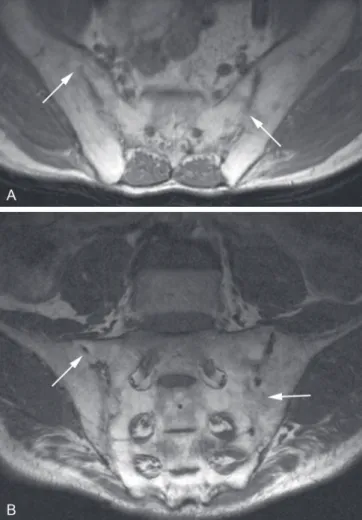
Figure 5. 49-year-old female patient diagnosed with seronegative spondyloarthropathy 7 years prior. A,B: Axial fast spin-echo T1-weighted sequence and coronal T1-weighted sequence showing subchondral sclerosis (white arrows) and fat deposits (black arrows).
Figure 4. 17-year-old male patient recently diagnosed with seronegative spondyloarthropathy. A,B: Intravenous contrast-enhanced coronal STIR and T1-weighted sequences with fat saturation, showing edema in the ibrous region of the right sacroiliac joint, characteristic of enthesitis.
image. The examination should be considered negative if the bone edema is not obvious(7).
Althoff et al. reported that contrast-enhanced MRI examinations are useful in the early diagnosis of inlam-matory sacroiliitis and could help boost the conidence of inexperienced radiologists. However, in patients with pre-existing disease or in patients undergoing follow-up ex-aminations, the use of contrast agents can be dispensed with(7).
Diffusion-weighted MRI seems to be an alternative means of using contrast agents in seronegative spondylo-arthropathies. Diffusion-weighted imaging shows higher apparent diffusion coeficient values in patients with ar-eas of bone marrow edema than in those with
mechani-cal low back pain, although further studies are needed in order to determine the relevance of this technique in imaging studies of seronegative spondyloarthropathies(8).
Knowledge of the diagnostic criteria for disease activity is essential for the radiologist, in order to facilitate the early diagnosis and treatment, as well as the follow-up, of spon-dyloarthropathies, thus reducing the associated morbidity and improving the quality of life of the affected patients.
Figure 6. 61-year-old male patient diagnosed with seronegative spondyloar-thropathy 11 years prior. A,B: Axial fast spin-echo T1-weighted sequence and coronal fast spin-echo T1-weighted sequence showing bone bridges/ankylosis (arrows).
REFERENCES
1. Dougados M, van der Linden S, Juhlin R, et al. The European Spon
-dylarthropathy Study Group preliminary criteria for the classiication of spondylarthropathy. Arthritis Rheum. 1991;34:1218–27.
2. Zochling J, Smith EU. Seronegative spondyloarthritis. Best Pract Res Clin Rheumatol. 2010;24:747–56.
3. Shinjo SK, Gonçalves R, Gonçalves CR. Medidas de avaliação clínica em pacientes com espondilite anquilosante. Revisão da literatura. Rev Bras Reumatol. 2006;46:340–6.
4. Torres TM, Ciconelli RM. Instrumentos de avaliação em espondilite anquilosante. Rev Bras Reumatol. 2006;46(Supl. 1):52–9.
5. Pertuiset E. Diagnosis of early spondyloarthritis. Rev Med Interne. 2008;29:596–605.
6. Rudwaleit M, Jurik AG, Hermann KG, et al. Deining active sacroi
-liitis on magnetic resonance imaging (MRI) for classiication of axial spondyloarthritis: a consensual approach by the ASAS/OMERACT MRI group. Ann Rheum Dis. 2009;68:1520–7.
7. Althoff CE, Feist E, Burova E, et al. Magnetic resonance imaging of active sacroiliitis: do we really need gadolinium? Eur J Radiol. 2009; 71:232–6.