Arq Bras Endocrinol Metab vol.58 número6
Texto
Imagem
Documentos relacionados
Computed tomography scan and magnetic resonance imaging showed a normal prostate and a 9.0 cm cystic tumor, replacing the left seminal vesicle.. The gross appearance and
Physical models of the foetus created using magnetic resonance imaging, computed tomography, and ultrasound data: history, description, and potential uses.. Werner H, Lopes dos
Amongst the available imaging methods (Doppler ultrasonography, computed tomography, magnetic resonance imaging and digital subtraction angiography), angiography is considered the
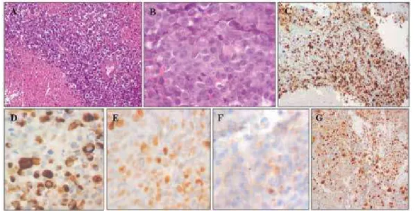
Recently, more stu- dies have reported that it is the newly uncovered gene associated with various autoimmune diseases (16-19). In present study, we performed a case-control study of
We report on a rare case of an adult with severe hypercalcemia secondary to the ectopic secre- tion of parathyroid-related peptide (PTH-rP) from a penile squamous cell cancer (PC)..
A novel nonstop mutation in the stop codon and a novel missen- se mutation in the type II 3beta-hydroxysteroid dehydrogenase (3beta-HSD) gene causing, respectively, nonclassic
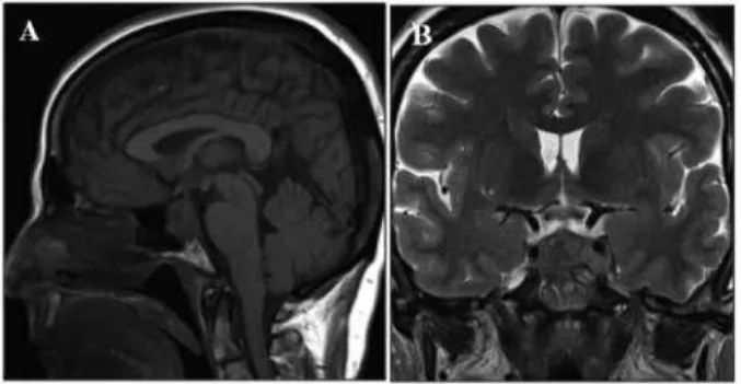
Therefore, although their occurrence is uncom- mon, a working knowledge of vascular lesions in the sella turcica or pituitary gland, such as a carotid internal artery aneurism
A computed tomography (CT) scan ( Figure 1 ) and magnetic resonance imaging (MRI) scan of the chest ( Figure 2 ) revealed the presence of a nodular lesion in the right pulmonary
