C A S E R E P O R T
Open Access
A stab wound to the axilla illustrating the
importance of brachial plexus anatomy in
an emergency context: a case report
Diogo Casal
1,2*, Teresa Cunha
1, Diogo Pais
2, Inês Iria
3,4, Maria Angélica-Almeida
1,2, Gerardo Millan
1,
José Videira-Castro
1and João Goyri-O
’
Neill
2Abstract
Background:Although open injuries involving the brachial plexus are relatively uncommon, they can lead to permanent disability and even be life threatening if accompanied by vascular damage. We present a case report of a brachial plexus injury in which the urgency of the situation precluded the use of any ancillary diagnostic
examinations and forced a rapid clinical assessment.
Case presentation:We report a case of a Portuguese man who had a stabbing injury at the base of his left axilla. On observation in our emergency room an acute venous type of bleeding was present at the wound site and, as a result of refractory hypotension after initial management with fluids administered intravenously, he was immediately carried to our operating room. During the course of transportation, we observed that he presented hypoesthesia of the medial aspect of his arm and forearm, as well as of the ulnar side of his hand and of the palmar aspect of the last three digits and of the dorsal aspect of the last two digits. Moreover, he was not able to actively flex the joints of his middle, ring, and small fingers or to adduct or abduct all fingers. Exclusively relying on our anatomical knowledge of the axillary region, the site of the stabbing wound, and the physical neurologic examination, we were able to unequivocally pinpoint the place of the injury between the anterior division of the lower trunk of his brachial plexus and the proximal portion of the following nerves: ulnar, medial cutaneous of his arm and forearm, and the medial aspect of his median nerve. Surgery revealed a longitudinal laceration of the posterior aspect of his axillary vein, and confirmed a
complete section of his ulnar nerve, his medial brachial and antebrachial cutaneous nerves, and an
incomplete section of the ulnar aspect of his median nerve. All structures were repaired microsurgically. Three years after the surgery he showed a good functional outcome.
Conclusions: We believe that this case report illustrates the relevance of a sound anatomical knowledge of the brachial plexus in an emergency setting.
Keywords: Brachial plexus, Brachial plexus injuries, Brachial plexus anatomy, Wounds and injuries, Peripheral nervous system, Neurological examination, Nerve repair, Case report
* Correspondence:[email protected]
1Plastic and Reconstructive Surgery Department and Burn Unit, Centro
Hospitalar de Lisboa Central, Lisbon, Portugal
2Anatomy Department, NOVA Medical School, Universidade NOVA de Lisboa,
Campo dos Mártires da Pátria, 130, 1169-056 Lisbon, Portugal Full list of author information is available at the end of the article
Background
Although open injuries involving the brachial plexus (BP) are relatively uncommon nowadays, not only can they lead to permanent severe limb dysfunction, but they also might be life threatening, since many of these injuries are accompanied by vascular damage and sometimes even by lung injury [1–7]. In such emer-gency situations, immediate surgical exploration is ne-cessary and there is consensus for simultaneous vascular and nerve repair [4, 8, 9]. Immediate nerve re-pair also minimizes the need for nerve grafts, flaps, or nerve reconstruction conduits [5].
Therefore, the only opportunity to assess and evalu-ate the patient is often during the transfer from the emergency department to the operating room. In these circumstances, clinical evaluation might be the only diagnostic tool and therefore plays a pivotal role in early diagnosis and surgical planning [4, 8, 9]. In fact, a summary medical history and a directed phys-ical examination are in most cases sufficient to iden-tify the level of injury, the nerves involved, and the severity of injury [8, 9]. However, it should be noted that in many cases of open wounds associated with major vascular bleeding, patients are too unstable for even a summary neurological examination to be made prior to transport to the operating room [10]. In fact, frequently patients are carried to the emergency room already under sedation and ventilated [8–10]. Depend-ing on the severity and degree of vascular involve-ment, the urgency of these situations may even preclude the use of any ancillary diagnostic methods and force a rapid clinical assessment based on a sound knowledge of BP anatomy [10].
Even though there have been reports of BP lesions since at least the eighth century BC in Homer’s Iliad
[11], even today the complexity, multiplicity, and po-tential anatomical variations of these structures make the study of the topographic anatomy of the axilla and that of the cervical-thoracic outlet a difficult sub-ject for health professionals in general [12–14].
Case presentation
A 40-year-old right-handed Portuguese man was brought to our Emergency Department 10 minutes after sustaining a stab wound to the base of his left axillary region after being mugged. His past medical his-tory was unremarkable.
On observation, a profuse acute venous type bleeding was present at the wound site. The wound was located in the middle of his left axilla. It measured approximately 3 cm in length and was oriented in an anterior–posterior axis. A compressive dressing was applied at the entry point of the stab wound. As a result of refractory hypotension
after initial management with vigorous fluidotherapy, he was immediately carried to our operating theatre.
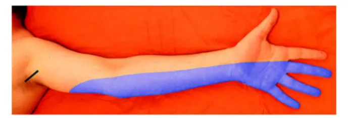
During the course of transportation, it was possible to clinically assess his left upper limb in a summary fashion. Pinprick and light touch sensory examination revealed hypoesthesia of the medial aspect of his arm and forearm from the axillary crease to the palmar wrist crease, as well as of the ulnar side of his hand and of the palmar aspect of the last three digits and of the dorsal aspect of the last two digits (Fig. 1). All other areas of his left upper limb showed a normal sensory response.
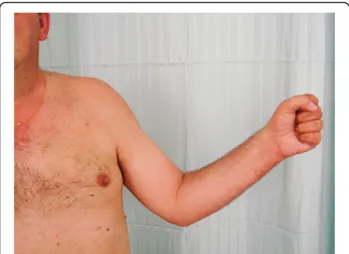
A motor examination revealed that he was not able to actively flex the joints of his middle, ring, and little fin-gers nor to adduct or abduct any of the finfin-gers of his left hand (Fig. 2). Moreover, he was not able to adduct his wrist. The remaining motor examination of his left upper limb showed no deficits.
The clinical presentation enabled us to promptly lo-cate the nerve injury between the anterior division of the lower trunk of his BP and the proximal portion of his following nerves: ulnar, medial cutaneous of his arm and forearm, and the medial aspect of his median nerve (Figs. 3 and 4).
Surgical exploration revealed a longitudinal lacer-ation of the posterior aspect of his axillary vein, as well as a complete section of his ulnar nerve, his medial brachial and antebrachial cutaneous nerves, and an incomplete section of the ulnar aspect of his median nerve (Fig. 5). A surgical approach was made under surgical loupes’ magnification. It began with vessel repair using an interrupted 8/0 Nylon suture, followed by direct end-to-end repair of the severed nerves using 8/0 Nylon simple stitches. Fibrin glue was applied around the repaired nerves.
His postoperative period was uneventful. He started an intensive physiotherapy program after hospital discharge, which occurred 3 days after surgery. The physiotherapy was aimed at maintaining joint mobility and at strengthening the paralyzed muscles, as rein-nervation occurred. Physiotherapy was performed daily for the first year after surgery and three times a
week for the following year. In the postoperative period, he also started swimming following the at-tending physician’s advice.
One year after surgery he resumed his employment. Three years after surgery, even though there was a slight atrophy of the intrinsic muscles of his hand, he presented a good overall function of his left upper limb (Figs. 6, 7, 8 and 9). At the last evaluation, 3 years after the accident, his motor function was M4 in all the previously paralyzed muscles according to the Medical Research Council Scale (muscle strength was reduced but muscle contraction could still move joints against resistance) [15]. Moreover, according to this scale [15], his sensory recovery was defined as S3 (return of superficial cutaneous pain and tactile sens-ibility without over-response) at the medial aspect of his arm and forearm, and as S2 (return of superficial cutaneous pain and some degree of tactile sensibility) at the ulnar side of his hand and at the palmar aspect of the last three digits and at the dorsal aspect of the last two digits.
This case report portrays a rare clinical situation in contemporary times: a major vascular lesion associated with a BP lesion in a conscious patient [16]. At present, this situation is rare because BP lesions are increasingly less frequent in most countries [16]. In addition, open BP injuries account for only a small percentage of all BP lesions [16–19]. In most cases of open BP wounds associated with major vascular bleeding, patients are too unstable for even a summary neurological examin-ation to be made prior to transportexamin-ation to the operat-ing room. Most commonly, patients are carried to the emergency room already under sedation and ventilated. The patient presented in this case report was fortunate
Fig. 2Photograph illustrating the motor deficit presented by the patient at admission. The patient was not able to flex joints of the middle, ring and little fingers of the left hand
enough to have been close to the hospital at the time of the lesion. Therefore, despite the severe vascular dam-age, he did not yet have changes to his consciousness when he arrived at the trauma room. All these improb-able events allowed a summary physical examination to be performed immediately before the emergency
surgery. This in turn permitted a prompt diagnosis of the location of the nerve lesions, based solely on the physical findings and knowledge of anatomy.
In 2002, Dubuisson and Kline described 23 open BP injuries in 100 consecutive cases of BP lesions [20]. In 2003, from a total of 1019 patients with BP injuries, Kim et al. reported only 19% with open injuries, of
Fig. 6Photograph of the patient's left upper limb three years after surgery. There is evidence of slight atrophy of the muscles innervated by the ulnar and median nerves, but its overall function is good. There is a slight limitation in the maximal extension of the metacarpal-phalangeal joint of the fifth finger
Fig. 5Photograph of the patient’s left axilla showing the intraoperative view of the axillary wound after control of bleeding and nerve repair. Longitudinal section of the posterior side of the axillary vein along with complete section of the ulnar, medial brachial cutaneous and medial antebrachial cutaneous nerves as well as partial section of the median nerve were found. 1, median nerve; 2, ulnar nerve; 3, axillary vein; 4, medial brachial cutaneous nerve; 5, medial antebrachial cutaneous nerve
which 7% involved lacerations and 12% were gunshot wounds [17]. Lacerations involving the BP may occur secondary to sharp instruments such as knives and glass, or from blunt trauma following animal bites or automobile accidents [5, 6, 17, 20–22]. These sources of injury most probably lead to neurotmesis (accord-ing to Seddon’s classification), which is the most se-vere type of injury to the peripheral nerves in which all the nerve layers are disrupted [7, 10].
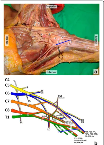
Figure 3 illustrates the BP and the muscles innervated by each of its nerve branches. In most cases the conver-gence of the anterior rami of the spinal nerve roots from C5 to T1 forms the spinal nerve roots, the trunks, the divisions, the cords, and the terminal branches of the BP [23]. The terminal branches of the BP are responsible
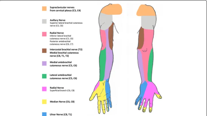
for most of the sensory, motor, and autonomic innerv-ation of the upper limb (Fig. 4).
A classical aphorism in neurological diagnosis is to try to attribute all signs and symptoms to a single lesion whenever possible [24].
As can be seen in Fig. 3, the fact that our patient’s stab wound was at the base of his axilla, thereby inferior to his clavicle, suggested that the lesion was probably lo-cated at the level of the divisions, cords, or terminal branches of his BP [25].
The hypoesthesia of the medial aspect of his arm, fore-arm, and hand (Fig. 1) could be explained by: (a) a sec-tion of the anterior division of the lower trunk of his BP; (b) a complete section of the medial cord of his BP; (c) a complete section of his medial brachial and medial ante-brachial cutaneous nerves, and his ulnar nerve, and a partial section of his median nerve (or medial root of his median nerve) [10, 12, 14, 26, 27].
Furthermore, paralysis of his flexor carpi ulnaris, med-ial part of his flexor digitorum profundus, third and fourth lumbricals, both palmar and dorsal interossei, ad-ductor pollicis, abad-ductor digiti minimi, flexor digiti minimi, and opponens digiti minimi muscles, indicates complete dysfunction of his entire ulnar nerve. The par-alysis of the muscle bellies of his flexor digitorum super-ficialis and flexor digitorum profundus for his third finger suggests partial median nerve dysfunction. Once more, this motor dysfunction could be caused by: (a) a section of the anterior division of the lower trunk of his BP; (b) a complete section of the medial cord of his BP; (c) a complete section of his ulnar nerve, and a partial section of his median nerve (or medial root of his me-dian nerve) [10, 12, 14, 26, 27].
A less likely cause of all these signs and symptoms could be either a lower trunk lesion or a lesion of the C8 and T1 roots of his BP. However, in either case, com-promise of the nerves arising from his dorsal cord, namely of his radial nerve, causing motor dysfunction and sensory changes in the territory of this nerve at the level of his forearm and hand would be present. In addition, sharp injury to the T1 root seemed unlikely, as this root is very close to the T1 sympathetic ganglion, whose lesion would produce Horner’s syndrome ipsilat-erally (meiosis, ptosis, enophthalmos, and facial anhy-drosis) [10, 28].
With all these data taken into consideration, the re-gion of the lesion of his BP could be safely pinpointed to the region between the anterior division of the lower trunk and the proximal portion of his ulnar, medial cuta-neous nerves of his arm and forearm, and the medial as-pect of his median nerve (Figs. 3 and 4). This in turn allowed a prompt planning of the surgical approach, and no doubt contributed to the good functional result ob-served 3 years after the surgery.
Fig. 8Photograph of the dorsum of the hands one year after surgery, showing slight atrophy of the muscles innervated by the ulnar and median nerve, as well as a mild ulnar claw
Conclusions
We present an increasingly rare clinical situation in present times: a major vascular lesion associated with a BP lesion in a conscious patient. In this clinical case, knowledge of the clinical anatomy of this region allowed a prompt diagnosis of the location of the nerve lesions. This, in turn, permitted repair not only of the vascular damage that was jeopardizing the patient’s life, but also his severed nerves, which no doubt played a major role in saving his life and achieving the good functional re-sults observed. We believe this case report eloquently demonstrates the clinical importance of a sound know-ledge of the anatomy of the BP in an emergency clinical setting.
Abbreviation
BP:Brachial plexus
Acknowledgements
We would like to express our gratitude to all the people who donated their body for medical research at our medical school, allowing us to obtain the dissection photograph of Fig. 3.
Funding
Diogo Casal received a grant from The Program for Advanced Medical Education, which is sponsored by Fundação Calouste Gulbenkian, Fundação Champalimaud, Ministério da Saúde e Fundação para a Ciência e Tecnologia, Portugal.
Availability of data and materials
All data generated or analyzed during this study are included in this published article.
Authors’contributions
DC, MAA, GM, and VC participated in the care of the patient. DC, TC, DP, II, and JGO drafted the manuscript. All authors have read and approved the manuscript.
Competing interests
The authors declare that they have no competing interests.
Consent for publication
Written informed consent was obtained from the patient for publication of this case report and any accompanying images. A copy of the written consent is available for review by the Editor-in-Chief of this journal.
Ethics approval and consent to participate
Not applicable.
Author details
1Plastic and Reconstructive Surgery Department and Burn Unit, Centro
Hospitalar de Lisboa Central, Lisbon, Portugal.2Anatomy Department, NOVA
Medical School, Universidade NOVA de Lisboa, Campo dos Mártires da Pátria, 130, 1169-056 Lisbon, Portugal.3UCIBIO, Life Sciences Department, Faculty of
Sciences and Technology, Universidade NOVA de Lisboa, Caparica, Portugal.
4CEDOC, NOVA Medical School, Universidade NOVA de Lisboa, Lisbon,
Portugal.
Received: 30 August 2016 Accepted: 30 November 2016
References
1. Terzis JK, Kostopoulos VK. The surgical treatment of brachial plexus injuries in adults. Plast Reconstr Surg. 2007;119:73e–92e.
2. Chuang DC. Brachial plexus reconstruction based on the new definition of level of injury. Injury. 2008;39 Suppl 3:S23–29.
3. Siqueira MG, Martins RS. Surgical treatment of adult traumatic brachial plexus injuries: an overview. Arq Neuropsiquiatr. 2011;69:528–35. 4. Sakellariou VI, Badilas NK, Mazis GA, Stavropoulos NA, Kotoulas HK,
Kyriakopoulos S, Tagkalegkas I, Sofianos IP. Brachial plexus injuries in adults: evaluation and diagnostic approach. ISRN Orthop. 2014;2014:726103. 5. Spinner RJ, Shin AY, Hébert-Blouin MN, Elhassan BT, Bishop AT. Traumatic
Brachial Plexus Injury. In: Wolfe SW, Hotchkiss RN, Pederson WC, Kozin SH, editors. Green's Operative Surgery, vol. 2. 6th ed. Philadelphia: Churchill Livingstone; 2011. p. 1235–92.
6. Sulaiman AR, Kline DG. Outcomes of treatment for adult brachial plexus injuries. In: Chung KC, Yang LJ, McGillicuddy JE, editors. Practical Management of Pediatric and Adult Brachial Plexus Palsies. 1st ed. USA: Elsevier; 2012. p. 344–65.
7. Dunkerton MC, Boome RS. Stab wounds involving the brachial plexus. A review of operated cases. J Bone Joint Surg Br. 1988;70:566–70. 8. Sinha S, Khani M, Mansoori N, Midha R. Adult brachial plexus injuries:
Surgical strategies and approaches. Neurol India. 2016;64:289–96. 9. Sinha S, Pemmaiah D, Midha R. Management of brachial plexus injuries in
adults: Clinical evaluation and diagnosis. Neurol India. 2015;63:918–25. 10. Gregory J, Cowey A, Jones M, Pickard S, Ford D. The anatomy, investigations
and management of adult brachial plexus injuries. Orthopaedics and Trauma. 2009;23:420–32.
11. Terzis JK, Papakonstantinou KC. The surgical treatment of brachial plexus injuries in adults. Plast Reconstr Surg. 2000;106:1097–124.
12. Aggarwal A, Puri N, Aggarwal AK, Harjeet K, Sahni D. Anatomical variation in formation of brachial plexus and its branching. Surg Radiol Anat. 2010;32:891–4. 13. Gutton C, Choquet O, Antonini F, Grossi P. [Ultrasound-guided interscalene block: Influence of anatomic variations in clinical practice]. Ann Fr Anesth Reanim. 2010;29:770–5.
14. Fetty LK, Shea J, Toussaint CP, McNulty JA. A quantitative analysis of variability in brachial plexus anatomy. Clin Anat. 2010;23:210–5. 15. Wang Y, Sunitha M, Chung KC. How to measure outcomes of peripheral
nerve surgery. Hand Clin. 2013;29:349–61.
16. Midha R. Epidemiology of brachial plexus injuries in a multitrauma population. Neurosurgery. 1997;40:1182–8. discussion 1188–9. 17. Kim DH, Cho YJ, Tiel RL, Kline DG. Outcomes of surgery in 1019 brachial
plexus lesions treated at Louisiana State University Health Sciences Center. J Neurosurg. 2003;98:1005–16.
18. Brooks DM. Open wounds of the brachial plexus. Spec Rep Ser Med Res Counc (G B). 1954;282:418–29.
19. Brooks DM. Open wounds of the brachial plexus. J Bone Joint Surg Br. 1949; 31B:17–33.
20. Dubuisson AS, Kline DG. Brachial plexus injury: a survey of 100 consecutive cases from a single service. Neurosurgery. 2002;51:673–82. discussion 682–3. 21. Birch R. The closed supraclavicular lesion. In: Surgical Disorders of the
Peripheral Nerves. Second edth ed. London: Springer; 2011. p. 375–427. 22. Hentz VR. Adult and Obstetrical Brachial Plexus Injuries. In: Slutsky DJ, Hentz
VR, editors. Peripheral Nerve Surgery: Practical Applications in the Upper Extremity. 1st ed. Philadelphia: Churchill Livingstone; 2006. p. 299–317. 23. Johnson EO, Vekris M, Demesticha T, Soucacos PN. Neuroanatomy of the
brachial plexus: normal and variant anatomy of its formation. Surg Radiol Anat. 2010;32:291–7.
24. Waxman SG, deGroot J. Introduction to clinical thinking: the relationship between neuroanatomy and neurology. In: Waxman SG, deGroot J, editors. Correlative neuroanatomy, vol. 1. Twenty-second edth ed. USA: Lange; 1995. p. 35–43.
25. Chen N, Yang LJ, Chung KC. Anatomy of the brachial plexus. In: Chung KC, Yang LJ, McGillicuddy JE, editors. Practical Management of Pediatric and Adult Brachial Plexus Palsies. 1st ed. USA: Elsevier; 2012. p. 3–12. 26. Pellerin M, Kimball Z, Tubbs RS, Nguyen S, Matusz P, Cohen-Gadol AA,
Loukas M. The prefixed and postfixed brachial plexus: a review with surgical implications. Surg Radiol Anat. 2010;32:251–60.
27. Gu YD. Functional motor innervation of brachial plexus roots. An intraoperative electrophysiological study. J Hand Surg Br. 1997;22:258–60. 28. Amonoo-Kuofi HS. Horner’s syndrome revisited: With an update of the