Review
Printed in Brazil - ©2014 Sociedade Brasileira de Química0103 - 5053 $6.00+0.00
*e-mail: [email protected]
Anti-
Trypanosoma cruzi
Compounds: Our Contribution for the Evaluation and
Insights on the Mode of Action of Naphthoquinones and Derivatives
Eufrânio N. da Silva Júnior,a Guilherme A. M. Jardim,a Rubem F. S. Menna-Barretob
and Solange L. de Castro*,b
aLaboratório de Química Sintética e Heterocíclica, Departamento de Química, Instituto de Ciências Exatas, Universidade Federal de Minas Gerais (UFMG), 31270-901 Belo Horizonte-MG, Brazil
bLaboratório de Biologia Celular, Instituto Oswaldo Cruz, Fiocruz, Av. Brasil, 4365, Manguinhos, 21045-900 Rio de Janeiro-RJ, Brazil
A doença de Chagas causada pelo Trypanosoma cruzi afeta cerca de oito milhões de pessoas
em países em desenvolvimento, sendo classificada como uma doença tropical negligenciada pela Organização Mundial da Saúde. A quimioterapia disponível para esta doença é baseada em dois nitro-heterocíclicos, nifurtimox e benznidazol, ambos com graves efeitos colaterais e eficácia variável, e assim novos medicamentos visando um tratamento mais eficiente são necessários com urgência. Nos últimos 20 anos, temos desenvolvido em colaboração com grupos focados em química medicinal, um programa de quimioterapia experimental da doença de Chagas, investigando a eficácia, seletividade, toxicidade, alvos celulares e mecanismos de ação de diferentes classes de
compostos sobre T. cruzi. Neste artigo, apresentamos uma visão geral desses estudos, enfocando
protótipos naftoquinoidais e derivados, examinando a sua síntese, a atividade e mecanismo de ação, o que foi realizado e o que precisa ser abordado, avaliando e discutindo possíveis melhorias. Esta mini-revisão discute nosso esforço continuado visando a caracterização biológica e a síntese de compostos naftoquinoidais, auxiliando no desenvolvimento de um novo arsenal de drogas candidatas com eficácia contra o T. cruzi.
Chagas disease is caused by the parasite Trypanosoma cruzi and affects approximately
eight million individuals in the developing world; it is also classified as a neglected tropical disease by the World Health Organization. The available therapy for this disease is based on two nitroheterocycles, nifurtimox and benznidazole, both of which exhibit severe side effects and variable efficacy; therefore, new drugs and better treatment schedules are urgently needed. For the past 20 years, we have been collaborating with groups focused on medicinal chemistry to produce experimental therapies for Chagas disease by investigating the efficacy, selectivity, toxicity,
cellular targets and mechanisms of action of different classes of compounds against T. cruzi. In
this report, we present an overview of these studies, focusing on naphthoquinonoid prototypes and discuss their synthesis, activity and mechanisms of action. Furthermore, we summarise the research that has been performed to date and suggest future research directions while assessing and discussing potential improvements. This mini-review discusses our continued efforts toward the biological characterisation and synthesis of naphthoquinoidal compounds, aiming to contribute
to the development of a new arsenal of candidate drugs that exhibit effective anti-T. cruzi activity
Keywords: naphthoquinones, β-lapachone, Trypanosoma cruzi, Chagas disease, chemotherapy
1. Introduction
Chagas disease (CD) is caused by the intracellular obligatory parasite Trypanosoma cruzi and was first described more than one hundred years ago, in 1909, by Carlos
Chagas.1 This disease has high morbidity and mortality
rates, affects approximately eight million individuals in the developing world and displays a limited response to therapy; it has also been classified as a neglected tropical disease by the World Health Organization (WHO).2,3 Chagas disease
contamination, through laboratory accidents and congenital routes. T. cruzi is a hemoflagellate protozoan (family Trypanosomatidae, order Kinetoplastida)4 that exhibits a
complex life cycle involving distinct morphological stages during its passage through vertebrate and invertebrate hosts. Briefly, after ingestion of bloodstream trypomastigotes by insect vectors, the parasites are converted to epimastigote forms, which proliferate and subsequently differentiate into metacyclic forms within the posterior intestine of the triatomine. These infective parasite forms are released in the faeces of the triatomine and can invade new vertebrate cells. The parasites then undergo another round of differentiation into intracellular amastigote forms, which proliferate and subsequently transform back into trypomastigotes, the form that disseminates the infection.
Although vector and transfusion transmissions have sharply declined over the past 20 years due to the Southern Cone countries policy, several challenges still need to be overcome including those related to sustainable disease control through the adoption of public policies in the endemic areas.5,6 In addition, despite effective efforts to
control vector and blood transmission, Chagas disease still presents many challenges including the following: (i) its peculiar epidemiology is characterised by a variety of risk factors (many potential vectors and reservoirs, different forms of transmission and diverse parasite isolates present in domiciliar, peridomiciliar and sylvatic environments); and importantly, (ii) the lack of prophylactic therapies and effective therapeutic treatments.7,8 Current major
concerns include disease transmission by the ingestion of contaminated food or liquids and the disease’s emergence in nonendemic areas such as North America and Europe, a phenomenon which is likely due to the immigration of infected individuals.9,10 This disease is also recognised as
an opportunistic infection in HIV-infected individuals.11
Outbreaks of acute Chagas disease associated with the ingestion of contaminated food and drink have been reported in South America,12,13 and are associated with a
high mortality rate mainly due to myocarditis.
Chagas disease is characterised by two clinical phases. The acute phase appears shortly after infection, and in some cases the individual may not even realise he/she is infected. Symptoms range from flu-like symptoms to intense myocarditis (in approximately 10% of infected people). If left untreated, symptomatic chronic disease develops in about one third of the individuals after a long latent period (10-30 years) that is known as the indeterminate form. The main clinical manifestations of Chagas disease include digestive and/or cardiac alterations, although disorders of the central and peripheral nervous system may also occur.14,15 In the chronic digestive form of the disease, the
clinical manifestations are caused by dysperistalsis of the oesophagus and colon, which are due to the destruction of the myenteric plexus and leads to mega syndromes.16 The
chronic cardiac form of the disease is the most significant clinical manifestation, and consequences include dilated cardiomyopathy, congestive heart failure, arrhythmias, cardioembolism and stroke.17 The pathogenesis of Chagas
disease is the result of a sustained inflammatory process due to an anti-parasitic and/or anti-self-immune response, which is associated with low-grade persistent parasite presence.18-22 Growing evidence shows that parasite
persistence within the target organs associated with an unregulated host immune response are involved in disease progression and clinical outcomes.19,23 Control of T. cruzi
infection depends on both the innate and acquired immune responses which are triggered during early infection and are critical for host survival. These responses involve macrophages, natural killer cells, T and B lymphocytes and the production of pro-inflammatory cytokines.24
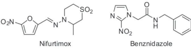
The available therapy for Chagas disease is based on two nitroheterocyclic agents that were developed over five decades ago (Figure 1).
Nifurtimox (Nif, 3-methyl-4-(5’-nitrofurfurylidene-amine)tetrahydro-4H-1,4-tiazine-1,1-dioxide) is a nitrofuran that was developed by Bayer in 1967 and marketed as Lampit®. It acts by reducing the nitro group
to generate nitro-anions that subsequently react with molecular oxygen to produce toxic superoxide and peroxide radicals. Today, Nif is produced by Bayer HealthCare at the Corporacion Bonima in El Salvador. Benznidazole (Bz, N-benzyl-2-nitroimidazole acetamide) is a nitroimidazole that was developed by Roche in 1972 and was formerly marketed as Rochagan® or Radanil®; it is currently
produced by the Laboratório Farmacêutico do Estado de Pernambuco, Brazil (www.pe.gov.br/orgaos/lafepe-laboratorio-farmaceutico-de-pernambuco/). This drug appears to act differently, as it produces metabolites that react with macromolecules such as DNA, RNA, proteins, and possibly lipids. In both cases, the antiparasitic activity of the drug is intimately linked with their inherent toxicity. Both drugs are effective against acute infections, but they show poor activity during the late chronic phase.16 Due
to their severe side effects and limited efficacy against
different parasitic isolates,25 these drugs are hardly the
best treatment options to offer patients. One of the major drawbacks of Nif is its high incidence of side effects, which is observed in up to 40% of patients and includes nausea, vomiting, abdominal pain, weight loss and severe anorexia. Furthermore, adverse neurological effects such as restlessness, paresthesia, twitching, insomnia and seizures have also been observed.21 In comparison to Nif, Bz has the
advantage of a lower incidence of side effects; however, its side effects include hypersensitivity (dermatitis, generalised oedema, ganglionic infarction and joint and muscle pains), bone marrow depletion and peripheral polyneuropathy.26
Because of the challenges regarding the efficacy vs. the toxicity of both nitro-heterocyclic compounds, the current recommendations for using either drug to treat Chagas disease suggest that all acute cases, including reactivations due to immunosuppression, recent chronic cases (including children up to 12 years of age), and indeterminate or benign chronic forms should be treated. In addition, cases should be treated at the discretion of the attending physician. In contrast, the contra-indications for specific treatment are pregnancy, liver and kidney failure, neurological diseases unrelated to CD, advanced CD with grade III or IV cardiopathy (Pan American Health Organization, PAHO)/(WHO), or other pathologies that may be worsened by treatment.26
Between 12 and 18% of patients who undergo treatment have to suspend their therapy prematurely because of side effects.27 Overall, the 2010 Latin American Guidelines for
Chagas cardiomyopathy indicate that unrestricted treatment for patients with chronic Chagas disease should not be regarded as standard therapy.28
Several new compounds are currently under preclinical development, and different approaches have been used to identify new drug leads including in vitro parasite phenotype screens and target-based drug discovery.29
Although many attempts have been made to treat the disease since its identification in 1912, only allopurinol and some antifungals have been used in clinical trials since the introduction of Nif and Bz.25,30 In 2009, the
Drugs for Neglected Diseases initiative (DNDi) and its partners launched the Chagas disease Clinical Research Platform (http://www.dndi.org/strenghtening-capacity/
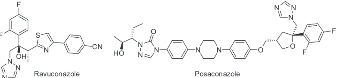
chagas-platform/publications.html), which aims to promote technical discussions, develop a critical mass of expertise, strengthen institutional research capacities, and link investigators through a collaborative network. As a result, three phase II clinical trials began in 2011 to investigate the potential uses of posaconazole (a structural analogue of itraconazole) (SCH 56592; Schering-Plough Research Institute, SPRI) and of a prodrug of ravuconazole (E1224; Eisai) (Figure 2).
Both drugs are triazole derivatives that inhibit fungal and protozoan cytochrome P-450-dependent enzyme CYP51 (C14α-lanosterol demethylase) (TcCYP51).31-33
Two clinical studies were performed with posaconazole: STOP-CHAGAS (in Argentina, Colombia, Mexico and Venezuela, funded by Merck) with results expected by 2014 and CHAGASAZOL (in Spain at University Hospital Vall d’Hebron Research Institute in Barcelona), which was completed in March 2013 (results were posted at http://clinicaltrials.gov/show/NCT01162967, accessed in July, 2014). Another study investigated the use of E1224 (DNDi/Eisai Pharmaceuticals) and was developed in Bolivia. It involved a total of 231 patients, and the drug exhibited a good safety profile and was effective at clearing the parasite; however, it had little to no sustained efficacy one year after treatment. The key disadvantages of novel azole derivatives (i.e., posaconazole) are their complexity and manufacturing costs.31
Among the drugs identified in preclinical studies, several of them have yielded valuable results. For example, CYP51 inhibitors such as tipifarnib (an anti-cancer drug that inhibits the human protein farnesyltransferase)32
and the fenarimol series show promise.33 In addition,
fexinidazole (a substituted 5-nitroimidazole that was rediscovered by the DNDi and is currently in phase II/III clinical study for the treatment of human African trypanosomiasis),34 diamidine analogues35 and a series of
oxaboroles (prototype AN4169) are promising new drugs for the treatment of T. cruzi infections.36 Other drug targets
under investigation include cysteine proteases because T. cruzi contains a cathepsin L-like enzyme (cruzipain) that is responsible for the majority of the proteolytic activity that occurs in all developmental forms. The vinyl
sulfone K777 is an irreversible cruzipain inhibitor that has shown efficacy in chronic rodent models and is also under preclinical development.29 Some of the most promising
targets identified in T. cruzi include protein prenylation, hypoxanthine-guanine phosphoribosyltransferase, cysteine proteases,29,37 and topoisomerases.38 The utility
of 14-demethylase inhibitors,39,40 squalene synthase
inhibitors,41 farnesyl pyrophosphate synthase inhibitors,42
farnesyl transferase inhibitors,43,44 dihydrofolate reductase
inhibitors45 and natural products such as canthinones,
quinolines, lignans, and naphthoquinones are also being explored.46-48 New and established pharmacophores based
on synthetic and natural product chemistry have been identified through improved screening technologies and have generated hits from libraries provided largely by the pharmaceutical industry and other entities.
Another approach aimed at the treatment of Chagas disease is the achievement of greater efficacy through the use of combinations of existing drugs that display different mechanisms of action. Combination therapy has been proven to be more effective than monotherapies for several infectious diseases and also minimises the risk of drug resistance. Several studies in animal models have examined the use of combinations of Bz and CYP51 inhibitors,49-52
the arylimidamide DB766,53 and allopurinol,54,55 and the
results were encouraging. Coura26 proposed the use of
combinations of [Nif + Bz], [Nif or Bz + allopurinol] and [Nif or Bz + ketoconazole, fluconazole or itraconazole] in specified treatment schemes that were adapted according to the side effects observed.
Based on current knowledge of parasite and host biological characteristics, a desired drug candidate for Chagas disease would include the following attributes: (i) high activity against the evolving forms of the parasite present in the mammalian hosts and different reservoirs of the parasite, (ii) efficacy against both acute and chronic infections, (iii) oral administration of only a few doses, (iv) low toxicity and an improved safety profile (including children and women of reproductive age), (v) low cost and high stability suitable for a long shelf life in tropical temperatures, and (vi) high levels of tissue accumulation and long terminal half-lives.55
Over the past 20 years, our group has been working on experimental chemotherapy for Chagas disease in collaboration with research groups focused on medicinal chemistry. We have been investigating the efficacy, selectivity, toxicity, cellular targets and mechanisms of action of different classes of compounds on T. cruzi. In this report, we present an overview of these studies, focusing on the development of novel naphthoquinonoid prototypes for the clinical treatment of Chagas disease. We also
describe their synthesis, activity and mechanisms of action. Furthermore, we summarise the current state of research in the field and suggest future directions while assessing and discussing potential improvements. This mini-review discusses our continued efforts toward the biological characterisation and synthesis of naphthoquinoidal compounds, aiming to contribute in the development of a new arsenal of candidate drugs that exhibit effective anti-T. cruzi activity.
2. Quinoidal Compounds and Derivatives
Quinoidal compounds can be found in various plant families or as synthetic substances.56-59 The structural
components of these compounds are the focus of many studies attempting to determine their activity against several parasites such as Leishmania,60T. cruzi61 and Plasmodium
falciparum.62 Quinones participate in multiple biological
oxidative processes due to their structural properties and their capacity to generate reactive oxygen species.63,64
The first report published in collaboration with Antonio V. Pinto’s group from the Federal University of Rio de Janeiro in 1994 described a series of natural and synthetic drugs that exhibited activity against T. cruzi.65 In this
work, we evaluated 45 compounds for activity against bloodstream forms of T. cruzi. From there, a fruitful partnership began, and several molecules were synthesised and screened for activity against this parasite.
Following this initial study, we dedicated our efforts to the identification of new quinoidal molecules. Lapachol (1) is an important natural naphthoquinone; we used it and its derivatives to explore the chemical reactivity of the drug class, and several heterocycles were obtained with good yields (Schemes 1-3). Their effects on the bloodstream forms of T. cruzi were evaluated, and the results are shown in Table 1. Some compounds that exhibited initial activity were identified as potential candidates for further studies due to comparable activity with crystal violet, a substance indicated for the sterilisation of chagasic blood.66 Unless
otherwise stated, all of the screening assays presented in this review were performed using bloodstream trypomastigotes of the Y strain obtained from infected albino mice at the peak of parasitaemia. These trypomastigotes were isolated by differential centrifugation and resuspended (107 cells mL-1) in Dulbecco’s modified Eagle medium
weekly transfers of liver infusion tryptose (LIT) medium and harvested during the exponential phase of growth (5-day-old culture forms). The assays were performed in 24-well microplates and were incubated up to 4 days at 28 °C in LIT medium. Cell counts were performed in a Neubauer chamber, and trypanocidal activity was expressed as an IC50 value corresponding to the concentration that
lyses 50% of the parasites.
Meanwhile, we reported the synthesis and evaluation of naphthoxazoles containing both electron donating and withdrawing groups (Figure 3).67,68 Heterocycles, as for
instance, indole and 1,3-benzodioxole, as substituent groups were also evaluated. The compounds were easily obtained from the reaction of β-lapachone or nor-β-lapachone and aromatic aldehydes in the presence of an ammonium salt. In general, these structures exhibited efficient anti-T. cruzi activity and represented an excellent starting point for the synthesis of new prototypes.
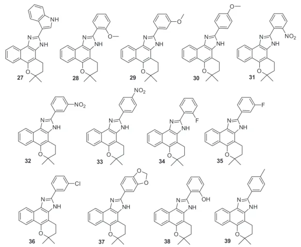
Another class of structures prepared from the same reaction were the naphthoimidazole derivatives
27-39 (Figure 4). The trypanocidal activities of the naphthoxazoles 19-26 and naphthoimidazoles 27-39 are displayed in Table 2. From these substances, compounds
18 (IC50 = 37.0 ± 0.7 µM), 27 (IC50 = 15.4 ± 0.2 µM) and
39 (IC50 = 15.5 ± 2.9 µM) were selected for further studies
of the trypanocidal mechanism of action.69
The naphthoimidazoles 18, 27 and 39 were also effective against the proliferative forms of T. cruzi
(intracellular amastigotes and epimastigotes), and the main ultrastructural targets identified were the mitochondrion and nuclear DNA.70 Electron microscopy analyses
revealed mitochondrial swelling, abnormal chromatin condensation, endoplasmic reticulum profiles surrounding organelles and autophagosome-like structures in treated parasites. We also observed reservosome disorganisation and trans-Golgi network cisternae disruption specifically in the epimastigote forms.70,71 Interestingly, the
pre-incubation of the parasites with the cysteine protease inhibitor E64 or calpain inhibitor I partially attenuated the trypanocidal effect of the naphthoimidazoles suggesting that the deactivation of cysteine proteases is involved in their mode of action.70 Because the reservosome is a
target in epimastigotes and is rich in cysteine proteases, disruption of this organelle could release proteases into the cytosol and initiate a proteolytic pathway, ultimately leading to parasite death. Alterations of mitochondrion, chromatin, and reservosomes and the detection of an autophagy process encouraged further studies regarding death pathways. The investigation of the apoptotic features demonstrated discrete phosphatidylserine exposure and strong DNA fragmentation by both electrophoresis and terminal deoxynucleotidyl transferase dUTP nick end labeling (TUNEL) techniques.70-72 Naphthoimidazoles
are planar in structure and could possibly interact with the parasite’s DNA to induce fragmentation, which is a decisive event during trypanocidal activity. In contrast,
Scheme 2. Lawsone 7 and its derivatives 8 and 9.66
the morphological evidence of autophagy induction after treatment with compounds 18, 27 and 39 stimulated a more detailed evaluation of this pathway. Strong labelling of monodansylcadaverine (an autophagosome probe) together with ATG (autophagic-related genes) overexpression and total abolition of the compounds’ effects by the well-known autophagic inhibitors wortmannin or 3-methyladenine in both treated epimastigotes and trypomastigotes supported the hypothesis that autophagy was involved in the naphthoimidazoles’ mode of action.72 However, further
proteomic analysis is needed to identify T. cruzi molecules involved in the mechanism of action of compounds 18,
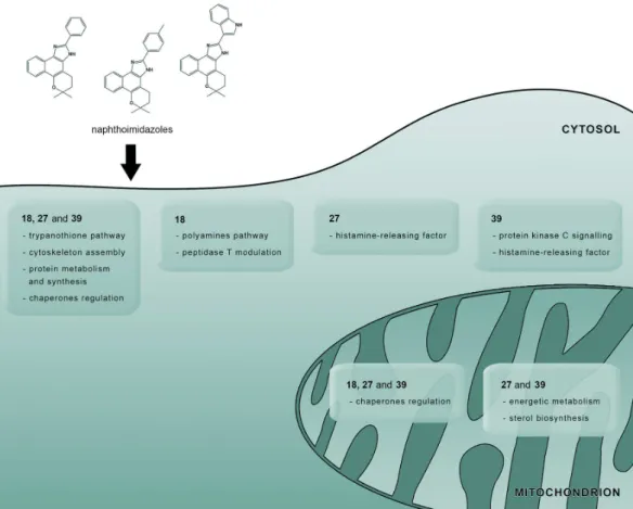
27 and 39. In 2010, the first assessment of the proteomic profile of naphthoimidazole-treated epimastigotes was performed. Multiple biochemical pathways were involved in their trypanocidal activity including redox metabolism, energy production, ergosterol biosynthesis, cytoskeleton
assembly, protein metabolism and chaperone modulation. An imbalance among these fundamental pathways could lead to the loss of homeostasis and culminate in T. cruzi death.73 Among the proteins modulated by the treatment, 26
proteins were downregulated, and only three proteins were overexpressed. Surprisingly, most of the modulated proteins were exclusive to each particular compound, indicating that differences in their modes of action existed (Figure 5).
Mitochondrial proteins were the most commonly modulated proteins, thus confirming the previous biochemical and ultrastructural evidence that described this organelle as the primary target of these compounds.70,71,73 Tubulin was
downregulated in parasites treated with compounds 18,
27 and 39. In trypanosomatids, different tubulin isoforms are present because each one is linked to the kinetics of microtubule assembly. Enzyme-linked immunosorbent assay (ELISA) data showed that the tyrosinated tubulin
pool decreased after treatment. This protein isoform was associated with labile microtubules, suggesting that these compounds interfered with intracellular vesicle traffic and/or mitotic spindle formation. This hypothesis was also supported by the absence of ultra-structural damage in subpellicular and flagellar microtubules and the blockage of mitosis in treated epimastigotes.70,71,73 Due to the results
obtained about the activity and mechanism of action of
18, 27 and 39 higher amounts of the compounds were synthesised and experiments are underway in our laboratory aiming the evaluation of nitroimidazoles in the treatment of experimentally T. cruzi-infected mice.74
To synthesise new heterocycles, Pinto and co-workers67
developed a methodology to produce pyran derivatives of β-lapachone (3) through a reaction using active methylene reagents under basic conditions. The resulting cyclopentenones and chromenes were evaluated for anti-T. cruzi activity in addition to the other heterocyclic compounds shown in Figure 6. The results of the trypanocidal activity studies are shown in Table 3. Unfortunately, this class of compounds did not exhibit trypanocidal activity comparable to that of the naphthoimidazole derivatives, with the exception of compound 45. Thus, these substances have not been the subject of subsequent studies.
In the same manner, we continued the search for trypanocidal heterocyclic compounds and obtained a phenazine derivative 50 (Figure 7) from β-lapachone (3), which was subsequently well characterised by crystallographic methods. This compound was almost twice as active as Bz, with an IC50 (24 h) of 61.3 ± 9.6 µM.
75
Despite its promising activity level, the yield for obtaining compound 50 from lapachone (3) was low (25% yield), which discouraged further studies. However, phenazines obtained from lapachones generally exhibited low levels of cytotoxicity,76 and this phenazine represents an important
prototype for the design of novel trypanocidal drugs. Over the last few years, our group has focused on synthesising and measuring the trypanocidal activity of nor-β-lapachones substituted with heterocyclic rings. In general, a molecular hybridisation strategy was used to design the new compounds,77 and the subject of our
study was the combination of a quinoidal moiety with a 1,2,3-triazole group. The first synthetic route we developed followed the principles of medicinal chemistry and
Table 1. Effects of the original quinones and their naphthoxazole and naphthoimidazole derivatives on T. cruzi
Compound IC50, 24 h / µMa
1 2 3 4 5 6 7 8 9 10 11 12 13 14 15 16 17 18
Crystal violet
410.8 ± 53.5 > 4800 391.5 ± 16.5 1280.6 ± 167.2
> 400 164.8 ± 30.5
> 2500 420.7 ± 71.2 330.7 ± 62.4
> 2500 49.5 ± 1.4 283.5 ± 25.0 171.9 ± 51.2 197.3 ± 25.8
> 2500 325.2 ± 21.3
> 4800 37.0 ± 0.7 536.0 ± 3.0
aMean ± standard deviation from three experiments performed in triplicate.
Figure 4. Naphthoimidazoles 27-39 obtained from β-lapachone (3).67,68
Table 2. Effects of naphthoxazoles and naphthoimidazoles on T. cruzi
Compound IC50, 24 h / µMa
19 20 21 22 23 24 25 26 27 28 29 30 31 32 33 34 35 36 37 38 39
Benznidazole
283.5 ± 25.0 > 9600 3502.5 ± 305.3 1641.3 ± 147.0 269.5 ± 46.5 351.4 ± 12.4
> 4800 > 2500 15.4 ± 0.2 6444.6 ± 483.7 3057.8 ± 836.7 259.3 ± 40.4 1858.1 ± 366.7
579.3 ± 52.5 303.6 ± 12.2
243.3 372.0 1064.2 1850.5 4455.5 ± 465.8
15.5 + 2.9 103.6 ± 0.6
aMean ± standard deviation from three experiments performed in triplicate.
produced lapachone-based 1,2,3-triazoles with global yields higher than 50%. Using the Hooker oxidation method,78
nor-lapachol (4) was prepared and used to obtain the key intermediate 3-azido nor-β-lapachone (51). Compound 51
was used to prepare the respective 1,2,3-triazole derivatives
52-61 by employing a 1,3-dipolar reaction catalysed by Cu(I), a type of reaction also known as “click chemistry” (Scheme 4).79 The results of the trypanocidal activity studies
are shown in Table 4.80,81
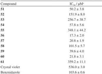
Overall, all compounds exhibited good trypanocidal activity, and several compounds were even more active than Bz. It was recently suggested in the Perspectives Section of the Journal of Medicinal Chemistry82 that a
triazolic naphthofuranquinone compound (56) represents an important trypanocidal prototype. Compound 56 was the most active with an IC50 (24 h) value of 17.3 ± 2.0 µM,
and this substance was chosen for further studies of its mechanism of action.83 This compound was also effective
against the epimastigote and intracellular amastigote forms of T. cruzi, with IC50 (24 h) values below 25 µM.
Figure 5. Similarities and differences among the mechanisms of action of each naphthoimidazole in T. cruzi epimastigotes. Most of the modulated proteins are mitochondrial proteins, indicating that this organelle is the main target of compounds 18, 27 and 39. These three compounds regulate the trypanothione pathway, cytoskeleton assembly, protein metabolism/synthesis and chaperone diversity. These alterations compromise different biological processes and lead to parasite death. Other proteins and/or pathways were also affected by the naphthoimidazoles including the polyamine pathway and peptidase T activity (18), ergosterol biosynthesis, energetic metabolism, histamine-releasing factor activity (27 and 39), and protein kinase C signalling (39).
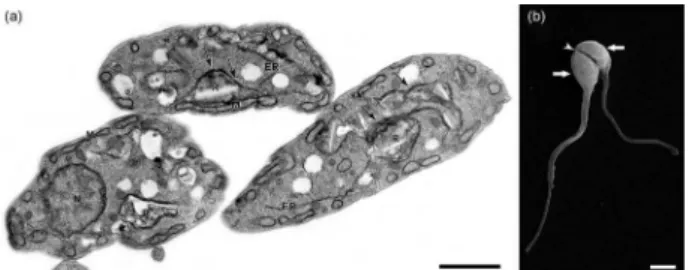
Cell cycle evaluations revealed a reduction in the number of parasites with duplicated genetic material, suggesting that the compound blocked cytokinesis. Transmission electron microscopy analyses of epimastigotes revealed the formation of well-developed endoplasmic reticulum profiles surrounding the reservosomes; these results suggest that there is close contact between both membranes. The
appearance of cytosolic concentric membrane structures was another morphological feature, suggesting that autophagy is a partial mode of action for compound 56. Fluorescence microscopy analyses reinforced these data and indicated that a high percentage of MDC-labelled epimastigotes was present after treatment. Morphological damage in Golgi cisternae and blebbing of the flagellar membrane were also frequent alterations induced by this triazolic quinone. Interestingly, ultra-structural and flow cytometry studies showed that the mitochondrion was not affected by the treatment, suggesting that this organelle is not a target of compound 56. The mechanism of action of this triazolic naphthofuranquinone differs from that of the other naphthoquinones studied because it involves autophagy (especially of the reservosomes) and cytokinesis impairment (Figure 8).83
Compound 56 was considered an important prototype for anti-T. cruzi activity, but its high level of cytotoxicity
Table 3. Effects of the heterocyclic compounds 40-49 on T. cruzi
Compound IC50, 24 h / µMa
40 > 4000
41 1216.7 ± 349.1
42 ndb
43 > 4000
44 > 4000
45 56.1 ± 15.5
46 > 4000
47 786.9 ± 80.0
48 ndb
49 > 4000
Benznidazole 103.6 ± 0.6
aMean ± standard deviation from three experiments performed in triplicate; bnot determined.
Figure 7. Phenazine derivative 50 obtained from β-lapachone (3).75
Scheme 4. Nor-β-lapachone-based 1,2,3-triazoles 52-61.80,81
Table 4. Effects of nor-β-lapachone-based 1,2,3-triazoles on T. cruzi
Compound IC50 / µMa
51 50.2 ± 3.8
52 151.9 ± 8.0
53 256.7 ± 38.7
54 57.8 ± 5.6
55 348.1 ± 44.2
56 17.3 ± 2.0
57 20.8 ± 1.9
58 101.5 ± 5.7
59 39.6 ± 4.0
60 21.8 ± 3.1
61 359.2 ± 11.1
Crystal violet 536.0 ± 3.0
Benznidazole 103.6 ± 0.6
in mammalian cells was an impediment for further studies. We believed that it was necessary to structurally modify this compound to obtain a substance with a higher selectivity index (SI) that corresponds to the ratio LC50 (concentration
that leads to damage of 50% of the mammalian cells)/IC50.
Another possibility would be to develop the compound within a controlled delivery system, which has been the focus of several studies aimed at solving drug toxicity issues. This important strategy can be used to optimise the therapeutic efficacy of the drug and reduce toxic side effects.84 In Scheme 5, the naphthoquinoidal compounds
designed to couple ortho-quinone to para-quinoidal structures are displayed. Our strategy was based on the combination of ortho- and para-quinoidal moieties that are able to generate high concentrations of reactive oxygen species, a property that is generally associated with the activity of this class of compounds. Based on the structural skeleton of compound 56, compounds 62-64 were designed to preserve the main group, the quinoidal pharmacophore. Our approach proved to be effective, and compounds 62, 63, and 64 exhibited IC50 (24 h) values of 80.8, 6.8 and 8.2 µM,
respectively (Table 5).85 We were pleasantly surprised when
heart muscle cell toxicity analyses produced LC50 (24 h)
values of 63.1 and 281.6 µM for compounds 63 and 64, respectively, which corresponded to SI of 9.3 and 34.3.85
Aiming the establishment of a panel of minimum standardised procedures to advance leading compounds to clinical trials, the workshop Experimental Models in Drug Screening and Development for Chagas Disease was held in Rio de Janeiro (Brazil) organised by the Fiocruz Program for Research and Technological Development on Chagas Disease (PIDC) and DNDi. During the meeting, the minimum steps, requirements and decision gates for the determination of the efficacy of lead compounds were evaluated by interdisciplinary experts and an in vitro and in vivo flowchart was designed to serve as a general and standardised protocol for drug screening.86 Based
on this flowchart and due to the high SI value attained, compound 64 will be assayed further for its effectiveness in T. cruzi-infected mice.
To obtain additional trypanocidal molecules with low toxicity in mammalian cells, new triazolic α- and nor-α-lapachones were synthesised and assayed for anti-T. cruzi activity based on a strategy we recently described involving C-ring modification.87
α-Lapachone-based 1,2,3-triazoles were synthesised as previously described (Scheme 6).88 4-Bromo-α-lapachone
was prepared from α-lapachone (2) by obtaining a key
Figure 8. Ultra-structural analysis of T. cruzi epimastigotes treated with compound 56. (a) Transmission electron microscopy revealed reservosome disorganisation (R) and endoplasmic reticulum (ER) profiles in close contact with this organelle’s membrane (black arrows). The nucleus and mitochondrion (M) exhibited typical morphologies. (b) Scanning electron microscopy examination revealed parasite body retraction (white thick arrows) and the impairment of mitosis (white arrowhead). Bar in (a): 0.2 µm. Bar in (b): 1 µm.
Scheme 5. Nor-β-lapachone 1,2,3-triazole coupled 1,4-naphthoquinones 62-64.85
Table 5. Effects of compounds 62-64 on T. cruzi
Compound IC50 / µMa
62 80.8 ± 6.5
63 6.8 ± 0.7
64 8.2 ± 0.7
Benznidazole 103.6 ± 0.6
Crystal violet 536.0 ± 3.0
intermediate, 4-azido-α-lapachone (65). Using the click chemistry method,89 several 1,2,3-triazoles 66-68 were
synthesised. Unfortunately, this class of compoundswas not active against trypomastigotes of T. cruzi and revealed IC50 (24 h) values greater than 500 µM for all derivatives.
Using the same methodology with one minor difference (in this case, the initial compound used was nor-α -lapachone (69)), we prepared compounds 71-74 with high yields (Scheme 7). These substances were evaluated under the same conditions for anti-T. cruzi activity and were also found to be inactive.85
To structurally modify β-lapachone (3), C-ring modification87 was used to synthesise compounds
that were more active and selective towards T. cruzi. Thus, we described the insertion of 1,2,3-triazoles into compound 3. The preparation of these derivatives was easily accomplished using the 3,4-dibromo-β-lapachone (75) obtained from compound 3. After two steps, the key intermediate 77 was isolated and used to prepare
β-lapachone-based 1,2,3-triazoles with moderate yields (Scheme 8).90Thesetriazoles were evaluated against the
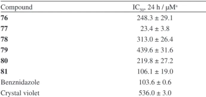
trypomastigote form of T. cruzi, and all of the substances were more effective than crystal violet. When compared to Bz, compound 77 was 4 times more active than the standard drug and compound 81 exhibited similar activity (Table 6).90
Scheme 6. Nor-α-lapachone 1,2,3-triazoles 66-68.88
Scheme 7. Nor-α-lapachone-based 1,2,3-triazole 71-74.88
Scheme 8.β-Lapachone-based 1,2,3-triazoles 78-81.90
Table 6. Activity of β-lapachone-based 1,2,3-triazoles 78-81 on T. cruzi
Compound IC50, 24 h / µMa
76 248.3 ± 29.1
77 23.4 ± 3.8
78 313.0 ± 26.4
79 439.6 ± 31.6
80 219.8 ± 27.2
81 106.1 ± 19.0
Benznidazole 103.6 ± 0.6
Crystal violet 536.0 ± 3.0
1,4-Naphthoquinone coupled to 1,2,3-triazole N-phthalimides (82-91) were recently prepared from brominated, chlorinated or unsubstituted quinones (Scheme 9).85 Compounds 82-91 were inactive against
T. cruzi and more studies regarding the mechanism of insertion of the 1,2,3-triazole ring into 1,4-naphthoquinone are necessary.
Meanwhile, 1,4-naphthoquinones with a direct insertion of a heterocyclic ring 1,2,3-triazole into the quinoidal structure were prepared, as shown in Scheme 10. Synthesis of the naphthoquinones coupled to 1,2,3-triazoles was initially reported by Nascimento et al. (Scheme 10).91 In
assays with trypomastigote forms of T. cruzi, the most active substances displayed IC50 values in the range of 10.9
to 80.2 µM (Table 7).85 Compounds 93 and 98 exhibited
IC50 values of 10.9 and 17.7 µM, respectively, and are thus
very promising structures. Further studies regarding their mechanism of action, cytotoxicity levels and in vivo activity are therefore necessary. It is important to note that the para-naphthoquinone 1,2,3-triazoles are easily obtained in only two steps from the starting material 1,4-naphthoquinone and both reactions have good to excellent yields.
Using the methodology described by the Pinto group,92
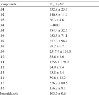
we prepared substituted nor-β-lapachones arylamino from nor-lapachol (4) at high yields (Figure 9), and these compounds were evaluated for anti-T. cruzi activity (Table 8).93,94 The trypanocidal activity of compounds 103,
108, 110, and 112-114 was higher than that of Bz, a drug commonly used to combat T. cruzi infections. Compound
112, which contained the bromine atoms, was the most active compound and exhibited an IC50 value of 24.9 µM.
Scheme 9. 1,4-Naphthoquinone-derived 1,2,3-triazoles 82-91.85
Scheme 10. Naphthoquinone-based 1,2,3-triazoles 93-100.91
Table 7. Effects of naphthoquinone-based 1,2,3-triazoles 93-100 on T. cruzi
Compound IC50 / µMa
93 10.9 ± 1.8
94 45.8 ± 5.1
95 492.2 ± 17.5
96 2005.7 ± 9.9
97 113.1 ± 5.7
98 17.7 ± 3.1
99 80.2 ± 5.4
100 67.6 ± 7.7
Benznidazole 103.6 ± 0.6
Crystal violet 536.0 ± 3.0
Table 10. Effects of the naphthoquinones 117-119 on epimastigote forms of T. cruzi (in µM)
Compounds IC50, 1 day IC50, 2 day IC50, 3 day IC50, 4 day
117 13.2 ± 2.2 12.4 ± 1.4 11.7 ± 1.5 12.7 ± 2.0
118 24.9 ± 1.8 21.8 ± 2.4 19.5 ± 2.4 18.3 ± 4.9
119 7.9 ± 1.3a 3.7 ± 0.3 3.0 ± 0.7 2.6 ± 0.3 aMean ± standard deviation from three independent experiments.
Table 9. Effects of the naphthoquinones 117-119 on T. cruzi
Compounds IC50 / µMa
117 641 ± 38
118 398 ± 56
119 158 ± 9
Benznidazole 103.6 ± 0.6
aMean ± standard deviation from three experiments performed in triplicate.
Table 8. Activity of nor-β-lapachone arylamino substituted compounds
101-116 on T. cruzi
Compounds IC50 / µMa
101 332.8 ± 23.3
102 140.8 ± 11.9
103 86.3 ± 4.6
104 > 4000
105 384.4 ± 52.5
106 952.5 ± 71.1
107 857.3 ± 96.4
108 88.2 ± 6.7
109 2517.9 ± 169.8
110 55.6 ± 4.6
111 1756.1 ± 91.8
112 24.9 ± 7.4
113 43.8 ± 7.4
114 59.6 ± 13.2
115 526.2 ± 80.5
116 156.2 ± 9.1
Benznidazole 103.6 ± 0.6
aMean ± standard deviation from three experiments performed in triplicate.
These structures represent an important starting point for the attainment of new trypanocidal compounds.
In a previous work,92 Silva et al. described the synthesis
of derivatives obtained from C-allyl lawsone, as shown in Scheme 11. These compounds exhibited activity against T. cruzi in both the bloodstream trypomastigote and epimastigote forms (Tables 9 and 10). The effects of compounds 117-119 on epimastigote proliferation were monitored for up to 4 days.
Compounds 117-119 derived from C-allyl lawsone were effective against the three forms of the parasite, and the intracellular amastigote was the most susceptible form.95 Transmission electron microscopy examination of
treated epimastigotes and bloodstream trypomastigotes revealed a drastic mitochondrial swelling with a washed-out matrix profile. Potent dose-dependent collapse of the mitochondrial membrane potential revealed by rhodamine 123 staining together with an inhibition of mitochondrial complex I-III activities and a reduction in succinate-induced oxygen consumption strongly corroborated the central role of the mitochondrion in these compounds’ mechanisms of action. Moreover, an
Scheme 11. Synthetic route for the attainment of methylated and iodinated naphthoquinones 117-119.92
increase in the production of hydrogen peroxide by this organelle in treated epimastigotes was also observed. However, some differences in the mode of action of naphthofuranquinones were apparent in epimastigotes and trypomastigotes. In the insect form, the trypanocidal effects of the compounds were a consequence of the parasite redox balance modulation, whereas in the bloodstream form, mitochondrial dysfunction affected energy transduction reactions, which compromised the protozoa’s bioenergy efficiency. Naphthoquinones interfere with electron flow at the inner mitochondrial membrane by diverting electrons away from ubiquinone. The oxidation of semiquinones back to quinones leads to the generation of reactive oxygen species that compromise the activity of complex I-III and oxygen consumption capability, which culminates in parasite death.95
In another set of experiments, the trypanocidal activity of sixteen 1,4-naphthoquinones was assessed on both T. cruzi trypomastigotes and epimastigotes (Figure 10 and Table 11).96 In the case of the naphthoquinones 120-134,
different assay conditions were used to analyse the effects on trypomastigotes. While all of the previous experiments were performed in the presence of 5% mouse blood and at 4 °C (Bz IC50 = 103.6 ± 0.6 µM) as previously mentioned,
Table 11. Effects of the naphthoquinones 120-134 on T. cruzi at 37 °C
Compound IC50 / µMa
120 0.79 ± 0.02
121 6.04 ± 0.35
122 63.02 ± 5.8
123 1.37 ± 0.03
124 2.17 ± 0.29
125 6.51 ± 0.48
126 0.16 ± 0.01
127 1.02 ± 0.29
128 2.15 ± 0.22
129 2.43 ± 0.50
130 1.25 ± 0.26
131 2.52 ± 0.37
132 0.85 ± 0.08
133 1.41 ± 0.15
134 1.38 ± 0.26
7 563.18 ± 83.28
Benznidazole 26.0 ± 4.0
aMean ± standard deviation from three experiments performed in triplicate.
Figure 10. Naphthoquinones 120-134 and lawsone (7).96
Four compounds were selected from this series for mode of action studies: the prototype naphthoquinone 120 and three juglone derivatives (126, 127 and 130).96 These four
compounds were effective against parasite proliferative forms (epimastigotes and intracellular amastigotes) and
endoplasmic reticulum surrounding reservosomes, which is indicative of autophagy. In addition, intense cytosolic vacuolisation, the formation of blebs in the flagellar membrane and the loss of cytosolic electron-density were also observed. The ultra-structural autophagic evidence suggests that the endoplasmic reticulum participates in the observed pre-autophagosomal formation.96
3. Conclusions
This review describes our efforts to develop an effective trypanocidal drug. Synthesis procedures and biological data regarding anti-T. cruzi activity were described and studies of the mechanism of action of these compounds were detailed to provide an overview of the progress made by our research group in collaboration with several researchers around the world. Among the quinones and derivatives investigated, naphthoimidazoles derived from β-lapachone presented promising biological activity together with low toxicity to the host cells, opening interesting perspectives for their investigation in vivo. On the other hand, naphthoquinones presenting different moieties in their structures showed distinct modes of action. It is well-known that quinones induce ROS production also in T. cruzi. Our previous data pointed to ROS generation as part of the naphtoquinones’ mechanism of action and the central role of the parasite mitochondrion, depending on the moiety linked to the quinoidal ring. In this scenario, as an example, a triazolic naphthoquinone led to discrete increase in ROS levels and
Figure 11. Transmission electron microscopy analysis of a T. cruzi epimastigote treated with compound 130. The treatment induced the appearance of membranous structures inside the mitochondrion (black thick arrows). N: nucleus; G: Golgi; FP: flagellar pocket; F: flagellum; K: kinetoplast. Bar: 0.5 µm.
did not compromise the mitochondrial functionality as well. The naphthofuranquinone and juglone derivatives strongly affected this organelle physiology interfering with the oxygen uptake and mitochondrial membrane potential. High amounts of ROS were produced by the mitochondrion of treated parasites culminating in T. cruzi death. Notwithstanding, many questions still remain unanswered about the molecular mechanisms involved in the trypanocidal effect of these compounds and their selectivity for different cellular structures in the protozoa, we hope that this review contributes to the development of new candidates for Chagas disease.
Acknowledgments
We wish to thank Conselho Nacional de Pesquisa (CNPq), Coordenadoria de Aperfeiçoamento de Pessoal de Nível Superior (CAPES), FAPEMIG and FAPERJ. Dr. E. N. da Silva Júnior thanks Programa Institucional de Auxílio à Pesquisa de Doutores Recém-Contratados and Universidade Federal de Minas Gerais. This paper is dedicated to the memory of our beloved Professor Antonio Ventura Pinto because of his intense commitment to the development of novel trypanocidal drugs. Prof. Ventura always believed in the potential of the quinoidal compounds, especially the structures obtained from lapachol. His passion for the study of the chemical reactivity of naphthoquinonoid compounds and discovering new reactions was a key point in our lives.
Eufrânio N. da Silva Júnior received his degree in chemistry from the Catholic University of Brasília (UCB). In 2007, he completed his MSc at the Fluminense Federal University (UFF) and in 2009 he concluded his PhD at the University of Brasilia (UnB). In 2010, he became Professor of Chemistry at the Federal University of Minas Gerais (UFMG). His research interests are focused on click chemistry reactions, asymmetric organocatalysis and on the synthesis of heterocyclic and naphthoquinoidal bioactive compounds. Currently, he is also interested in obtaining fluorescent substances for the study of pharmacological and DNA-binding properties.
on the synthesis and biological study of heterocyclic compounds besides the preparation of biosensors with application in molecular biology.
Rubem F. S. Menna-Barreto received his degree in Biology in Santa Ursula University (2003). In 2008, he completed his PhD in Cell and Molecular Biology at the Oswaldo Cruz Institute (FIOCRUZ) and after a postdoctoral period at the Federal University of Rio de Janeiro in Biochemistry at the Medical Biochemistry Institute, he became an associate researcher at the Oswaldo Cruz Foundation. His research interests are focused on parasitology, specially animal protozoology, Trypanosoma cruzi, chemotherapy, electron microscopy, mitochondrion, cell death, autophagy and naphthoquinones.
Solange L. de Castro received her degree in Industrial Chemistry from the Federal University of Rio de Janeiro (UFRJ). In 1991, she completed her PhD at the Oswaldo Cruz Institute (FIOCRUZ) in experimental chemotherapy of Chagas disease. She is a senior researcher at FIOCRUZ. Her research interests are focused on chemotherapy, with special interest in the studies about the trypanocidal activity and mechanism of action of naphthoquinones and derivatives.
References
1. Chagas, C.; Mem. Inst. Oswaldo Cruz1909, 1, 159.
2. World Health Organization (WHO); Sustaining the Drive to Overcome the Global Impact of Neglected Tropical Diseases, Second WHO Report on Neglected Tropical Diseases; WHO Press: Geneva, 2013.
3. http://www.who.int/neglected_diseases/diseases/chagas/en/ accessed in July 2014.
4. Hoare, C. A.; Wallace, F. G.; Nature1966, 244, 69.
5. Schofield, C. J.; Jannin, J.; Salvatella, R.; Trends Parasitol.
2006, 22, 583.
6. Dias, J. C.; Prata, A.; Correia, D.; Rev. Soc. Bras. Med. Trop.
2008, 41, 193.
7. Coura, J. R.; de Castro, S. L.; Mem. Inst. Oswaldo Cruz2002, 97, 3.
8. Coura, J. R.; Mem. Inst. Oswaldo Cruz2007, 102, 113. 9. Schmunis, G.; Yadon, Z. E.; Acta Trop.2010, 115, 14. 10. Coura, J. R.; Viñas, P. A.; Nature2010, 465, S6.
11. Hotez, P. J.; Dumonteil, E.; Woc-Colburn, L.; Serpa, J. A.; Bezek, S.; Edwards, M. S.; Hallmark, C. J.; Musselwhite, L. W.;
Flink, B. J.; Bottazzi, M. E.; PLoS Neglected Trop. Dis.2012, 6, e1498.
12. Bastos, C. J.; Aras, R.; Mota, G.; Reis, F.; Dias, J. P.; de Jesus, R. S.; Freire, M. S.; de Araújo, E. G.; Prazeres, J.; Grassi, M. F.; PLoS Neglected Trop. Dis.2010, 4, e711.
13. Noya, B. A.; Diaz-Bello, Z.; Colmenares, C.; J. Infect. Dis.
2010, 201, 1308.
14. Rassi Júnior, A.; Rassi, A.; Marin-Neto, J. A.; Lancet2010, 375, 1388.
15. Rassi Júnior, A.; Rassi, A.; Rezende, J. M.; Infect. Dis. Clin. North. Am.2012, 26, 275.
16. Coura, J. R.; Borges-Pereira, J.; Mem. Inst. Oswaldo Cruz2011, 106, 641.
17. Machado, F. S.; Jelicks, L. A.; Kirchhoff, L. V.; Shirani, J.; Nagajyothi, F.; Mukherjee, S.; Nelson, R.; Coyle, C. M.; Spray, D. C.; de Carvalho, A. C.; Guan, F.; Prado, C. M.; Lisanti, M. P.; Weiss, L. M.; Montgomery, S. P.; Tanowitz, H. B.; Cardiol. Rev.
2012, 20, 53.
18. Tarleton, R. L.; Trends Parasitol. 2003, 19, 447.
19. Higuchi, M. L.; Benvenuti, L. A.; Martins-Reis, M.; Metzger, M.; Cardiovasc. Res.2003, 60, 96.
20. Rocha, M. O.; Teixeira, M. M.; Ribeiro, A. L.; Expert Rev. Anti-Infect. Ther.2007, 5,727.
21. Marin-Neto, J. A.; Rassi Júnior, A.; Avezum Júnior, A.; Mattos, A. C.; Rassi, A.; Morillo, C. A.; Sosa-Estani, S.; Yusuf, S.; Mem. Inst. Oswaldo Cruz2009, 104, 319.
22. Rassi Júnior, A.; Rassi, A.; Marin-Neto, J. A.; Mem. Inst. Oswaldo Cruz.2009, 104, 152.
23. Marino, A. P.; Silva, A. A.; Santos, P. V.; Pinto, L. M.; Gazinelli, R. T.; Teixeira, M. M.; Lannes-Vieira, J.; Mem. Inst. Oswaldo Cruz2005, 100, 93.
24. Machado, F. S.; Dutra, W. O.; Esper, L.; Gollob, K. J.; Teixeira, M. M.; Factor, S. M.; Weiss, L. M.; Nagajyothi, F.; Tanowitz, H. B.; Garg, N. J.; Semin. Immunopathol.2012, 34, 753. 25. Brener, Z.; Cançado, J. R.; Galvão, L. M.; da Luz, Z. M.; Filardi,
L. S.; Pereira, M. E.; Santos, L. M.; Cançado, C. B.; Mem. Inst. Oswaldo Cruz1993, 88, 149.
26. Coura, J. R.; Mem. Inst. Oswaldo Cruz2009, 104, 549. 27. Viotti, R.; Vigliano, C.; Lococo, B.; Expert Rev. Anti-Infect.
Ther.2009, 7, 157.
28. Sarli, I. V.; Bocchi, A. E.; Lancet2010, 376, 768.
29. McKerrow, J. H.; Doyle, P. S.; Engel, J. C.; Podust, L. M.; Robertson, S. A.; Ferreira, R.; Saxton, T.; Arkin, M.; Kerr, I. D.; Brinen, L. S.; Craik, C. S.; Mem. Inst. Oswaldo Cruz 2009, 104, 263.
30. Apt, W.; Arribada, A.; Zulantay, I.; Solari, A.; Sánchez, G.; Mundaca, K.; Coronado, X.; Rodríguez, J.; Gil, L. C.; Osuna, A.; Ann. Trop. Med. Parasitol.2005, 99, 733. 31. Urbina, J. A.; Acta Trop. 2010, 115, 55.
Laydbak, J. U.; Chatelain, E.; Scandale, I.; Verlinde, C. L.; Charman, S. A.; Lepesheva, G. I.; Gelb, M. H.; Antimicrob. Agents Chemother.2012, 56, 4914.
33. Keenan, M.; Chaplin, J. H.; Alexander, P. W.; Abbott, M. J.; Best, W. M.; Khong, A.; Botero, A.; Perez, C.; Cornwall, S.; Thompson, R. A.; White, K. L.; Shackleford, D.; Koltun, M.; Chiu, F. C.; Morizzi, J.; Ryan, E.; Campbell, M.; von Geldern, T. W.; Scandale, I.; Chatelain, E.; Charman, S. A.; J. Med. Chem.
2013, 56, 10158.
34. Bahia, M. T.; de Andrade, I. M.; Martins, T. A.; do Nascimento, A. F.; Diniz, L. F.; Caldas, I. S.; Talvani, A.; Trunz, B. B.; Torreele, E.; Ribeiro, I.; PLoS Neglected Trop. Dis.2012, 6, 1870.
35. Soeiro, M. N.; Werbovetz, K., Boykin, D. W.; Wilson, W. D.; Wang, M. Z.; Hemphill, A.; Parasitology2013, 140, 929. 36. Bahia, M. T.; Nascimento, A. F.; Mazzeti, A. L.; Marques,
L. F.; Gonçalves, K. R.; Mota, L. W.; Diniz, L. D.; Caldas, I. S.; Talvani, A.; Shackleford, D. M.; Koltun, M.; Saunders, J.; White, K. L.; Scandale, I.; Charman, S. A.; Chatelain, E.; Antimicrob. Agents Chemother.2014, 4362.
37. Sajid, M.; Robertson, S. A.; Brinen, L. S.; McKerrow, J. H.; Adv. Exp. Med. Biol.2011, 712, 100.
38. Jonckers, T. H.; van Miert, S.; Cimanga, K.; Bailly, C.; Colson, P.; J. Med. Chem. 2002, 45, 3497.
39. Buckner, F.; Yokoyama, K.; Lockman, J.; Aikenhead, K.; Ohkanda, J.; Proc. Natl. Acad. Sci. U. S. A. 2003, 100, 15149. 40. Buckner, F. S.; Adv. Exp. Med. Biol. 2008, 625, 61.
41. Sealey-Cardona, M.; Cammerer, S.; Jones, S.; Ruiz-Perez, L. M.; Brun, R.; Antimicrob. Agents Chemother.2007, 51, 2123. 42. Szajnman, S. H.; Ravaschino, E. L.; Docampo, R.; Rodriguez,
J. B.; Bioorg. Med. Chem. Lett.2005, 15, 4685.
43. Hucke, O.; Gelb, M. H.; Verlinde, C. L.; Buckner, F. S.; J. Med. Chem.2005, 48, 5415.
44. Kraus, J. M.; Verlinde, C. L.; Karimi, M.; Lepesheva, G. I.; Gelb, M. H.; J. Med. Chem.2009, 52, 1639.
45. Schormann, N.; Senkovich, O.; Walker, K.; Wright, D. L.; Anderson, A. C.; Proteins2008, 73, 889.
46. Urbina, J. A.; Curr. Pharm. Des.2002, 8, 287.
47. Duschak, V. G.; Couto, A. S.; Recent Pat. Anti-Infect. Drug Discovery2007, 2, 19.
48. Ioset, J. R.; Curr. Org. Chem.2008, 12, 643.
49. Araujo, M. S.; Martins-Filho, O. A.; Pereira, M. E.; Brener, Z.; J. Antimicrob. Chemother.2000, 5, 819.
50. Diniz, L. F.; Caldas, I. S.; Guedes, P. M.; Crepalde, G.; de Lana, M.; Carneiro, C. M.; Talvani, A.; Urbina, J. A.; Bahia, M. T.; Antimicrob. Agents Chemother. 2010, 54, 2979.
51. Bustamante, J. M.; Craft, J. M.; Crowe, B. D.; Ketchie, S. A.; Tarleton, R. L.; J. Infect. Dis. 2014, 209, 150.
52. Cencig, S.; Coltel, N.; Truyens, C.; Carlier, Y.; Int. J. Antimicrob. Agents2012, 40, 527.
53. Batista, D. G.; Batista, M. M.; Oliveira, G. M.; Britto, C. C.;
Rodrigues, A. C.; Stephens, C. E.; Boykin, D. W.; Soeiro, M. N. C.; PLoS One2011, 6, e22155.
54. Grosso, N. L.; Alarcon, M. L.; Bua, J.; Laucella, S. A.; Riarte, A.; Fichera, L. E.; Parasitology2013, 140, 1225. 55. Nwaka, S.; Hudson, A.; Nat. Rev. Drug Discovery2006, 5, 941. 56. Arenas, P.; J. Ethnopharmacol.1987, 21, 279.
57. Bastien, J. W.; J. Ethnopharmacol.1983, 8, 97.
58. Hazra, B.; Das Sarma, M.; Sanyal, U.; J. Chromatogr. B: Anal. Technol. Biomed. Life Sci.2004, 812, 259.
59. Nawrat, C. C.; Moody, C. J.; Angew. Chem., Int. Ed.2014, 53, 2056.
60. Lima, N. M. F.; Correia, C. S.; Leon, L. L.; Machado, G. M. C.; Madeira, M. F.; Santana, A. E. G.; Goulart, M. O. F.; Mem. Inst. Oswaldo Cruz2004, 99, 757.
61. Ramírez-Macías, I.; Marín, C.; Es-Samti, H.; Fernández, A.; Guardia, J. J.; Zentar, H.; Agil, A.; Chahboun, R.; Alvarez-Manzaneda, E.; Sánchez-Moreno, M.; Parasitol. Int.2012, 61, 405.
62. Grellier, P.; Maroziene, A.; Nivinskas, H.; Sarlauskas, J.; Aliverti, A.; Cenas, N.; Arch. Biochem. Biophys.2010, 494, 32. 63. Powis, G.; Pharmacol. Ther.1987, 35, 57.
64. O’Brien, P. J.; Chem.-Biol. Interact.1991, 80, 1.
65. Castro, S. L.; Pinto, M. C. F. R.; Pinto, A. V.; Microbios1994, 78, 83.
66. Pinto, A. V.; Neves Pinto, C.; Pinto, M. C. F. R.; Santa Rita, R. M.; Pezzella, C.; de Castro, S. L.; Arzneim. Forsch.1997, 47, 74.
67. Neves-Pinto, C.; Dantas, A. P.; Moura, K. C. G.; Emery, F. S.; Polequevitch, P. F.; Pinto, M. C. F. R.; de Castro, S. L.; Pinto, A. V.; Arzneim. Forsch.2000, 50, 1120.
68. de Moura, K. C. G.; Emery, F. S.; Pinto, C. N.; Pinto, M. C. F. R.; Dantas, A. P.; Salomão, K.; de Castro, S. L.; Pinto, A. V.; J. Braz. Chem.Soc.2001, 12, 325.
69. de Moura, K. C. G.; Salomão, K.; Menna-Barreto, R. F. S.; Emery, F. S.; Pinto, M. C. F. R.; Pinto, A. V.; de Castro, S. L.; Eur. J. Med. Chem.2004, 39, 639.
70. Menna-Barreto, R. F.; Corrêa, J. R.; Pinto, A. V.; Soares, M. J.; de Castro, S. L.; Parasitol. Res. 2007, 101, 895.
71. Menna-Barreto, R. F.; Henriques-Pons, A.; Pinto, A. V.; Morgado-Diaz, J. A.; Soares, M. J.; de Castro, S. L.; J. Antimicrob. Chemother. 2005, 56, 1034.
72. Menna-Barreto, R. F.; Corrêa, J. R.; Cascabulho, C. M.; Fernandes, M. C.; Pinto, A. V.; Soares, M. J.; de Castro, S. L.; Parasitology2009, 136, 499.
73. Menna-Barreto, R. F.; Beghini, D. G.; Ferreira, A. T.; Pinto, A. V.; de Castro, S. L.; Perales, J.; J. Proteomics2010, 73, 2306. 74. Salomão, K.; de Souza, E. M.; Carvalho, A. S.; Silva, E. F.;
Fraga, C. A. M.; Barbosa, H. S.; de Castro, S. L.; Antimicrob. Agents Chemother. 2010, 54, 2023.
76. Carneiro, P. F.; Pinto, M. C. F. R.; Coelho, T. S.; Cavalcanti, B. C.; Pessoa, C.; de Simone, C. A.; Nunes, I. C. K.; de Oliveira, N. M.; de Almeida, R. G.; Pinto, A. V.; de Moura, K. C. G.; da Silva, P. A.; da Silva Júnior, E. N.; Eur. J. Med. Chem.2011, 46, 4521.
77. Viegas Júnior, C.; Danuello, A. C.; Bolzani, V. S.; Barreiro, E. J.; Fraga, C. A. M.; Curr. Med. Chem.2007, 14, 1829. 78. Fieser, L. F.; Fieser, M.; J. Am. Chem. Soc.1948, 70, 3215. 79. Rostovtsev, V. V.; Green, L. G.; Fokin, V. V.; Sharpless, K. B.;
Angew. Chem., Int. Ed. 2002, 41, 2596.
80. da Silva Júnior, E. N.; Menna-Barreto, R. F. S.; Pinto, M. C. F. R.; Silva, R. S. F.; Teixeira, D. V.; de Souza, M. C. B. V.; de Simone, C. A.; de Castro, S. L.; Ferreira, V. F.; Pinto, A. V.; Eur. J. Med. Chem.2008, 43, 1774.
81. da Silva Júnior, E. N.; de Melo, I. M. M.; Diogo, E. B. T.; Costa, V. A.; de Souza Filho, J. D.; Valença, W. O.; Camara, C. A.; de Oliveira, R. N.; de Araujo, A. S.; Emery, F. S.; dos Santos, M. R.; de Simone, C. A.; Menna-Barreto, R. F. S.; de Castro, S. L.; Eur. J. Med. Chem.2012, 52, 304.
82. Cavalli, A.; Bolognesi, M. L.; J. Med. Chem. 2009, 52, 7339. 83. Fernandes, M. C.; da Silva, E. N.; Pinto, A. V.; de Castro, S. L.;
Menna-Barreto, R. F.; Parasitology2012, 139, 26. 84. Salomon, C. J.; J. Pharm. Sci.2012, 101, 888.
85. Diogo, E. B. T.; Dias, G. G.; Rodrigues, B. L.; Guimarães, T. T.; Valença, W. O.; Camara, C. A.; de Oliveira, R. N.; da Silva, M. G.; Ferreira, V. F.; de Paiva, Y. G.; Goulart, M. O. F.; Menna-Barreto, R. F. S.; de Castro, S. L.; da Silva Júnior, E. N.; Bioorg. Med. Chem. 2013, 21, 6337.
86. Romanha, A. J.; de Castro, S. L.; Soeiro, M. N.; Lannes-Vieira, J.; Ribeiro, I.; Talvani, A.; Bourdin, B.; Blum, B.; Olivieri, B.; Zani, C.; Spadafora, C.; Chiari, E.; Chatelain, E.; Chaves, G.; Calzada, J. E.; Bustamante, J. M.; Freitas-Júnior, L. H.; Romero, L. I.; Bahia, M. T.; Lotrowska, M.; Soares, M.; Andrade, S. G.; Armstrong, T.; Degrave, W.; Andrade, Z. A.; Mem. Inst. Oswaldo Cruz2010, 105, 233.
87. de Castro, S. L.; Emery, F. S.; da Silva Júnior, E. N.; Eur. J. Med. Chem.2013, 69, 678.
88. Guimarães, T. T.; Pinto, M. C. F. R.; Lanza, J. S.; Melo, M. N.; do Monte-Neto, R. L.; de Melo, I. M. M.; Diogo, E. B. T.; Ferreira, V. F.; Camara, C. A.; Valença, W. O.; de Oliveira, R. N.; Frézard, F.; da Silva Júnior, E. N.; Eur. J. Med. Chem.
2013, 63, 523.
89. Kolb, H. C.; Finn, M. G.; Sharpless, K. B.; Angew. Chem., Int. Ed. 2001, 40, 2004.
90. da Silva Júnior, E. N.; Guimarães, T. T.; Menna-Barreto, R. F. S.; Pinto, M. C. F. R.; de Simone, C. A.; Pessoa, C.; Cavalcanti, B. C.; Sabino, J. R.; Andrade, C. K. Z.; Goulart, M. O. F.; de Castro, S. L.; Pinto, A. V.; Bioorg. Med. Chem.2010, 18, 3224.
91. do Nascimento, W. S.; Camara, C. A.; de Oliveira, R. N.; Synthesis2011, 20, 3220.
92. Silva, R. S. F.; Costa, E. M.; Trindade, U. L. T.; Teixeira, D. V.; Pinto, M. C. F. R.; Santos, G. L.; Malta, V. R. S.; de Simone, C. A.; Pinto, A. V.; de Castro, S. L.; Eur. J. Med. Chem.2006, 41, 526.
93. da Silva Júnior, E. N.; de Souza, M. C. B. V.; Pinto, A. V.; Pinto, M. C. F. R.; Goulart, M. O. F.; Barros, F. W. A.; Pessoa, C.; Costa-Lotufo, L. V.; Montenegro, R. C.; de Moraes, M. O.; Ferreira, V. F.; Bioorg. Med. Chem.2007, 15, 7035.
94. da Silva Júnior, E. N.; de Souza, M. C.; Fernandes, M. C.; Menna-Barreto, R. F.; Pinto, M. C. F. R.; de Assis, L. F.; de Simone, C. A.; Andrade, C. K.; Pinto, A. V.; Ferreira, V. F.; de Castro, S. L.; Bioorg. Med. Chem.2008, 16, 5030.
95. Menna-Barreto, R. F.; Goncalves, R. L.; Costa, E. M.; Silva, R. S.; Pinto, A. V.; Oliveira, M. F.; de Castro, S. L.; Free Radical Biol. Med. 2009, 47, 644.
96. Salomão, K.; Santana, N. A.; Molina, M. T.; de Castro, S. L.; Menna-Barreto, R. F. S.; BMC Microbiol.2013, 13, 196.
Submitted: March 5, 2014