ISSN/$–see front matter © 2013 Sociedade Brasileira de Ortopedia e Traumatologia. Published by Elsevier Editora Ltda. All rights reserved. Rev Bras Ortop. 2013;48(1):108-110
www.rbo.org.br/
*Corresponding author at: Av. Itália, 2556, Bairro Cariru, Ipating, MG, Brazil. Phone/fax: (+55 31) 38252529. CEP: 35160-115.
E-mail: [email protected]; [email protected]
article info
Article history:
Received August 25 2011 Approved December 21 2011
Keywords:
Acromioclavicular articulation /surgery
Clavicle/injuries Dislocations
Case Report
Acromioclavicular dislocation type VI associated with
diaphyseal fracture of the clavicle
Evander Azevedo Grossi,
1*Roberto Araújo Macedo
21Orthopedist and Shoulder Surgeon at Hospital Márcio Cunha and Hospital Unimed Vale do Aço, Ipatinga, MG, Brazil. 2Orthopedist and Shoulder/Elbow Surgeon at Hospital Márcio Cunha and Hospital Unimed Vale do Aço, Ipatinga, MG, Brazil.
Work performed at the São Francisco Xavier Foundation, Hospital Márcio Cunha, Ipatinga, MG, Brazil.
a b s t r a c t
The purpose is to present a very unusual case of the acromioclavicular joint inferior dislocation associated with the clavicle fracture. It concerns to a young patient who had a bike fall and had this type of pathology, had been operated and obtained excellent clinic result. The literature mentions many cases of subcoracoide dislocation, but there are only two subacromial similar to ours. The case is described, a literary revision is done and discussed and the treatment is discussed.
© 2013 Sociedade Brasileira de Ortopedia e Traumatologia. Published by Elsevier Editora Ltda. All rights reserved.
Introduction
Acromioclavicular dislocation is one of the most ancient traumatic pathological conditions recorded in the literature, and its frequency is 5 to 10 times greater among males.1 The
most common cause of its occurrence is a fall on the shoulder with the arm adducted, but indirect trauma may also injure this joint. 1
Acromioclavicular dislocations are classified into six types, according to Rockwood et al.,2 and type VI is divided into
subacromial and subcoracoid.
The first case of inferior dislocation of the clavicle (subcoracoid) was described by Patterson in 1967.3 In this, the
mechanism for subcoracoid dislocation comes from forced hyperabduction of the arm in association with retraction of the scapula. This mechanism may injure the accessory nerve. Injuries of this type generally occur in multiple trauma cases, and may also be associated with fractures of the acromion, clavicle, scapula and/or ribs.4
For subacromial dislocation, there is no specific description of a trauma mechanism, but from the characteristics of lesions that occur in fractures of the clavicular diaphysis, associated with inferior dislocation of the acromioclavicular joint, it can be suggested that subacromial dislocations are caused by segmental fractures, in which there would be several traumatic events affecting the clavicle.5
Rev Bras Ortop. 2013;48(1):108-110
109
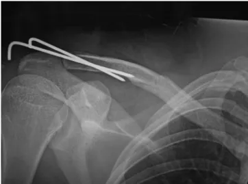
Fig. 2 - Reduction of the acromioclavicular dislocation and Fig. 1A - Incomplete diaphyseal fracture of the right clavicle
and inferior acromioclavicular dislocation.
Fig. 1B - Image obtained via radioscopy, showing the deviation.
We report a rare case of a young adult who presented subacromial acromioclavicular dislocation in association with an incomplete diaphyseal fracture, with deviation of the clavicle, which was treated surgically with a satisfactory result.
Case report
The patient was a 19-year-old male who suffered a fall from a bicycle and was taken to an emergency service with a single complaint of pain in the right clavicle. Clinical examination did not show any local neurovascular deficit or associated pathological conditions. Digital radiographic examination of the clavicle showed an incomplete fracture of the diaphysis with posteroinferior deviation and consequent dislocation of the lateral extremity of the clavicle to the subacromial space and posteriorly (Figs. 1A and 1B). Clinically, there was a prominence in the medial
region of the acromion and an inferior deviation of the distal extremity of the clavicle. The patient complained about a lot of local pain and significant limitation of movements.
Surgical treatment was chosen, and surgical reduction was performed on the acromioclavicular dislocation and on the clavicular fracture, with a manual maneuver. Since the diaphyseal fracture was of greenstick type, the reduction was easy and stable. The coracoclavicular ligaments were complete, while the acromioclavicular ligament was injured. The acromioclavicular ligament was sutured and this joint was then fixed using two Steinmann wires (Fig. 2). These wires were subsequently removed, seven weeks later, and the patient was referred for physiotherapy. The digital radiological examination facilitated the evaluation of the two pathological conditions, and there was no need for a different examination for each condition.5 An excellent clinical and radiographic
result was presented after 12 months (Fig. 3).
110
Rev Bras Ortop. 2013;48(1):108-110Discussion
In reviewing the bibliography, we found 11 articles reporting on acromioclavicular dislocation type VI, of which five cases presented dislocation inferior to the coracoid3,4,6-8 and six were
subacromial. Of the latter, three articles related to recurrent acromioclavicular dislocation,8-10 two presented associations
with diaphyseal fractures of the clavicle11,12 and one showed an
association with incarcerated subacromial dislocation.1
Koka and D’Arcy11 described the presence of subacromial
acromioclavicular dislocation associated with complete diaphyseal fracture, with deviation. Surgical treatment similar to what is described in our report was performed, with an excellent functional result.
Juhn and Simonian12 described a case of a greenstick
diaphyseal fracture of the clavicle with good evolution after clinical treatment. On the day after the trauma, the patient already presented good range of motion, and for this reason conservative treatment was chosen.
Like Juhn and Simonian,12 we agree that acromioclavicular
dislocations should be classified as type VI-A for subacromial and type VI-B for subcoracoid.
Therefore, like some authors,1,2,4,8,10,11 we believe that
surgical treatment for inferior lesions of the acromioclavicular joint ensures a satisfactory functional result.
Conflicts of interest
The authors declare that there was no conflict of interests in conducting this study.
R E F E R E N C E S
1. Singh B, Singh S, Saraf N, Farooque K , Sharma V. Unusual mechanism of injury with segmental fracture clavicle. J Orthop Surg. 2007;6(1):7.
2. Rockwood Jr CA, Williams GR, Young DC. Disorders of the acromioclavicular joint. In: Rockwood Jr CA, Matsen FA. The shoulder. Philadelphia: Saunders; 1998. p. 483-553.
3. Patterson WR. Inferior dislocation of the distal end of the clavicle: a case report. J Bone Joint Surg.1967;49(6):1184-6. 4. McPhee IB. Inferior dislocation of the outer end of the
clavicle. J Trauma. 1980;20(8):709-10.
5. Grossi EA. Fratura segmentar da clavícula. Rev Bras Ortop. 2011;46(6):733-5.
6. Torrens C, Mestre C, Perez P, Marin M. Subcoracoid dislocation of the distal end of the clavicle. Clin Orthop. 1998;(348)121-3. 7. Schwarz N, Kuderna H. Inferior acromioclavicular
separation report of an unusual case. Clin Orthop Relat Res. 1988;(234):28-30.
8. Sage J. Recurrent inferior dislocation of the clavicle at the acromioclavicular joint: a case report. Am J Sports Med. 1982;10(8)145-6.
9. Naumann T. Der seltene Fall einer habituellen lateralen Claviculaluxation nach dorsal subacromial (Fallbericht). Z Orthop. 1986;124(1):34-5.
10. Leppilahti J, Jalovaara P. Recurrent inferior dislocation of the acromioclavicular joint: a report of two cases. J Shoulder Elbow Surg. 2001;10(4):387-8.
11. Koka SR, D’Arcy JC. Inferior (subacromial) dislocation of the outer end of the clavicle. Injury. 1993; 24(3)210-1.
12. Juhn MS, Simonian PT. Type VI acromioclavicular separation with middle-third clavicle fracture in an ice hockey player. Clin J Sport Med. 2002;12(5):315-7.